

EOS (3D Skeletal System Imaging)
Diagnosis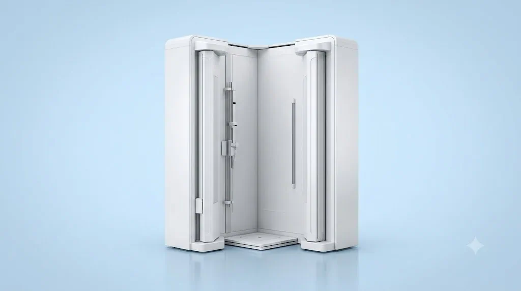
Quick answer
EOS 3D Skeletal System Imaging is a low-dose X-ray technology that captures standing, whole-body frontal and side images to assess spinal and lower-limb alignment in three dimensions. At Acibadem in Turkey, it supports orthopedic evaluation, treatment planning, and follow-up for posture, deformity, and alignment-related conditions.
EOS (3D Skeletal System Imaging)
EOS is an advanced X-ray imaging system designed to provide detailed views of the spine, pelvis and lower limbs while a patient is standing in a natural, weight-bearing position. This is important because many orthopedic conditions, including spinal curvature and leg alignment differences, can look different when the body is supporting its own weight.
Unlike conventional imaging that may focus on one area at a time, EOS can capture frontal and side views of a large part of the skeleton in a single examination. The images can also support three-dimensional measurements, helping Orthopedics and Traumatology specialists evaluate alignment with greater precision.
At Acibadem International, EOS imaging may be used as part of a personalised diagnostic and treatment-planning pathway. It can help specialists understand how the spine, hips, knees and lower limbs work together, supporting earlier detection, clearer clinical decisions and more informed follow-up care.
What Is EOS Imaging in Plain Language?
EOS is a low-dose, X-ray-based imaging technology for assessing the skeleton in an upright position. During the examination, the patient stands inside an open imaging cabin while images are taken from the front and side at the same time.
The system is particularly useful for evaluating posture, spinal curves and the alignment of the legs. It can provide full-length images of the body in a functional position, meaning the position people commonly use when standing and moving through daily life.
For selected patients, EOS software can use the acquired images to create three-dimensional models and measurements. These measurements may help specialists assess spinal balance, pelvic position, limb length and lower-limb alignment. The exact information obtained depends on the clinical question and the patient’s individual assessment.
EOS does not replace every type of X-ray, MRI or CT scan. Instead, it is a valuable diagnostic option when specialists need a detailed overview of skeletal alignment, especially under weight-bearing conditions.
How EOS 3D Skeletal System Imaging Works
EOS uses two X-ray imaging systems positioned at right angles to each other. As the imaging units move vertically, they capture simultaneous frontal and side images of the patient. This approach allows the specialist to assess the body’s alignment in both planes from one examination.
Because the scan is performed while standing, the images can show how gravity and body weight affect the spine and lower limbs. This can be particularly meaningful in conditions where posture, balance or joint loading is part of the clinical picture.
The images are reviewed by qualified specialists, who may use dedicated planning software to take measurements and, where appropriate, build a three-dimensional representation of the skeleton. These findings are considered together with your symptoms, physical examination, medical history and any previous scans or reports.
Low-Dose Imaging Approach
EOS is designed to use a lower radiation dose than many conventional radiographic imaging approaches for comparable full-body skeletal examinations. Radiation exposure still needs to be considered carefully for every patient, particularly for children, adolescents, people requiring repeat imaging and pregnant patients.
Your medical team will recommend imaging only when it is clinically appropriate. If EOS is suitable, specialists will select the examination protocol that best supports your diagnostic needs while following radiation-safety principles.
Weight-Bearing Assessment
Many musculoskeletal concerns are best assessed while the body is upright. A standing examination can help reveal spinal balance, pelvic tilt, knee alignment and the relationship between the hips, knees and ankles under normal loading conditions.
This whole-body perspective can be especially valuable when treatment planning requires more than an isolated image of one joint or spinal segment.
Clinical Applications
EOS imaging supports the work of Orthopedics and Traumatology teams caring for children, adolescents and adults with a range of spinal and lower-limb concerns. The technology may be used for diagnosis, treatment planning, surgical planning or ongoing monitoring, depending on the patient’s needs.
| Clinical application | How EOS may help | Conditions or concerns commonly assessed |
|---|---|---|
| Spinal alignment assessment | Provides frontal and side views of the spine while standing, helping evaluate overall posture and balance. | Scoliosis, kyphosis, lordosis, spinal imbalance and posture-related concerns. |
| Scoliosis monitoring | Supports repeat assessment of spinal curvature and alignment when ongoing imaging is clinically needed. | Adolescent idiopathic scoliosis, adult scoliosis and other spinal curvature conditions. |
| Whole-body balance evaluation | Helps specialists assess the relationship between the head, spine, pelvis and lower limbs in an upright position. | Forward-leaning posture, compensatory posture changes and complex spinal deformity. |
| Lower-limb alignment | Creates full-length, weight-bearing views that can support measurement of leg axes and joint alignment. | Knock knees, bow legs, varus or valgus alignment, limb malalignment and gait-related concerns. |
| Limb-length assessment | May help measure and compare lower-limb lengths as part of orthopedic evaluation. | Suspected leg-length discrepancy, developmental differences and post-traumatic changes. |
| Pre-treatment and surgical planning | Provides measurements that can support personalised planning for selected orthopedic interventions. | Spinal deformity surgery, corrective osteotomy planning and selected lower-limb procedures. |
| Post-treatment follow-up | Helps clinicians monitor alignment after treatment when imaging follow-up is appropriate. | Brace treatment, spinal surgery, limb correction and orthopedic rehabilitation follow-up. |
Not every patient with back pain, joint pain or postural concerns needs EOS imaging. Your specialist will decide whether EOS, conventional X-ray, MRI, CT or another diagnostic method is most appropriate for your condition.
Conditions EOS May Help Specialists Evaluate
EOS can be particularly useful when specialists need to understand the relationship between several areas of the musculoskeletal system rather than assessing a single body part in isolation. For example, a spinal curve may affect pelvic position, while pelvic position can influence lower-limb mechanics and standing balance.
In children and adolescents, EOS may be considered for selected patients who require careful spinal monitoring during growth. In adults, it may support the assessment of degenerative spinal changes, complex posture concerns, lower-limb deformities or planning for orthopedic treatment.
Potential uses may include the evaluation of:
- Scoliosis and other spinal deformities
- Kyphosis, including excessive rounding of the upper back
- Lordosis and changes in the curve of the lower back
- Spinal imbalance or changes in upright posture
- Pelvic tilt and pelvic asymmetry
- Leg-length discrepancy
- Varus and valgus knee alignment
- Bow-leg or knock-knee patterns
- Lower-limb mechanical axis concerns
- Complex musculoskeletal alignment before or after selected orthopedic procedures
EOS findings are never interpreted in isolation. Your care team considers the images alongside your symptoms, mobility, pain level, clinical examination and medical history. This complete approach helps ensure that recommendations are based on your individual needs rather than imaging results alone.
Benefits and Advantages for Patients
One of the main strengths of EOS is its ability to provide a detailed, full-body perspective while the patient is standing. For patients with spinal or lower-limb alignment concerns, this can provide clinically useful information that may not be available from an image taken while lying down.
EOS can also be beneficial when repeat imaging is required over time, such as during scoliosis monitoring or postoperative follow-up. Its low-dose approach may help reduce radiation exposure compared with certain traditional full-body radiographic studies, while still providing the information needed for orthopedic decision-making.
| Patient benefit | What it can mean for your care |
|---|---|
| Weight-bearing imaging | Your spine and lower limbs are assessed while you are standing, helping show alignment under everyday body weight. |
| Whole-body perspective | Specialists can evaluate how the spine, pelvis, hips, knees and ankles relate to one another. |
| Simultaneous front and side views | Images from two directions can support a more complete assessment during one examination. |
| Three-dimensional planning support | For selected cases, specialised software can help clinicians obtain detailed alignment measurements and plan care. |
| Low-dose technology | EOS is designed to support diagnostic imaging with lower radiation exposure than many conventional full-body X-ray approaches. |
| Non-invasive experience | No injections, sedation or surgical preparation are usually required for a standard EOS examination. |
| Useful for follow-up | May support consistent monitoring when your specialist recommends repeated assessment of skeletal alignment. |
| Personalised treatment planning | Clear measurements can help your orthopedic team discuss treatment options that fit your anatomy, symptoms and goals. |
What to Expect During an EOS Examination
Your EOS journey begins with a clinical review. Your specialist may ask about your symptoms, previous injuries, family history, growth history in children and adolescents, earlier scans, prior treatments and any surgery you have had. Please bring previous imaging reports and medical records whenever possible.
Before the examination, you may be asked to remove metal objects or clothing items that could affect image quality, such as belts, jewellery or garments with metal fasteners. The imaging team will explain what you need to do and help you prepare comfortably.
During the scan, you will stand inside the EOS imaging area in a specific position. Depending on the study requested, your feet, hands or arms may need to be placed in a particular way. It is important to remain still for the short time required to obtain clear images.
The EOS system is open rather than tunnel-shaped. Many patients find this reassuring, especially those who feel uncomfortable in enclosed spaces. A trained radiology professional will guide you throughout the examination and can answer practical questions before the scan begins.
The image acquisition itself is usually brief. In most cases, you can return to your usual activities immediately afterwards. There is generally no recovery time, no contrast injection and no need for anesthesia for a standard EOS scan.
After the Examination
Your images are reviewed by the appropriate radiology and orthopedic specialists. Depending on the clinical pathway, your treating doctor may discuss the findings with you during a follow-up consultation or incorporate them into your wider treatment plan.
If the examination identifies an alignment concern, your specialist may recommend observation, physiotherapy, bracing, additional diagnostic tests, medication for related symptoms or a surgical consultation. The best next step depends on the diagnosis, severity, age, symptoms, activity level and personal treatment goals.
Safety, Radiation Awareness and Accreditation
EOS is an X-ray-based technology, so it involves ionising radiation. However, it is designed to provide low-dose imaging for appropriate orthopedic applications. Your clinical team will only recommend the examination when its expected diagnostic value outweighs any potential risk.
Radiation safety is especially important for children, adolescents and patients who may need imaging more than once. Our teams follow established safety protocols and use imaging principles aimed at keeping exposure as low as reasonably achievable while obtaining clinically useful images.
It is essential to tell your care team if you are pregnant, think you may be pregnant or are trying to conceive. Your doctors and radiology team will assess whether the examination should be postponed, modified or replaced with another imaging method.
Acibadem International coordinates care through JCI-accredited hospitals, where imaging services are delivered by trained professionals under established quality, patient-safety and infection-prevention standards. Your examination is performed as part of a coordinated clinical process, with appropriate communication between radiology, Orthopedics and Traumatology, and other relevant specialties.
As with any diagnostic examination, image quality and usefulness can depend on the patient’s ability to maintain the required standing position. If standing is difficult due to pain, balance concerns or mobility limitations, please tell the team in advance so they can assess the safest and most suitable option.
Why EOS Matters for International Patients
Travelling abroad for orthopedic care can involve many practical questions, particularly when you need diagnostic imaging, specialist consultations and treatment planning within a limited timeframe. EOS can support an efficient evaluation pathway by providing extensive skeletal alignment information during a single, non-invasive imaging visit when clinically appropriate.
At Acibadem International, our international patient team helps coordinate your journey before, during and after your visit. You can share your existing reports and imaging in advance for preliminary review. This allows our specialists to understand your history and advise whether an EOS assessment may be relevant to your evaluation.
During your visit, international patient coordinators can help organise appointments, support communication in your preferred language and guide you through the clinical process. Your team will explain what the examination involves, what information it may provide and how the results may influence your treatment plan.
For patients who require a multidisciplinary assessment, EOS results can be shared with the relevant specialists involved in your care. This may include orthopedic surgeons, spine specialists, pediatric orthopedists, physiotherapists, rehabilitation specialists and radiologists, depending on your condition.
After your consultation, the team can help you understand the recommended next steps and provide documentation to support continuity of care when you return home. If follow-up imaging is needed, your specialist will explain the preferred timing and whether follow-up can be coordinated locally, remotely or during a future visit.
Is EOS Right for You?
EOS may be an appropriate option if you have a known or suspected spinal deformity, concerns about posture or balance, lower-limb alignment differences, leg-length discrepancy or a need for detailed weight-bearing skeletal assessment. It may also be recommended as part of planning or follow-up for selected orthopedic treatments.
Suitability always depends on your individual medical assessment. Your specialist will consider your symptoms, age, diagnosis, prior imaging, ability to stand safely and the specific information needed to guide your care.
If you are planning treatment in Türkiye, you can submit your available medical reports, X-rays, MRI or CT results through a consultation request. Our international patient team will coordinate review by the appropriate specialist and help determine whether EOS 3D Skeletal System Imaging may be part of your personalised orthopedic evaluation.
Related Treatments
Speak with our medical team
Find out how this technology could support your diagnosis or treatment.