3 Tesla MRI: High-Resolution Imaging and Patient Safety
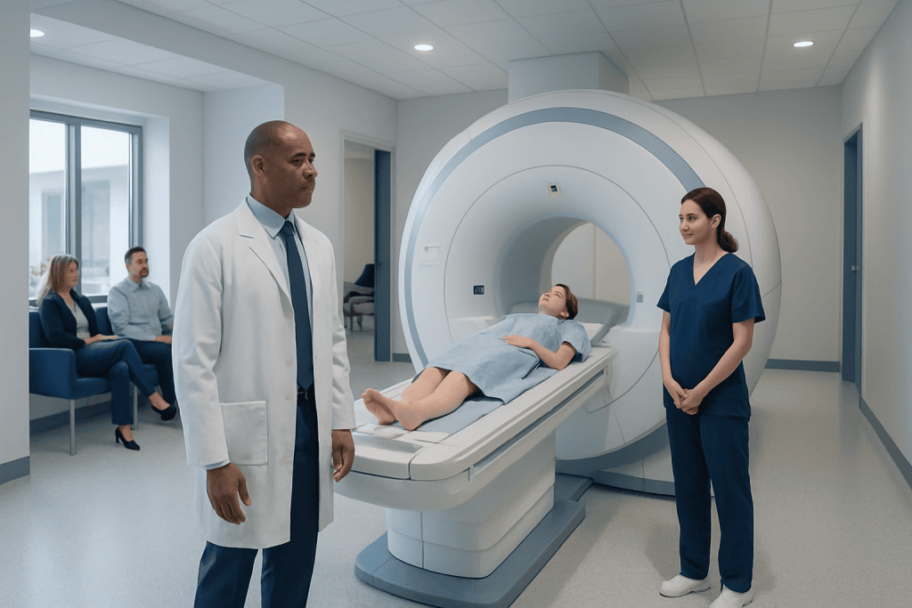
3 Tesla MRI provides high-resolution images that can help doctors detect subtle changes in the brain, spine, joints, blood vessels, and internal organs. MRI does not use ionizing radiation; it relies on a magnetic field and radiofrequency energy.
Key Takeaways
- 3 Tesla MRI provides high-resolution images that can help doctors detect subtle changes in the brain, spine, joints, blood vessels, and internal organs.
- MRI does not use ionizing radiation; it relies on a magnetic field and radiofrequency energy.
- Safety screening is essential because some implants, devices, metal fragments, or medical conditions may affect whether a 3T MRI is appropriate.
- Some MRI exams use gadolinium-based contrast material, which is generally well tolerated but requires extra caution in certain patients.
- The scan is painless, but patients may hear loud sounds and may need to remain still for clear images.
- Patients should inform the radiology team about implants, allergies, kidney disease, pregnancy, prior surgeries, and any concerns about confined spaces.
3 Tesla MRI is an advanced imaging technology that uses a strong magnetic field to create detailed images of organs, tissues, blood vessels, joints, and the nervous system. With proper screening and expert supervision, it is a safe and valuable diagnostic tool for many patients.
Overview: What Is 3 Tesla MRI?
3 Tesla MRI, often called 3T MRI, is a type of magnetic resonance imaging that uses a magnetic field strength of 3 tesla. A tesla is a unit used to measure magnetic field strength. Compared with many standard MRI scanners that operate at 1.5 tesla, a 3T MRI scanner can produce a stronger signal, which may allow more detailed images or shorter scan times depending on the examination.
MRI creates images without using ionizing radiation such as X-rays. Instead, it uses a strong magnet, radiofrequency pulses, and advanced computer processing to show the structure of organs and tissues. This makes MRI especially useful for viewing soft tissues such as the brain, spinal cord, muscles, ligaments, cartilage, blood vessels, and many abdominal and pelvic organs.
The main advantage of 3T MRI is image clarity. Higher resolution can help radiologists and treating physicians evaluate small anatomical structures and subtle abnormalities more confidently. However, the best scanner is not always the strongest one; the most appropriate MRI depends on the clinical question, the patient’s medical history, and the expertise of the imaging team.
How 3T MRI Works
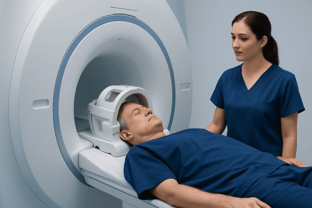
During an MRI scan, the patient lies on a table that moves into the scanner. The magnetic field temporarily aligns hydrogen atoms in the body, especially those in water and fat. Radiofrequency energy is then applied, and when the atoms return to their usual state, they release signals that are captured by the scanner and converted into images.
A 3T MRI scanner receives a stronger signal than lower-field systems. This stronger signal can be used in different ways. In some exams, it helps create thinner slices and sharper images. In others, it may help reduce the total time needed for the scan. The radiology team selects the imaging protocol based on the body part being examined and the reason for the scan.
Special coils are placed around or near the body area being imaged. These coils act like antennas, receiving the MRI signal. For example, a head coil is used for brain MRI, while dedicated coils may be used for the knee, shoulder, breast, spine, or abdomen. The combination of scanner strength, coil design, software, and radiologist expertise determines the final quality and usefulness of the exam.
Common Uses of 3 Tesla MRI
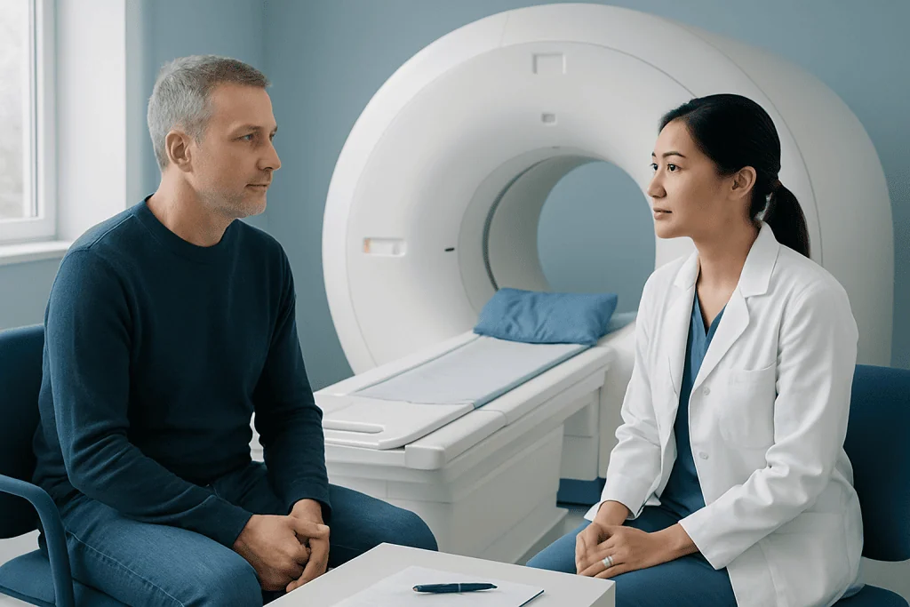
3 Tesla MRI may be recommended when doctors need detailed information about soft tissues or complex anatomy. It is widely used in neurological imaging, including examinations of the brain, pituitary gland, inner ear structures, or spine. In some cases, it may help evaluate conditions such as tumors, inflammation, stroke-related changes, multiple sclerosis, epilepsy-related abnormalities, disc disease, or nerve compression.
In orthopedics and sports medicine, 3T MRI can provide detailed images of cartilage, ligaments, tendons, muscles, and bone marrow. It may be used to assess knee injuries, shoulder problems, hip conditions, wrist and ankle injuries, or unexplained joint pain. In some joint examinations, high-resolution imaging can help identify small tears or early cartilage changes.
3T MRI is also used in body imaging and vascular imaging. Depending on the clinical need, it may support evaluation of the liver, prostate, uterus, ovaries, kidneys, heart, breasts, or blood vessels. Examples of common uses include:
- Brain and spine imaging for neurological symptoms
- Musculoskeletal imaging for joint or tendon injuries
- Prostate MRI for assessment of prostate abnormalities
- Breast MRI in selected patients when clinically appropriate
- MR angiography to visualize blood vessels
- Abdominal and pelvic MRI for organ and soft tissue evaluation
Patient Safety and Screening
MRI is considered a safe imaging method for many patients because it does not use ionizing radiation. However, the strong magnetic field requires careful safety screening. Before the scan, patients are asked about implants, surgeries, metal exposure, medical devices, allergies, kidney disease, pregnancy, and other health conditions. This screening is an essential part of the MRI process.
Some devices and implants are labeled as MRI-safe or MRI-conditional, meaning they may be scanned only under specific conditions. Examples include certain pacemakers, defibrillators, cochlear implants, neurostimulators, insulin pumps, aneurysm clips, surgical clips, stents, and joint replacements. Patients should provide device cards, operative notes, or manufacturer information whenever possible so the radiology team can verify safety.
Loose metal objects must not enter the MRI room because they can move toward the magnet with force. Patients are usually asked to remove jewelry, watches, hearing aids, hairpins, glasses, removable dental appliances, body piercings, coins, keys, and phones. Clothing with metallic fibers or fasteners may also need to be replaced with a gown. People who have worked with metal, such as welders or metalworkers, may require additional checks for tiny metal fragments, especially near the eyes.
Pregnancy should be reported before an MRI. MRI may be used during pregnancy when the expected medical benefit outweighs potential concerns, but the decision is individualized. Contrast material is generally avoided during pregnancy unless it is considered essential. The patient’s doctor and radiology team can explain the safest approach for the specific situation.
Contrast Material: When It Is Used
Some 3T MRI examinations are performed without contrast, while others use a gadolinium-based contrast agent injected into a vein. Contrast can help highlight blood vessels, inflammation, tumors, infection, scar tissue, or areas with abnormal blood flow. The decision to use contrast depends on the body area being examined and the clinical question.
Gadolinium-based contrast agents are generally well tolerated. Some patients may experience mild, temporary effects such as a cool feeling during injection, nausea, headache, or discomfort at the injection site. Allergic-like reactions are uncommon, but patients should tell the radiology team if they have had a previous reaction to MRI contrast or any severe allergies.
Kidney function is important when contrast is considered. Patients with significant kidney disease, those on dialysis, or those with certain medical conditions may need blood tests or a different imaging plan. The radiology team balances the benefit of contrast-enhanced images with the patient’s overall safety. Patients should never hesitate to ask why contrast is recommended and whether a non-contrast exam would be sufficient.
What Patients Can Expect During the Scan
A 3T MRI scan is painless. The patient lies on a movable table, and the technologist helps position the body comfortably. Cushions, straps, or supports may be used to reduce movement and improve image quality. Depending on the exam, a coil is placed around the head, joint, chest, abdomen, or other body part.
MRI scanners make loud tapping, knocking, or buzzing sounds while images are being taken. Patients are given ear protection, such as earplugs or headphones. The technologist monitors the patient throughout the scan and can communicate through an intercom. Many scanners also have a call button that allows the patient to alert the team if assistance is needed.
Remaining still is very important because movement can blur the images. Some scans require brief breath-holds, particularly for chest or abdominal imaging. The total scan time varies depending on the body area and whether contrast is used, but many MRI examinations take between about 20 and 60 minutes. If a patient feels anxious in confined spaces, this should be discussed before the appointment; options may include extra preparation, relaxation techniques, a support person when permitted, or medication prescribed by a doctor.
Benefits and Limitations of 3T MRI
The main benefit of 3T MRI is the potential for very detailed imaging. This can be especially helpful when doctors need to evaluate small structures, early disease changes, complex joints, or delicate neurological anatomy. In selected cases, the increased signal can improve diagnostic confidence and help guide treatment planning.
However, 3T MRI is not automatically the best choice for every patient or every condition. Higher-field MRI can be more sensitive to certain artifacts, such as movement, metal-related distortion, or changes near air-filled spaces. Some implants that are permitted in a 1.5T scanner may not be approved for 3T scanning, or they may require special conditions. The radiology team determines whether 3T MRI is appropriate and safe.
Image interpretation also depends on the clinical context. MRI findings must be considered together with symptoms, physical examination, laboratory tests, and other imaging when needed. A detailed MRI report is a tool that helps the treating physician make decisions, but it does not replace a full medical evaluation.
Preparing for a 3 Tesla MRI and When to Speak With a Doctor
Preparation depends on the type of MRI. For many scans, patients can eat, drink, and take regular medications unless instructed otherwise. For some abdominal or pelvic exams, fasting or bowel preparation may be requested. Patients should bring prior imaging reports, laboratory results, device information, and a list of medications when available.
Before the appointment, patients should inform the imaging center if they have any implanted device, metal fragment, kidney disease, pregnancy, breastfeeding, severe claustrophobia, or a history of contrast reaction. They should also mention recent surgery or any implanted ports, pumps, clips, or stimulators. Clear communication helps the team plan a safe and efficient examination.
A doctor should be consulted if a patient is unsure whether MRI is the right test, if symptoms are worsening, or if previous imaging results need explanation. After the scan, the radiologist interprets the images and sends a report to the referring physician, who discusses the results and next steps with the patient. At Acibadem International, multidisciplinary specialists and JCI-accredited hospitals diagnose and treat a wide range of conditions using advanced imaging, including 3T MRI, for international patients when clinically appropriate.
Frequently asked questions
Is 3 Tesla MRI safe?
For many patients, 3 Tesla MRI is safe when proper screening is completed. It does not use ionizing radiation, but the strong magnetic field can interact with certain implants, devices, or metal fragments. Patients should answer all safety questions carefully and provide device information whenever possible.
How is 3T MRI different from 1.5T MRI?
A 3T MRI scanner has a stronger magnetic field than a 1.5T scanner. This can provide a stronger signal, which may allow higher-resolution images or shorter scan times in some examinations. However, 3T MRI is not always suitable for every patient, especially if an implanted device is approved only under specific MRI conditions.
Does a 3 Tesla MRI hurt?
The scan itself is painless. Patients may feel some pressure from positioning aids or hear loud knocking sounds during imaging, but ear protection is provided. If contrast is used, there may be a brief needle stick for the intravenous injection.
Can patients with implants have a 3T MRI?
Some implants are compatible with 3T MRI, while others are not or require special conditions. Examples include pacemakers, neurostimulators, cochlear implants, aneurysm clips, and some pumps or surgical devices. The radiology team must verify the exact device model and MRI conditions before the scan.
Is contrast always needed for 3T MRI?
No. Many MRI scans are performed without contrast. Contrast is used when it can provide important additional information, such as showing blood flow, inflammation, tumors, or infection. The decision depends on the medical question and the patient’s health history.
What should a patient do if they are claustrophobic?
Patients who feel anxious in enclosed spaces should tell the doctor or imaging center before the appointment. The team may suggest practical comfort measures, additional explanation, scheduling adjustments, or prescribed medication when appropriate. Good preparation often helps patients complete the scan successfully.
How soon are 3T MRI results available?
The timing varies by facility, urgency, and complexity of the examination. A radiologist reviews the images and prepares a report for the referring doctor. The referring doctor then explains the results in the context of the patient’s symptoms and overall medical evaluation.
References
- American College of Radiology
- Radiological Society of North America
- U.S. Food and Drug Administration
- European Society of Radiology
- International Society for Magnetic Resonance in Medicine
This article is for general information only and is not a substitute for professional medical advice. Please consult a qualified doctor about your individual situation.
Joint and spine care in Turkey — expert assessment & treatment
JCI-accredited · board-certified surgeons · reply within 24h
Add us as a Preferred Source to see more of our trusted health content across Google Search, AI Overviews and Discover.