

Cerebral Angiography: Step-by-Step Guide to Brain Vessel Imaging

Cerebral angiography uses contrast dye and X-ray imaging to show brain blood vessels clearly. It is often used when other scans do not provide enough detail or when treatment may be needed.
Key Takeaways
- Cerebral angiography uses contrast dye and X-ray imaging to show brain blood vessels clearly.
- It is often used when other scans do not provide enough detail or when treatment may be needed.
- The procedure is usually performed through a small catheter placed in the wrist or groin.
- Most people go home the same day, but brief monitoring afterward is important.
- Doctors use cerebral angiography to investigate aneurysms, vessel narrowing, malformations, and some strokes.
Cerebral angiography is a specialized imaging procedure that creates detailed pictures of the blood vessels in the brain and neck. It helps doctors diagnose and sometimes treat conditions affecting blood flow, vessel shape, or bleeding risk.
Overview of Cerebral Angiography
Cerebral angiography, also called a cerebral angiogram, is a medical imaging procedure used to examine the blood vessels of the brain, head, and neck. It provides highly detailed, real-time images that can show how blood flows through arteries and veins. For many vascular brain conditions, it remains one of the most precise diagnostic tools available.
During the procedure, a doctor inserts a thin tube called a catheter into a blood vessel, most often in the wrist or groin, and guides it toward the arteries supplying the brain. A contrast dye is then injected so the vessels appear clearly on X-ray images. This allows specialists to look for narrowing, blockages, abnormal connections, bulges, or bleeding sources.
Cerebral angiography may be recommended after other imaging tests such as CT, MRI, or ultrasound suggest a possible blood vessel problem but do not answer every question. It is especially useful when exact vessel anatomy matters for treatment planning. In some cases, the same session can lead directly to minimally invasive treatment through interventional neuroradiology procedures.
Why It Is Done
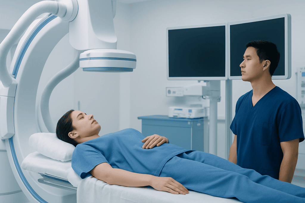
Doctors order cerebral angiography when they need a close look at the brain’s circulation. The test can help confirm a diagnosis, define the size and location of a blood vessel abnormality, or guide a treatment decision. Because it shows fine vessel detail, it can reveal problems that may not be fully visible on standard noninvasive imaging.
It is commonly used to investigate suspected aneurysms, arteriovenous malformations, blood vessel narrowing, inflammation of vessels, and some causes of bleeding in or around the brain. It may also be used to assess blood flow after a stroke or before surgery. If a patient has symptoms such as sudden severe headache, unexplained neurological problems, or findings concerning for brain aneurysm, angiography may help clarify the next steps.
In selected cases, cerebral angiography is not only diagnostic but therapeutic. Through the same catheter, a specialist may be able to close an abnormal vessel, treat a vascular malformation, or support treatment of an aneurysm. This combined diagnostic-and-treatment approach can reduce delays and may avoid open surgery for some patients.
How to Prepare and What Happens Step by Step

Before the procedure, the care team reviews the person’s medical history, medications, allergies, kidney function, and any past reactions to contrast dye. Patients are often asked not to eat or drink for several hours beforehand. Blood-thinning medicines may need to be adjusted, but only under medical guidance. The team also checks whether the patient is pregnant or may be pregnant, since X-rays are involved.
On arrival, an intravenous line is placed, and the patient changes into a hospital gown. Monitoring equipment is used to track heart rate, blood pressure, and oxygen levels. A mild sedative may be given to help the patient relax, though many people remain awake and can communicate during the test. The skin over the access site, usually the wrist or groin, is cleaned and numbed with local anesthetic.
The doctor then inserts the catheter into the artery and carefully guides it toward the vessels leading to the brain. Contrast dye is injected while a series of X-ray images is taken. Some patients notice a brief warm sensation, a metallic taste, or a flushed feeling when the dye enters the bloodstream. These sensations usually pass quickly. The imaging part often takes less than an hour, though total time can be longer depending on complexity.
After imaging is complete, the catheter is removed and pressure or a closure device is used to help prevent bleeding. The patient is monitored for a period of time to make sure the access site is stable and there are no immediate complications. Many people return home the same day, while others may stay longer if observation or additional treatment is needed.
What Conditions Cerebral Angiography Can Help Diagnose
Cerebral angiography is especially valuable for conditions involving the shape, flow, or integrity of brain blood vessels. It can show aneurysms, which are weak or bulging areas in an artery wall, and help doctors understand their exact size and neck shape. That information is important if treatment such as aneurysm treatment is being considered.
The test can also identify arteriovenous malformations and fistulas, where arteries and veins connect abnormally. These conditions may increase the risk of bleeding, seizures, or neurological symptoms. Angiography helps map the abnormal vessels in detail, which is essential for treatment planning. It may also be used when there is concern for arteriovenous malformation (AVM).
Another important use is evaluating narrowing or blockage in arteries that supply the brain. This may be relevant in stroke evaluation, repeated transient ischemic attacks, or reduced blood flow to certain brain regions. In some situations, angiography is part of planning stroke treatment or assessing whether a vessel problem contributed to symptoms.
Less commonly, the procedure can help investigate inflammation of blood vessels, unusual vascular tumors, or unexplained bleeding seen on other scans. Because angiography gives dynamic, real-time information about blood flow, it can answer questions that static images cannot always resolve.
Risks, Safety, and Recovery
Cerebral angiography is generally safe when performed by an experienced team, but like all invasive procedures it carries some risks. Possible complications include bruising or bleeding at the catheter site, allergic reaction to contrast dye, temporary changes in kidney function, infection, or damage to the blood vessel. There is also a small risk of stroke because the catheter is used inside the arterial system.
Healthcare teams reduce these risks by using careful technique, monitoring throughout the procedure, and checking the patient’s suitability beforehand. Patients should tell their doctor about kidney disease, diabetes, contrast allergy, bleeding disorders, or medicines that affect clotting. Being open about the full medical history helps the team plan safely.
Recovery instructions usually include resting for the remainder of the day, drinking fluids if advised, and avoiding heavy lifting or strenuous activity for a short period. Mild soreness or bruising at the access site can happen and often improves on its own. The care team explains how to keep the puncture site clean and what symptoms should prompt urgent medical attention.
Patients are typically told to seek help right away if they develop significant bleeding, worsening swelling, severe headache, weakness, trouble speaking, chest pain, shortness of breath, or signs of infection such as fever and increasing redness at the access site. Although serious complications are uncommon, quick evaluation is important if warning signs appear.
Understanding Results and Treatment Options
After the procedure, a specialist reviews the images to look at vessel shape, blood flow patterns, and any signs of abnormality. Results may be discussed the same day in urgent situations, while full interpretation in other cases may take longer. The findings help determine whether the blood vessels are normal or whether further care is needed.
If the angiogram shows an aneurysm, narrowing, clot, or abnormal vessel connection, treatment recommendations depend on the exact diagnosis, location, symptom pattern, and overall health of the patient. Some people only need monitoring and follow-up imaging. Others may benefit from medicines, lifestyle changes, or a minimally invasive catheter-based procedure.
In some situations, the doctor may proceed directly to treatment during the same session if this has been planned in advance and consent has been given. Examples include closing a bleeding-prone vessel or reducing blood flow to an abnormal connection. This can be especially useful in complex cases involving stroke or other urgent vascular conditions.
For patients who need further evaluation or treatment, care is often coordinated by neurologists, neurosurgeons, and interventional specialists. Acibadem International’s multidisciplinary specialists and JCI-accredited hospitals diagnose and treat cerebral vascular conditions for international patients, with treatment plans tailored to the individual’s findings and needs.
When to See a Doctor and Common Questions to Ask
A person may be referred for cerebral angiography if they have symptoms or scan findings suggesting a blood vessel problem in the brain. These may include sudden severe headache, unexplained neurological symptoms, recurrent transient weakness or numbness, or evidence of bleeding, aneurysm, or vascular malformation on CT or MRI. The test is also used when doctors need a more exact map of blood vessels before surgery or catheter-based treatment.
It is helpful for patients to ask why the test is needed, what alternatives exist, whether treatment might be done during the same procedure, and how long recovery will take. They may also want to discuss risks related to kidney disease, blood thinners, or prior contrast reactions. Good communication can reduce anxiety and help patients prepare.
Patients should contact their healthcare team after the procedure if they have questions about medicines, returning to exercise, driving, or wound care. If the access site starts bleeding or if new neurological symptoms develop, they should seek immediate medical care. Timely follow-up helps ensure a smooth recovery and clear planning for any next steps.
Frequently asked questions
Is cerebral angiography the same as a CT angiogram or MR angiogram?
No. CT angiography and MR angiography are noninvasive imaging tests, while cerebral angiography uses a catheter inserted into an artery. Cerebral angiography usually provides more detailed and dynamic images, which can be important when diagnosis is uncertain or treatment is being planned.
Does cerebral angiography hurt?
Most people do not feel significant pain during the procedure. They may feel a brief sting from the local anesthetic and temporary warmth when contrast dye is injected. Mild soreness or bruising at the wrist or groin afterward is common and usually improves within a few days.
How long does a cerebral angiogram take?
The imaging procedure itself often takes less than an hour, but the full visit is longer because of preparation and recovery monitoring. In more complex cases, especially if treatment is performed at the same time, the procedure can take longer. The care team can give a more personalized estimate beforehand.
Will the patient be asleep during cerebral angiography?
Many patients stay awake but receive medication to help them relax. Local anesthetic is used to numb the catheter insertion site. In certain situations, deeper sedation or anesthesia may be recommended, depending on the patient's condition and the planned procedure.
What are the main risks of cerebral angiography?
Possible risks include bleeding or bruising at the catheter site, contrast reaction, kidney strain, and blood vessel injury. There is also a small risk of stroke because the procedure involves the arteries that supply the brain. The team takes careful precautions to reduce these risks as much as possible.
Can someone go home the same day after cerebral angiography?
Yes, many people go home the same day after a period of observation. Some patients may stay longer if they had a more complex procedure, need additional treatment, or have other medical concerns. The decision depends on the procedure details and the person's overall condition.
References
- American Heart Association
- National Institute of Neurological Disorders and Stroke
- Radiological Society of North America
- Society of NeuroInterventional Surgery
- National Health Service
This article is for general information only and is not a substitute for professional medical advice. Please consult a qualified doctor about your individual situation.
Explore treatments in Turkey — costs, top hospitals & a free quote
JCI-accredited · board-certified surgeons · reply within 24h
Add us as a Preferred Source to see more of our trusted health content across Google Search, AI Overviews and Discover.
Check your numbers in seconds
BMI, calories, due date, blood pressure and 30+ more clinical calculators — free, instant, doctor-reviewed ranges.

More from the Health Library



Related Specialists

Dr. Ersan Şensoy
Anesthesiology
Dyt. Pakize Gizem Akgül
Nutrition & Diet
Prof. Dr. Ufuk Aydınlı
Orthopedic Surgery & Traumatology
Prof. Dr. Tuğçin Bora Polat
Pediatric Cardiology