Cancerous Moles: Signs That a Mole May Be Melanoma

A mole that changes in size, shape, color, or texture should be checked by a doctor. The ABCDE rule can help identify warning signs of possible melanoma.
Key Takeaways
- A mole that changes in size, shape, color, or texture should be checked by a doctor.
- The ABCDE rule can help identify warning signs of possible melanoma.
- Melanoma may also appear as a new dark spot, a sore that does not heal, or a lesion that itches or bleeds.
- Diagnosis usually involves a skin examination and a biopsy of suspicious tissue.
- Sun protection and regular self-checks can help lower risk and support early detection.
Cancerous moles may be an early sign of melanoma, a serious type of skin cancer that often begins as a new spot or a change in an existing mole. Knowing what changes to watch for can help people seek medical advice early, when treatment is often most effective.
Overview
Cancerous moles are skin lesions that may represent melanoma, the most serious form of skin cancer. Not every unusual mole is melanoma, and many moles are harmless. However, a mole that looks different from others or begins to change over time deserves medical attention.
Melanoma develops in melanocytes, the cells that produce pigment in the skin. It can arise in an existing mole or appear as a completely new spot. Although melanoma is less common than some other skin cancers, it is more likely to spread if not found early.
Early recognition is important because melanoma is often highly treatable when detected at an early stage. A patient-friendly way to think about this is simple: a stable mole is usually less concerning, while a mole that evolves should be assessed by a qualified doctor, often a dermatologist.
Symptoms and Warning Signs
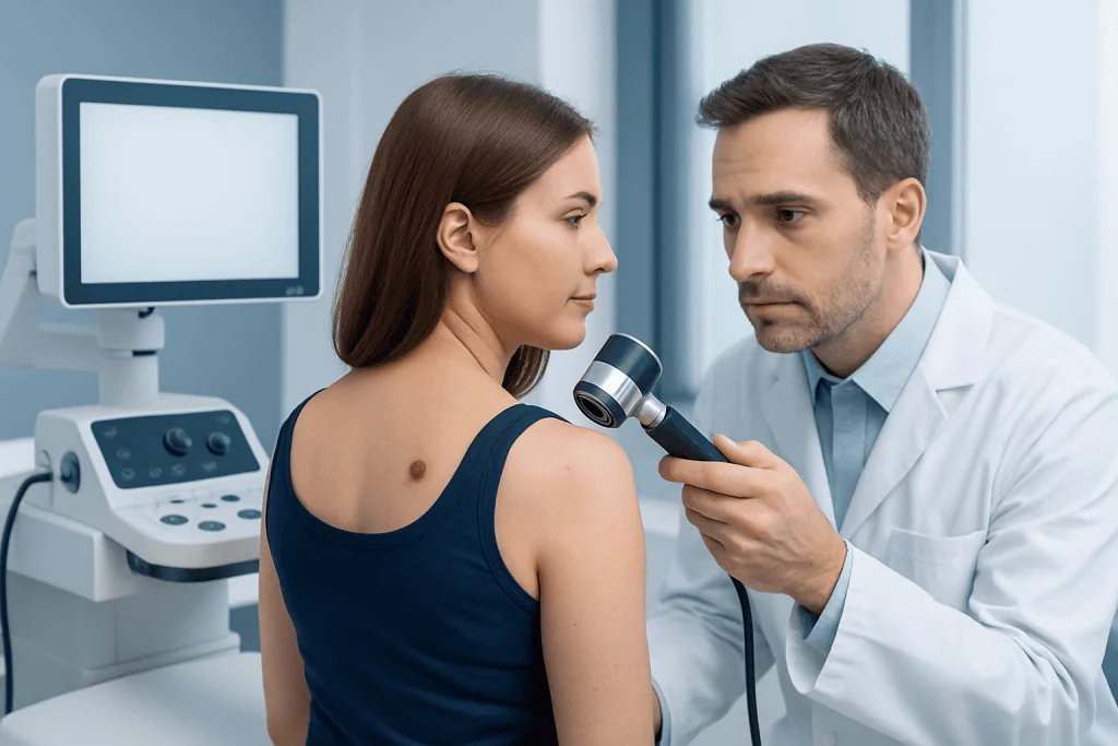
The most widely used guide for spotting possible melanoma is the ABCDE rule. This helps people notice changes that may suggest a cancerous mole rather than a benign one.
- A – Asymmetry: one half does not match the other.
- B – Border: edges are irregular, blurred, or ragged.
- C – Color: the mole has multiple shades such as brown, black, tan, red, white, or blue.
- D – Diameter: the spot is larger than about 6 mm, although melanomas can be smaller.
- E – Evolving: the mole changes in size, shape, color, height, or symptoms.
Another useful clue is the “ugly duckling” sign. This means one mole looks noticeably different from a person’s other moles. Because most people have a pattern to their moles, a lesion that stands out can be worth checking even if it does not meet every ABCDE feature.
Other symptoms can include itching, tenderness, crusting, oozing, bleeding, or a sore that does not heal. Melanoma may also appear under a nail, on the scalp, on the soles of the feet, or in other less visible areas. These spots can be easy to miss, which is why regular skin checks are helpful.
Causes and Risk Factors

Melanoma develops when pigment-producing skin cells grow in an abnormal and uncontrolled way. The exact cause is often complex, but ultraviolet (UV) radiation from sunlight or tanning beds is a major risk factor. UV exposure can damage the DNA in skin cells, increasing the chance of cancerous change over time.
Some people have a higher risk than others. Risk factors include fair skin, light eyes, red or blond hair, a history of sunburns, frequent intense sun exposure, or use of indoor tanning devices. Having many moles, large congenital moles, or atypical moles can also raise risk.
Family and personal medical history matter as well. A person who has had melanoma before, or who has a close relative with melanoma, may be more likely to develop it. A weakened immune system may also increase vulnerability. Even so, melanoma can occur in people without obvious risk factors, which is why attention to changing moles remains important for everyone.
How Doctors Diagnose a Suspicious Mole
Diagnosis usually begins with a careful skin examination. A dermatologist or another qualified doctor asks when the mole appeared, whether it has changed, and whether there are symptoms such as itching or bleeding. The doctor also checks the rest of the skin for other unusual lesions.
Dermoscopy is often used during the exam. This is a noninvasive tool that magnifies the skin and helps the doctor see patterns and structures not visible to the naked eye. While dermoscopy can improve assessment, it does not replace tissue testing when a lesion looks suspicious.
If melanoma is suspected, the mole is usually removed fully or partly for a biopsy. A pathologist then examines the tissue under a microscope to confirm whether cancer cells are present. If melanoma is diagnosed, additional tests may be recommended depending on the depth and features of the tumor and whether there is concern that it has spread as part of a broader melanoma evaluation.
Treatment Options for Melanoma
Treatment depends on how deep the melanoma is, whether it has spread, and the person’s overall health. For early melanoma, surgery is usually the main treatment. The goal is to remove the cancer completely along with a margin of normal-looking skin around it. This is commonly done with skin cancer surgery.
If there is concern that melanoma may have spread to nearby lymph nodes, a doctor may recommend additional staging procedures or surgery. In more advanced cases, treatment may include medicines that target cancer cells or help the immune system attack them. These approaches can be planned within a broader medical oncology program.
Some patients may also need radiation therapy in selected situations, such as when cancer has spread or when there is a specific need to reduce the risk of local recurrence. This may be part of care guided by radiation oncology. Treatment plans are individualized, and doctors often use a multidisciplinary approach to choose the safest and most appropriate option.
Patients commonly worry that every suspicious mole means advanced cancer, but this is not the case. Many removed moles are benign, and many melanomas found early are treated successfully with prompt care. The most helpful step is timely assessment rather than waiting to see if a changing spot settles on its own.
Prevention and Self-Care
Prevention focuses mainly on protecting the skin from UV damage. This includes seeking shade when the sun is strongest, wearing protective clothing, and using a broad-spectrum sunscreen with appropriate sun protection. Tanning beds are best avoided because they expose the skin to concentrated UV radiation.
Regular self-checks can support early detection. Many doctors suggest examining the skin once a month in good light, using a mirror for hard-to-see areas such as the back, scalp, and soles of the feet. Taking photos of moles can make it easier to notice subtle changes over time.
People with many moles, atypical moles, a strong family history, or previous melanoma may benefit from regular professional skin examinations. A doctor can advise how often checkups are needed. Self-care is not a substitute for medical evaluation, but it is a valuable habit that can help people identify concerning changes sooner.
When to See a Doctor
A doctor should be consulted if a mole changes in color, size, shape, or texture, or if a new spot looks unusual. Bleeding, crusting, itching, pain, or a sore that does not heal are also good reasons to arrange an appointment. It is especially important to seek medical advice if one lesion looks clearly different from all the others.
Urgency does not mean panic. Most skin changes are not melanoma, but it is safest to have a suspicious lesion assessed promptly rather than monitored without guidance. Early evaluation can bring reassurance when a mole is harmless and support faster treatment when it is not.
People who have many moles or a personal or family history of skin cancer may wish to ask about regular dermatology follow-up. Near the end of the care pathway, international patients may also choose centers such as Acibadem International, where multidisciplinary specialists in JCI-accredited hospitals evaluate and treat melanoma and other skin cancers.
Frequently asked questions
How can a person tell if a mole may be cancerous?
A mole may be suspicious if it changes over time or shows ABCDE features: asymmetry, irregular borders, uneven color, larger diameter, or evolution. A lesion that bleeds, itches, or looks different from other moles should also be checked by a doctor.
Are all dark or irregular moles melanoma?
No. Many dark, raised, or uneven-looking moles are benign. Even so, only a qualified medical assessment, and sometimes a biopsy, can reliably determine whether a mole is harmless or cancerous.
Can melanoma develop in a new spot rather than an old mole?
Yes. Melanoma can arise in an existing mole, but it can also appear as a completely new spot on the skin. That is why both new lesions and changing moles deserve attention.
What happens if a doctor thinks a mole could be melanoma?
The doctor usually examines the lesion closely and may use dermoscopy to look at skin structures in more detail. If the mole is suspicious, a biopsy is commonly recommended so the tissue can be examined under a microscope.
Can melanoma be cured if it is found early?
Early melanoma is often treated very effectively, most commonly with surgery. Outcomes are generally better when diagnosis happens before the cancer has spread, which is why prompt evaluation of changing moles matters.
Who should have regular skin checks?
People with many moles, atypical moles, fair skin, frequent sun exposure, a history of severe sunburns, or a personal or family history of melanoma may benefit from regular professional skin exams. A doctor can recommend an appropriate follow-up schedule based on individual risk.
References
- World Health Organization
- American Academy of Dermatology
- National Cancer Institute
- American Cancer Society
- National Health Service
This article is for general information only and is not a substitute for professional medical advice. Please consult a qualified doctor about your individual situation.
Melanoma in Turkey — costs, top hospitals & a free quote
JCI-accredited · board-certified surgeons · reply within 24h
Add us as a Preferred Source to see more of our trusted health content across Google Search, AI Overviews and Discover.

Medical Oncology Department
Medical treatment of cancer with chemotherapy, immunotherapy and targeted therapies under a multidisciplinary tumor board.
60 specialists in this unitMore from the Health Library

Related Specialists
Prof. Dr. Abdullah Büyükçelik
Medical Oncology
Dr. Kadir Yavaşoğlu
Orthopedic Surgery & Traumatology
Dr. Zeki Ermiş
Ophthalmology
Assoc. Prof. Dr. Berkin Toker
Orthopedic Surgery & Traumatology