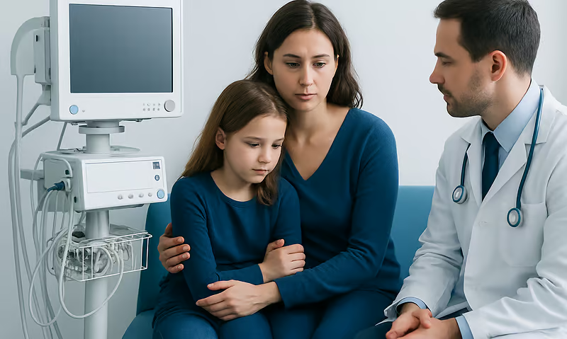
EEG Test: What Brain Wave Monitoring Shows and When It Is Used
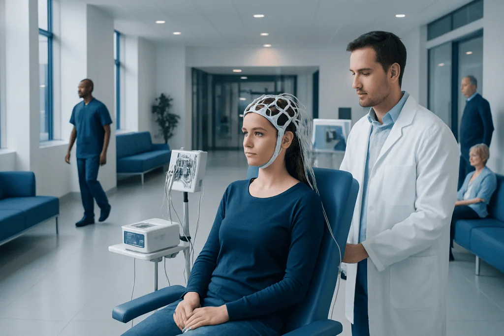
An EEG test measures brain wave patterns using painless electrodes placed on the scalp. It is commonly used to help assess seizures, epilepsy, sleep disorders, and unexplained episodes of confusion or fainting.
Key Takeaways
- An EEG test measures brain wave patterns using painless electrodes placed on the scalp.
- It is commonly used to help assess seizures, epilepsy, sleep disorders, and unexplained episodes of confusion or fainting.
- The test itself does not usually diagnose a condition on its own; doctors interpret it together with symptoms, history, and other tests.
- Routine, sleep-deprived, ambulatory, and video EEGs are different formats chosen for different clinical needs.
- Most people can return to normal activities right after the test unless their doctor gives different instructions.
An EEG test, or electroencephalogram, records the brain’s electrical activity through small sensors placed on the scalp. It is a safe, noninvasive test that helps doctors investigate seizures, sleep problems, changes in awareness, and other neurological symptoms.
Overview: What Is an EEG Test?
An EEG test, short for electroencephalogram, is a way to record the brain’s electrical activity. Nerve cells in the brain communicate through tiny electrical signals, and an EEG detects these signals using small metal discs called electrodes placed on the scalp. The recording appears as wave-like patterns that trained specialists analyze for normal and abnormal activity.
The test is noninvasive, meaning it does not require surgery or needles into the brain. It is also painless. During the test, the electrodes only detect activity; they do not send electricity into the body. For many patients, this makes EEG brain wave monitoring a reassuring first-step tool when neurological symptoms need further evaluation.
An EEG test does not directly show the structure of the brain the way CT or MRI scans do. Instead, it shows how the brain is functioning in real time. Because of this, it can offer valuable information when symptoms suggest that brain activity may be temporarily altered, such as in seizures, sudden confusion, or some sleep-related events.
What Brain Wave Monitoring Can Show
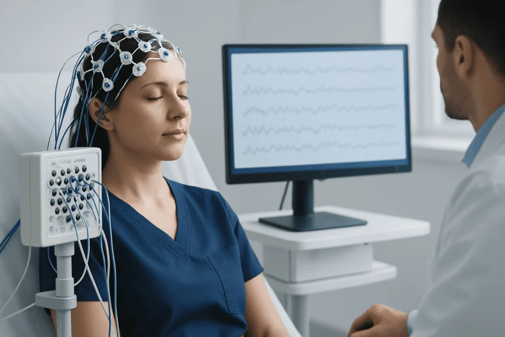
Brain wave monitoring can reveal patterns that suggest whether electrical activity is typical or unusual. Doctors may look for slowing, sharp waves, spike patterns, or changes linked to sleep and wakefulness. These findings can help support the diagnosis of conditions that affect how the brain works, especially when symptoms happen in episodes.
One of the best-known uses of an EEG test is in the assessment of seizures and epilepsy. An abnormal EEG may show patterns associated with a tendency toward seizures, although a normal EEG does not completely rule them out. Because some abnormalities happen only occasionally, the timing and type of EEG can affect what the test captures.
EEG findings may also help in the evaluation of sleep disorders, unexplained blackouts, memory lapses, confusion, brain inflammation, or changes in consciousness. In hospitals, EEG monitoring can be used to assess patients after head injury, during intensive care, or when doctors need to look for seizures that are not visible from the outside.
Importantly, EEG results are interpreted in context. A pattern on the recording is only one piece of the overall picture. Doctors consider symptoms, medical history, physical and neurological examination, and other investigations before making a diagnosis or treatment plan.
When an EEG Test Is Used
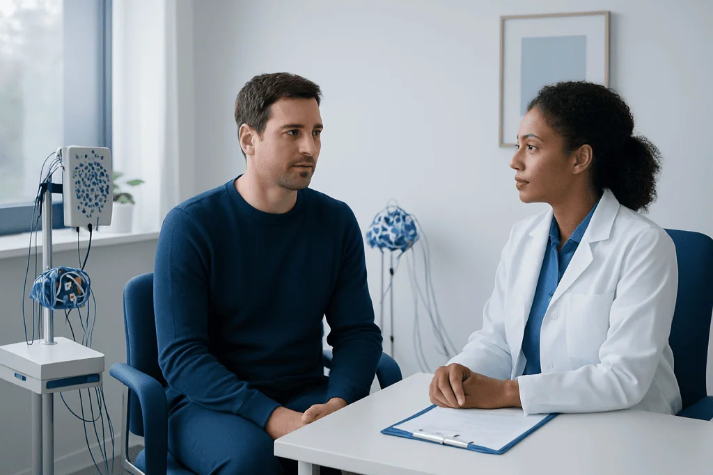
Doctors may recommend an EEG test when someone has symptoms that could be related to altered brain activity. These may include suspected seizures, staring spells, episodes of unresponsiveness, sudden confusion, unusual movements, fainting that is difficult to explain, or changes in awareness. Children and adults can both have EEG testing when clinically appropriate.
EEG may also be useful in evaluating certain sleep-related concerns, such as unusual nighttime behaviors or possible sleep-related seizures. In some cases, it is combined with other sleep assessments if the doctor wants to understand whether events during sleep are neurological, behavioral, or due to another sleep disorder.
Other situations include monitoring brain function during serious illness, checking for seizure activity after brain injury, and supporting the evaluation of conditions such as encephalopathy or inflammation affecting the brain. When symptoms suggest a broader neurological issue, doctors may also combine EEG with MRI scanning or other neurological assessments for a more complete picture.
Different forms of EEG are available depending on the goal of testing:
- Routine EEG: a short recording, often completed within about 20 to 40 minutes of active tracing.
- Sleep-deprived EEG: performed after reduced sleep to increase the chance of detecting certain abnormalities.
- Ambulatory EEG: recorded over many hours or days while the patient goes about normal activities.
- Video EEG: combines brain wave recording with video to compare physical events with electrical activity.
How to Prepare and What Happens During the Procedure
Preparation for an EEG test is usually simple. Patients are often asked to wash their hair the night before or the day of the test and avoid hair sprays, oils, gels, or conditioners that could make it harder for electrodes to stick. A doctor may also advise whether to continue regular medicines, since this depends on the reason for the test.
In some cases, the care team may ask the patient to sleep less than usual before the appointment, especially for a sleep-deprived EEG. The purpose is to make certain brain wave changes more likely to appear during the recording. Patients should follow instructions carefully and ask questions in advance if anything is unclear.
During the procedure, a technician measures the scalp and places several electrodes using a special paste or cap. The patient then rests in a chair or lies on a bed while the machine records brain activity. They may be asked to open and close their eyes, breathe deeply for several minutes, or look at flashing lights. These activation steps can sometimes bring out patterns that are not seen at rest.
The test itself is generally comfortable, though staying still can feel mildly inconvenient. For longer recordings, the setup may differ, especially with ambulatory or video EEG. Once the test is finished, the electrodes are removed and any paste is cleaned off. Most people can return to usual activities right afterward unless sedation or special instructions apply.
Understanding EEG Results
EEG results are usually reviewed by a neurologist or neurophysiology specialist with experience in reading brain wave patterns. A normal EEG shows expected rhythms that change with wakefulness, drowsiness, sleep, age, and other factors. Abnormal results may include slowing, asymmetry, spikes, sharp waves, or rhythmic patterns that suggest irritation or instability in certain brain areas.
Even so, EEG interpretation is nuanced. Some people with epilepsy can have a normal routine EEG between seizures, while some EEG changes may be nonspecific and not point to one exact diagnosis. This is why the test is rarely used in isolation. The final interpretation depends on the patient’s symptoms, age, medications, and the circumstances in which the recording was made.
Doctors may recommend repeating the test or using a longer format if the first EEG does not answer the clinical question. In some situations, additional tests such as CT scanning, blood tests, or sleep studies may be used alongside EEG to clarify the cause of symptoms. When events are infrequent, prolonged monitoring can be especially helpful because it increases the chance of capturing an episode.
Patients should feel comfortable asking what the results mean in practical terms. Useful questions include whether the EEG was normal or abnormal, whether it changes the diagnosis, and what next steps are recommended. Clear discussion helps patients understand both the value and the limits of the test.
Benefits, Limitations, and Safety
The main benefits of an EEG test are that it is safe, painless, and able to measure brain function in real time. It can provide important clues that structural scans cannot show, particularly when symptoms come and go. It is also widely used in both outpatient and hospital settings, making it an accessible tool for many neurological evaluations.
However, EEG also has limitations. It records activity from the scalp, so very deep or brief abnormalities may not always be detected. A normal result does not always rule out a seizure disorder or another neurological condition. Likewise, an abnormal EEG does not automatically confirm a diagnosis without supporting clinical evidence.
For most people, EEG is very safe. Rarely, activation methods such as flashing lights or deep breathing may trigger a seizure in someone who is already prone to seizures, which is why testing is supervised by trained professionals. The team is prepared to respond if this happens. For the great majority of patients, the experience is straightforward and uneventful.
When more advanced assessment is needed, doctors may combine EEG findings with other evaluations such as neurology assessment or long-term monitoring plans. At the appropriate stage of care, multidisciplinary specialists at Acibadem International and its JCI-accredited hospitals also evaluate and treat patients who need EEG-based neurological assessment.
When to See a Doctor and Practical Self-care Tips
A person should seek medical advice if they experience unexplained fainting, repeated episodes of staring or confusion, unusual movements, sudden memory gaps, nighttime events that are hard to explain, or suspected seizures. Prompt evaluation is especially important if symptoms are new, happen repeatedly, or occur after a head injury or serious illness.
Urgent medical attention is needed for prolonged seizures, repeated seizures without full recovery between them, serious injury during an episode, or a first seizure associated with difficulty breathing, high fever, pregnancy, or diabetes. These situations need immediate professional care rather than home observation.
Before an EEG appointment, practical self-care steps include following sleep instructions carefully, taking medications exactly as directed unless a doctor says otherwise, and arranging transport if sleep deprivation may make driving unsafe. It can also help to keep a symptom diary noting when episodes happen, how long they last, what was observed, and whether there were triggers such as lack of sleep.
Family members or caregivers can be helpful in describing events, since many neurological episodes are difficult for the patient to remember clearly. Good preparation supports a more accurate diagnosis and helps the doctor choose whether routine EEG, video EEG, or another test is most appropriate.
Frequently asked questions
What does an EEG test diagnose?
An EEG test does not always diagnose a condition by itself, but it helps doctors assess abnormal brain activity. It is commonly used when evaluating seizures, epilepsy, sleep-related events, confusion, and changes in consciousness.
Is an EEG test painful?
No, an EEG test is painless. The electrodes placed on the scalp only record electrical activity and do not send electricity into the body.
How long does an EEG test take?
A routine EEG is usually fairly short, while longer forms such as ambulatory or video EEG can continue for many hours or even several days. The exact duration depends on why the test is being done and how likely symptoms are to occur during recording.
Can an EEG be normal if someone still has seizures?
Yes, a person can have a normal EEG between seizure episodes. This is one reason doctors may repeat the test or recommend longer-term monitoring if symptoms strongly suggest seizures.
Do patients need to fast before an EEG?
Fasting is not usually required for a standard EEG test unless the care team gives specific instructions. In most cases, patients should eat normally and continue medications as directed by their doctor.
What should someone avoid before an EEG test?
Patients are often advised to avoid hair products such as sprays, gels, oils, or conditioners because these can interfere with electrode attachment. If a sleep-deprived EEG is planned, they should also follow instructions about reduced sleep carefully.
References
- World Health Organization
- National Institute of Neurological Disorders and Stroke
- International League Against Epilepsy
- American Clinical Neurophysiology Society
- NHS
This article is for general information only and is not a substitute for professional medical advice. Please consult a qualified doctor about your individual situation.
Hair transplant in Turkey — costs, graft counts & a free assessment
JCI-accredited · board-certified surgeons · reply within 24h
Add us as a Preferred Source to see more of our trusted health content across Google Search, AI Overviews and Discover.