

Nerve Biopsy: When It Is Used in Neuromuscular Disease Diagnosis

Nerve biopsy is not a routine first-line test and is used selectively. It can help diagnose some inflammatory, vasculitic, infiltrative, hereditary, or amyloid-related neuropathies.
Key Takeaways
- Nerve biopsy is not a routine first-line test and is used selectively.
- It can help diagnose some inflammatory, vasculitic, infiltrative, hereditary, or amyloid-related neuropathies.
- The procedure most often samples a sensory nerve, commonly the sural nerve near the ankle.
- Doctors usually combine biopsy results with symptoms, examination, blood tests, imaging, and nerve conduction studies.
- Possible risks include numbness, pain, infection, bleeding, and scar formation at the biopsy site.
- Patients should discuss benefits, limits, and alternatives with a neurologist before the procedure.
A nerve biopsy is a procedure in which a small piece of peripheral nerve is removed and examined to help diagnose certain nerve and muscle-related conditions. It is usually considered when symptoms, examination findings, blood tests, and nerve studies do not provide a clear answer.
Overview
A nerve biopsy is a diagnostic procedure used to examine a small sample of peripheral nerve tissue under a microscope. It can provide detailed information about how a nerve is affected, including whether there is inflammation, loss of myelin, damage to nerve fibers, abnormal protein deposits, or blood vessel-related injury. In neuromuscular medicine, this information may help clarify the cause of weakness, numbness, pain, tingling, or balance problems.
Most people with suspected neuropathy do not need a nerve biopsy. Doctors usually begin with a medical history, neurological examination, blood tests, and electrodiagnostic testing such as nerve conduction studies and electromyography. A biopsy is generally considered only when these tests do not fully explain the condition or when tissue confirmation is important for treatment decisions.
The test is most often used in selected cases of peripheral neuropathy rather than disorders affecting the brain or spinal cord. It may be especially helpful when there is concern for vasculitic neuropathy, amyloidosis, leprosy in relevant settings, certain inflammatory neuropathies, or infiltrative diseases. Because it is an invasive procedure and can leave an area of permanent numbness, the decision to perform it is made carefully.
When a Nerve Biopsy Is Used
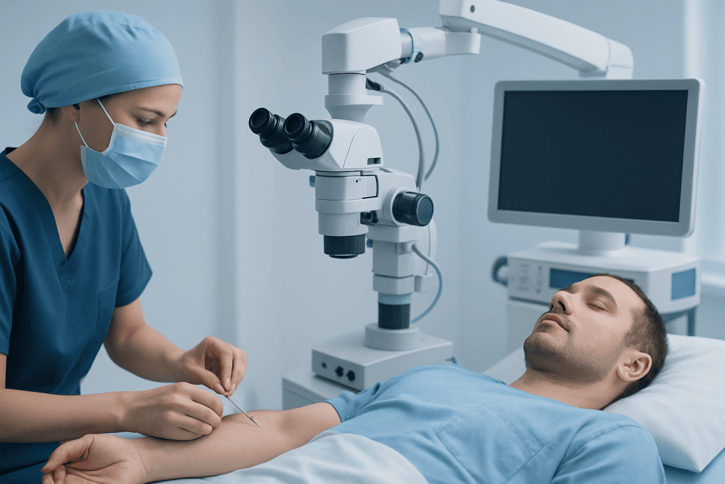
Doctors may recommend a nerve biopsy when symptoms suggest a nerve disorder, but the exact cause remains uncertain after standard testing. It is often considered when a patient has progressive sensory loss, burning pain, weakness, loss of reflexes, or an asymmetric pattern of nerve involvement. In some cases, the biopsy helps distinguish between different types of neuropathy that can look similar at first.
A nerve biopsy may be particularly useful when there is suspicion of inflammation affecting the blood vessels that supply the nerves, known as vasculitic neuropathy. In this setting, tissue examination can show vessel wall inflammation and nerve fiber injury, which may support the need for immunosuppressive treatment. It can also help identify amyloid deposits in some patients with suspected amyloid neuropathy, although less invasive tissue sampling may be tried first.
Sometimes the aim is to confirm a diagnosis that has been strongly suspected from other findings. For example, if a patient has symptoms and test results consistent with a severe or unusual peripheral neuropathy, a biopsy may provide the microscopic detail needed to guide further care. In selected patients with systemic inflammatory disease, connective tissue disorders, or unexplained rapidly worsening neuropathy, it may also help rule in or rule out specific causes.
Even when a biopsy is being considered, not every patient will benefit from it. If the likely diagnosis is already clear from blood work, genetic testing, imaging, or electrodiagnostic studies, a biopsy may not add meaningful information. In hereditary neuropathies, for instance, genetic testing has reduced the need for nerve biopsy in many cases.
How the Procedure Is Performed

A nerve biopsy is usually done as a minor surgical procedure under local anesthesia. The nerve most often sampled is the sural nerve, a sensory nerve near the outer part of the ankle and lower leg. This nerve is commonly chosen because removing a small segment usually causes numbness in a limited area rather than major weakness.
During the procedure, the skin is cleaned and numbed, and a small incision is made to identify the nerve. A short piece of the nerve is removed and sent to the laboratory for specialized analysis. The incision is then closed with stitches and covered with a dressing. In some cases, a muscle biopsy may be performed at the same time if the doctor suspects vasculitis or another disorder affecting both nerve and muscle.
After the biopsy, the tissue is examined using several techniques. These may include routine microscopy, special stains, immunohistochemistry, electron microscopy, and testing for abnormal deposits or infectious organisms when indicated. Because the value of the biopsy depends greatly on expert interpretation, it is usually best done in centers with experience in neuromuscular pathology.
Patients may also undergo related diagnostic tests before or after biopsy, depending on the clinical picture. These can include electromyography and nerve conduction studies, blood tests, imaging, or targeted evaluation for autoimmune, metabolic, or hereditary conditions. The biopsy is only one part of the overall diagnostic process.
What a Nerve Biopsy Can Show
A nerve biopsy can reveal several patterns of nerve injury. One major question is whether the problem primarily affects axons, the long fibers that carry nerve signals, or myelin, the insulating covering around them. This distinction can help narrow the list of possible causes and explain why symptoms are progressing in a particular way.
Pathologists also look for signs of inflammation, blood vessel damage, granulomas, abnormal protein deposits, or infiltrative cells. In vasculitic neuropathy, biopsy may show inflammation in or around blood vessel walls with evidence of ischemic nerve injury. In amyloid neuropathy, the sample may contain amyloid deposits that can be highlighted with special staining.
Another benefit is that the biopsy can sometimes help exclude certain conditions. If a doctor is considering inflammatory neuropathy but the tissue does not show expected findings, attention may shift to metabolic, toxic, infectious, or hereditary causes. This can help avoid treatments that may not be helpful and guide more targeted testing.
Still, nerve biopsy has limitations. A small sample may miss patchy disease, and some abnormalities are not specific to one diagnosis. Results are most useful when they are interpreted alongside a neurological examination, electrodiagnostic studies, and, when appropriate, MRI or other imaging. A normal or non-specific biopsy does not always rule out disease.
Risks, Limitations, and Recovery
Like any procedure that involves an incision, nerve biopsy carries some risks. The most common expected effect is numbness in the area supplied by the sampled sensory nerve. Because the tissue is removed, that numbness may be permanent, although it is often limited to a small patch of skin. Some people also notice tenderness, altered sensation, or shooting discomfort during healing.
Other possible risks include bleeding, infection, delayed wound healing, scarring, and pain at the biopsy site. Rarely, symptoms such as persistent neuropathic pain can be more troublesome than expected. For this reason, the potential value of the biopsy should always be balanced against the possibility of lasting sensory change.
Recovery is usually straightforward. Patients are often advised to keep the area clean and dry, limit strenuous activity for a short period, and follow instructions about dressing changes or stitch removal. Mild discomfort can often be managed with simple pain relief recommended by the treating team. Patients should contact their doctor if they develop increasing redness, swelling, fever, drainage, or severe pain.
There are also diagnostic limitations to consider. A biopsy may not identify the cause if the disease process is unevenly distributed or if the sampled nerve is not the most affected one. In many situations, less invasive tests can provide enough information, which is why neurologists reserve nerve biopsy for carefully selected cases.
Alternative and Complementary Tests
Before recommending a nerve biopsy, neurologists usually complete a broad evaluation. This often includes blood tests for diabetes, vitamin deficiencies, thyroid disorders, autoimmune disease, infections, kidney or liver disease, and paraproteinemias. Depending on the person’s symptoms and family history, doctors may also consider genetic testing, cerebrospinal fluid analysis, or screening for systemic illnesses.
Electrodiagnostic testing is one of the most important companion tools. EMG and nerve conduction studies can show whether the neuropathy is axonal or demyelinating, whether it is generalized or focal, and whether muscles are also involved. These findings help determine whether a biopsy is likely to be useful and which nerve, if any, should be sampled.
Imaging and tissue sampling from other sites may also reduce the need for nerve biopsy in some patients. For example, skin biopsy can measure small nerve fiber density in suspected small fiber neuropathy, and abdominal fat pad, salivary gland, or other tissue biopsies may sometimes detect amyloid without removing nerve tissue. In selected cases, genetic testing can identify inherited neuropathies and spare patients an invasive procedure.
If a broader neuromuscular disorder is being considered, the evaluation may also include tests for related conditions such as myasthenia gravis or muscle disease. The key point is that nerve biopsy is rarely a stand-alone answer; it is part of a carefully planned diagnostic strategy.
Treatment Planning After Biopsy
A nerve biopsy does not treat the condition by itself, but it can influence treatment decisions. If the biopsy shows vasculitic neuropathy or another inflammatory process, doctors may recommend medications that reduce immune system activity. If amyloid, infection, toxin-related damage, or an inherited disorder is identified, management can be adjusted to address the underlying cause as directly as possible.
Supportive treatment remains important regardless of the biopsy result. This may include pain management, physical therapy, occupational therapy, foot care, fall prevention, and treatment of contributing conditions such as diabetes or nutritional deficiency. For patients with weakness, rehabilitation and assistive devices can help maintain safety and independence.
In some cases, the biopsy result helps confirm that aggressive treatment is not needed, which can be just as valuable. Avoiding unnecessary immune therapies or repeated testing may reduce side effects, cost, and uncertainty. Follow-up with a neurologist is important so the patient’s symptoms, function, and any new findings can be reassessed over time.
For people seeking specialized assessment, multidisciplinary neurology teams can coordinate pathology review, electrodiagnostic testing, imaging, and rehabilitation. Acibadem International’s multidisciplinary specialists and JCI-accredited hospitals diagnose and treat neuromuscular conditions for international patients when advanced evaluation is needed.
When to See a Doctor
A person should seek medical evaluation for numbness, tingling, burning pain, unexplained weakness, muscle wasting, balance problems, or loss of coordination that is persistent, progressive, or affecting daily life. Symptoms that begin suddenly, worsen quickly, or affect one side more than the other also deserve prompt attention. Early assessment can help identify treatable causes before nerve damage becomes more advanced.
Urgent medical care is especially important if nerve symptoms occur with breathing difficulty, problems swallowing, severe back pain, facial weakness, new bladder or bowel dysfunction, or rapidly worsening weakness. These symptoms may point to a more serious neurological problem that requires immediate evaluation rather than outpatient testing.
If a doctor has suggested a nerve biopsy, patients may wish to ask why it is being recommended, what diagnosis is being considered, what information the biopsy may add, and what alternatives exist. Understanding the expected benefits and possible side effects can help patients make an informed decision that fits their situation.
Frequently asked questions
Is a nerve biopsy always needed to diagnose neuropathy?
No. Most neuropathies are diagnosed with a combination of medical history, neurological examination, blood tests, and nerve conduction studies. A nerve biopsy is usually reserved for cases where the cause remains unclear or when tissue confirmation could change treatment decisions.
What nerve is usually biopsied?
The sural nerve near the ankle is the most commonly biopsied nerve. It is a sensory nerve, so removing a small segment usually causes a limited patch of numbness rather than weakness.
Is a nerve biopsy painful?
The procedure is typically done under local anesthesia, so the area is numbed during the biopsy. Some soreness, tenderness, or altered sensation can occur afterward, but the treating team usually provides advice on how to manage recovery comfortably.
What conditions can a nerve biopsy help diagnose?
It may help diagnose vasculitic neuropathy, amyloid neuropathy, certain inflammatory or infiltrative neuropathies, and some infections in the right clinical setting. It can also provide clues about whether nerve damage is primarily axonal or demyelinating.
Can a nerve biopsy be normal even if symptoms are real?
Yes. Some nerve disorders are patchy, affect very small fibers, or are better detected by other tests, so a biopsy can sometimes be non-specific or miss the abnormal area. That is why doctors interpret biopsy results together with the full clinical picture.
How long does it take to recover from a nerve biopsy?
Initial wound healing is often relatively quick, but the exact timeline varies from person to person. A small area of numbness is expected and may be long-lasting, while temporary soreness usually improves over days to weeks with proper care.
References
- National Institute of Neurological Disorders and Stroke
- American Academy of Neurology
- National Institute of Diabetes and Digestive and Kidney Diseases
- Mayo Clinic
- MedlinePlus
This article is for general information only and is not a substitute for professional medical advice. Please consult a qualified doctor about your individual situation.
Oncology care in Turkey — second opinion and treatment plan
JCI-accredited · board-certified surgeons · reply within 24h
Add us as a Preferred Source to see more of our trusted health content across Google Search, AI Overviews and Discover.

More from the Health Library



Related Specialists

Dr. Tuğçe Baykara
Gynecology & Obstetrics
Assoc. Prof. Dr. M. Murat Sezer
Pulmonology
Dr. Nuray Keçecioğlu
Pediatrics
Prof. Dr. Hakan Ağır
Aesthetic Plastic & Reconstructive Surgery