Neurointerventional Radiology for Stroke: Who May Benefit From Thrombectomy?

Thrombectomy is a minimally invasive procedure used to remove a blood clot from a blocked brain artery. It is mainly used for certain types of ischemic stroke, not for bleeding strokes.
Key Takeaways
- Thrombectomy is a minimally invasive procedure used to remove a blood clot from a blocked brain artery.
- It is mainly used for certain types of ischemic stroke, not for bleeding strokes.
- Fast evaluation is essential because brain tissue can be damaged within minutes of reduced blood flow.
- Advanced brain and vessel imaging helps doctors decide who may benefit most from treatment.
- Treatment decisions depend on symptoms, clot location, timing, imaging findings, and overall health.
Neurointerventional radiology for stroke focuses on restoring blood flow to the brain when a clot blocks an artery. In carefully selected patients with ischemic stroke, thrombectomy may improve outcomes when performed quickly by a specialized team.
Overview: What Neurointerventional Radiology for Stroke Means
Neurointerventional radiology for stroke is a highly specialized field that uses imaging guidance and tiny catheters to treat blocked blood vessels in the brain. In acute ischemic stroke, a blood clot prevents oxygen-rich blood from reaching part of the brain. The goal of treatment is to reopen the blocked artery as safely and quickly as possible.
One of the most important procedures in this field is thrombectomy, also called mechanical thrombectomy. During this treatment, a specialist guides a thin tube through an artery, often starting in the groin or wrist, to reach the clot in the brain and remove it. This can restore circulation and help limit permanent brain injury.
Thrombectomy is not appropriate for every stroke. It is mainly used for selected patients with large artery blockages causing significant symptoms. Doctors combine the physical examination, the time symptoms started, and imaging results to decide whether this treatment may help.
Who May Benefit From Thrombectomy
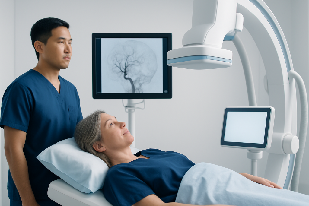
Thrombectomy may benefit people who have an acute ischemic stroke caused by a blockage in a large blood vessel supplying the brain. These blockages often affect major arteries such as the internal carotid artery or the middle cerebral artery. Patients usually have sudden, noticeable neurological symptoms, such as weakness on one side, trouble speaking, facial drooping, or vision changes.
The best candidates are often those who can be treated soon after symptoms begin, but in some cases thrombectomy may still be helpful later if brain imaging shows that salvageable brain tissue remains. For this reason, even patients who wake up with stroke symptoms or arrive after several hours may still be assessed urgently rather than assumed to be too late.
Doctors do not decide based on time alone. They also consider the severity of symptoms, the size and location of the clot, the amount of already damaged brain tissue, and the patient’s general medical condition. A person may receive intravenous clot-busting medicine first, if eligible, and then proceed to thrombectomy if a large vessel remains blocked.
Thrombectomy is generally not used for every mild stroke, every small-vessel stroke, or strokes caused by bleeding. Rapid testing is important because stroke treatment depends on the exact type and cause.
Symptoms That Need Emergency Evaluation

Stroke symptoms usually begin suddenly. Warning signs include weakness or numbness of the face, arm, or leg, especially on one side of the body; difficulty speaking or understanding speech; sudden confusion; loss of vision in one or both eyes; dizziness; trouble walking; and a severe, unexplained headache. Any of these symptoms should be treated as a medical emergency.
A helpful public message is the FAST test: Face drooping, Arm weakness, Speech difficulty, Time to call emergency services. Even if symptoms improve quickly, urgent evaluation is still necessary. A brief episode may represent a transient ischemic attack, which can be an important warning sign of a more serious stroke.
Not every person with these symptoms will be a candidate for thrombectomy, but the only way to know is to seek emergency care immediately. Delays at home can reduce treatment options and may worsen the chance of recovery. In stroke care, early action helps preserve brain function.
Causes, Risk Factors, and Why Large Vessel Blockage Matters
Ischemic stroke happens when a clot blocks blood flow in the brain. The clot may form in an artery already narrowed by atherosclerosis, or it may travel from another part of the body, especially the heart. A common example is atrial fibrillation, which can allow clots to form and then move to the brain.
Several factors increase the risk of stroke, including high blood pressure, diabetes, high cholesterol, smoking, obesity, sleep apnea, heart rhythm problems, older age, and a prior history of stroke or transient ischemic attack. Some people also have disease of the neck arteries, such as the carotid arteries, which can contribute to clot formation. In some cases, this is linked with carotid artery disease.
Large vessel occlusions are especially serious because they cut off blood supply to a wider area of the brain. They are more likely to cause severe symptoms and long-term disability if blood flow is not restored quickly. This is why thrombectomy can be so valuable in carefully selected patients: it directly targets the clot in a major artery.
Although risk factors increase the chance of stroke, a stroke can still occur unexpectedly. Prevention and emergency treatment both matter. Ongoing care after the acute event is also important to reduce the risk of another stroke in the future.
How Doctors Diagnose Stroke and Decide on Thrombectomy
Stroke diagnosis begins with a rapid clinical assessment. Emergency teams check when symptoms started or when the patient was last known to be well, measure blood pressure and blood sugar, and perform a neurological examination. At the same time, they look for other possible causes of symptoms, because some conditions can mimic stroke.
Brain imaging is essential. A non-contrast CT scan is often the first test because it can quickly identify bleeding in the brain. Since thrombectomy is used for ischemic stroke rather than hemorrhagic stroke, excluding bleeding is a key first step. Additional tests such as CT angiography or MR angiography can show whether a major artery is blocked.
In many centers, advanced imaging is also used to estimate how much brain tissue is already permanently injured and how much may still be saved. This information helps identify patients who may benefit from thrombectomy, including some who present later than the traditional early time window.
Because stroke treatment is time-sensitive and highly specialized, care is often delivered by a multidisciplinary team. This may include emergency physicians, neurologists, neuroradiologists, anesthesiology specialists, and critical care teams. In appropriate cases, patients may be transferred urgently to a center able to provide interventional radiology-guided stroke treatment.
What Happens During Thrombectomy and What Treatment Involves
During thrombectomy, the specialist inserts a thin catheter into an artery and guides it to the blocked vessel in the brain using real-time imaging. Very small devices are then used to capture or suction out the clot. Once the clot is removed and blood flow improves, the catheter is withdrawn. The procedure is designed to be minimally invasive compared with open surgery.
Some patients receive intravenous thrombolytic medicine before thrombectomy if they arrive within the appropriate treatment window and meet eligibility criteria. This medicine helps dissolve clots, but it may not be enough for larger vessel blockages. Mechanical thrombectomy can be used as an additional treatment when imaging shows a suitable target clot.
As with any urgent procedure, thrombectomy has potential risks. These can include bleeding, vessel injury, reaction to contrast dye, or failure to fully reopen the artery. Even so, for properly selected patients, the potential benefit of restoring brain blood flow can outweigh these risks. The treatment team explains the situation as clearly as possible to the patient or family.
After the procedure, patients are monitored closely in a stroke unit or intensive care setting. Recovery needs vary and may include stroke rehabilitation to address movement, speech, swallowing, and daily functioning. Follow-up care also focuses on finding the cause of the stroke and preventing recurrence.
Recovery, Prevention, and Self-care After Stroke
Recovery after stroke is different for each person. Some improve quickly after blood flow is restored, while others need longer-term rehabilitation. Early therapy can support recovery of speech, movement, balance, and independence. Family support, patience, and a structured rehabilitation plan can make a meaningful difference.
Preventing another stroke is a major part of care. Doctors may recommend medicines to reduce clotting risk, control blood pressure, lower cholesterol, or manage diabetes, depending on the cause of the stroke. If an irregular heart rhythm is present, treatment of that condition is also important. Lifestyle measures such as stopping smoking, eating a heart-healthy diet, staying physically active, and maintaining a healthy weight can further reduce risk.
People who have had a stroke should attend follow-up appointments and report any new neurological symptoms immediately. If a person has swallowing difficulties, weakness, memory changes, or mood symptoms, these issues deserve attention and treatment. Ongoing care often involves neurology, rehabilitation, and sometimes vascular or cardiac specialists.
In some situations, related conditions such as brain aneurysm or other vascular abnormalities may be identified during neurovascular assessment, though these are different from clot-related ischemic stroke. For international patients needing specialized assessment, Acibadem International’s multidisciplinary specialists and JCI-accredited hospitals provide diagnosis and treatment in neurovascular care.
When to See a Doctor
Anyone with sudden stroke symptoms should seek emergency medical help immediately, not wait for symptoms to pass. Calling emergency services is safer than traveling by private car because treatment can begin sooner and the patient can be taken to the most appropriate stroke center. Minutes matter when brain blood flow is blocked.
People should also speak with a doctor if they have risk factors such as uncontrolled high blood pressure, atrial fibrillation, diabetes, smoking, or a prior transient ischemic attack. Managing these problems can lower the chance of a future stroke. If warning signs have occurred in the past, even briefly, they should not be ignored.
After a stroke, urgent follow-up is needed if there is new weakness, worsening speech, severe headache, confusion, chest pain, shortness of breath, or repeated falls. These may signal complications or another vascular event. A qualified doctor can guide both emergency treatment and long-term prevention.
Frequently asked questions
What is thrombectomy for stroke?
Thrombectomy is a minimally invasive procedure used to remove a blood clot from a blocked artery in the brain. It is most often used for certain ischemic strokes caused by large vessel blockage. The aim is to restore blood flow quickly and reduce brain damage.
Who is eligible for thrombectomy?
Eligibility depends on several factors, including the type of stroke, the location of the clot, symptom severity, and brain imaging findings. Time from symptom onset is important, but some people may still qualify later if imaging shows brain tissue that can be saved. A stroke specialist makes this decision urgently based on the full clinical picture.
Is thrombectomy the same as clot-busting medicine?
No. Clot-busting medicine is given through a vein and works by chemically dissolving a clot. Thrombectomy uses catheters and tiny devices to physically remove the clot from the artery. Some patients receive both treatments if they meet the criteria.
How fast must treatment happen?
Treatment should happen as quickly as possible because brain cells can be injured within minutes of reduced blood flow. Some patients benefit within the first few hours, while others may still be candidates later based on advanced imaging. Even if there has been a delay, emergency evaluation is still essential.
What are the risks of thrombectomy?
Possible risks include bleeding, blood vessel injury, allergic reaction to contrast material, and incomplete clot removal. Not every patient improves, especially if significant brain injury has already occurred before treatment. The medical team weighs these risks against the possible benefit of restoring circulation.
Can someone recover fully after thrombectomy?
Some people recover very well, especially when treatment is performed early and blood flow is restored successfully. Others may still have weakness, speech problems, or other neurological deficits and need rehabilitation. Recovery depends on the size and location of the stroke, general health, and how quickly treatment begins.
References
- World Stroke Organization
- American Stroke Association
- National Institute of Neurological Disorders and Stroke
- Society of NeuroInterventional Surgery
- European Stroke Organisation
This article is for general information only and is not a substitute for professional medical advice. Please consult a qualified doctor about your individual situation.
Explore treatments in Turkey — costs, top hospitals & a free quote
JCI-accredited · board-certified surgeons · reply within 24h
Add us as a Preferred Source to see more of our trusted health content across Google Search, AI Overviews and Discover.
Check your numbers in seconds
BMI, calories, due date, blood pressure and 30+ more clinical calculators — free, instant, doctor-reviewed ranges.

Neurosurgery
Surgical treatment of the brain, spine and nervous system, including minimally invasive and functional procedures.
70 specialists in this unit