PET-CT Scan Results: How to Understand the Report and Next Steps
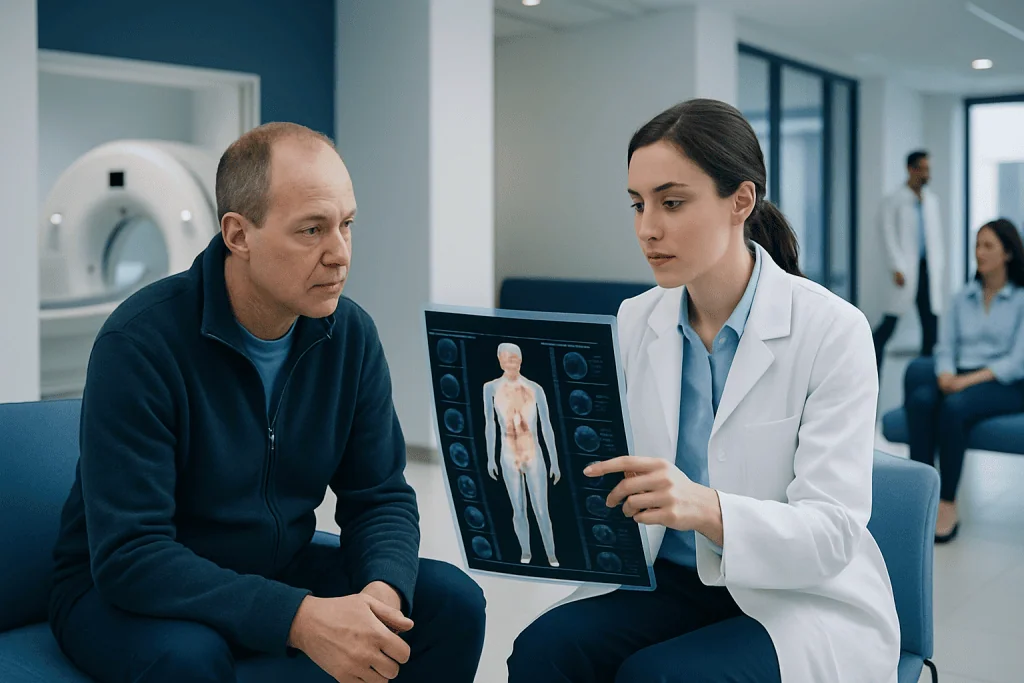
A PET-CT scan shows both anatomy and metabolic activity. Terms like uptake, lesion, and SUV describe patterns, not a final diagnosis by themselves.
Key Takeaways
- A PET-CT scan shows both anatomy and metabolic activity.
- Terms like uptake, lesion, and SUV describe patterns, not a final diagnosis by themselves.
- High uptake can be seen in cancer, but also in infection, inflammation, and normal tissues.
- Doctors use PET-CT results together with medical history, lab tests, and sometimes biopsy.
- Next steps may include follow-up imaging, additional tests, treatment planning, or reassurance when findings are benign.
PET-CT scan results combine structural imaging from CT with metabolic information from PET to help doctors understand what may be happening inside the body. Reading the report can feel technical, but the key findings usually need to be interpreted together with symptoms, examination, and other tests.
Overview: What PET-CT Scan Results Mean
PET-CT scan results are designed to answer two related questions: what a part of the body looks like and how active it is. The CT portion provides detailed pictures of organs, bones, and tissues. The PET portion shows how cells are using a small amount of radiotracer, which often reflects metabolism or other biological activity.
Because it combines these two types of information, PET-CT can help doctors evaluate suspected disease, stage cancer, check response to treatment, or look for recurrence. It is also used in some non-cancer settings, such as infection, inflammation, and certain heart or brain conditions. The report often sounds highly technical, but its main purpose is to help the care team decide whether the findings are concerning, stable, improving, or likely harmless.
Importantly, a PET-CT report rarely stands alone. A finding that appears significant on imaging may turn out to be benign when viewed in context, while a subtle finding may deserve more attention if it matches a person’s symptoms or previous test results. This is why the ordering doctor’s explanation is so valuable.
How a PET-CT Report Is Structured
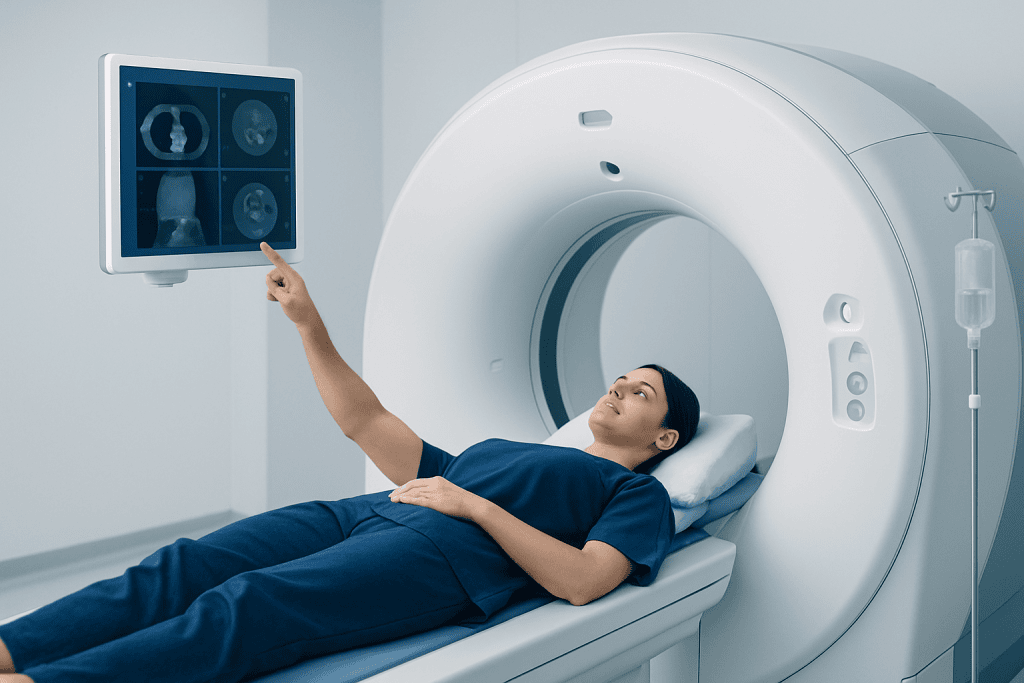
Most PET-CT reports follow a similar format. They usually begin with the reason for the scan, such as assessing a known cancer, investigating unexplained symptoms, or monitoring treatment response. The report may also mention the type of radiotracer used, whether blood glucose was checked beforehand, and technical details about the scan.
Next comes the findings section. This describes where uptake is seen, whether any masses or enlarged lymph nodes are present, and how those findings compare with prior scans. The impression section at the end is especially important because it summarizes the radiologist or nuclear medicine physician’s overall interpretation in a few key points.
Common report terms include:
- Uptake: how much radiotracer a tissue absorbs.
- Hypermetabolic: an area showing more activity than expected.
- Hypometabolic: an area showing less activity than expected.
- Lesion or focus: a spot or area of interest.
- Lymphadenopathy: enlarged lymph nodes.
- No evidence of disease: no clear imaging signs of active disease at the time of the scan.
The wording can sound worrying, but many terms are descriptive rather than diagnostic. For example, “increased uptake” means a pattern was seen; it does not automatically mean cancer.
Understanding Uptake, SUV, and Common PET-CT Terms
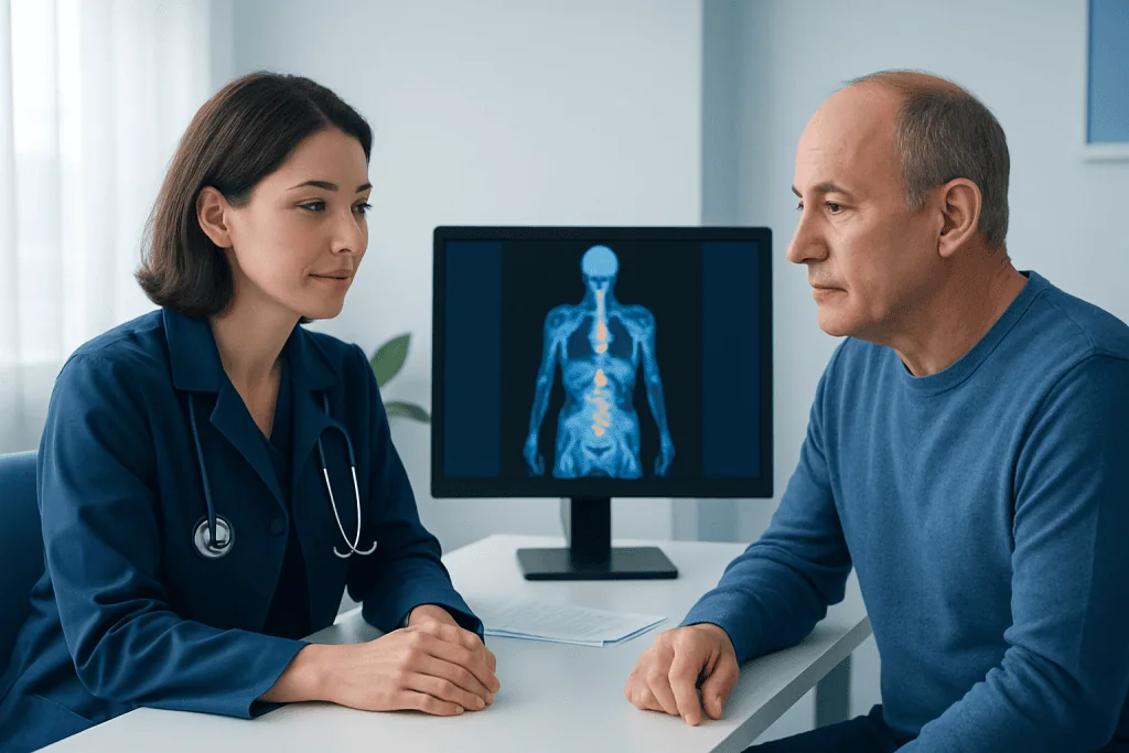
One of the most discussed parts of PET-CT scan results is the SUV, or standardized uptake value. SUV is a way to estimate how much radiotracer has collected in a particular area. In general, higher SUV values may suggest higher metabolic activity, but there is no single number that proves a tissue is cancerous or benign.
Many factors can affect SUV. These include the type of tissue, the radiotracer used, blood sugar level, body composition, timing of the scan, recent treatment, and the imaging technique itself. Some cancers have only mild uptake, while some infections, inflammatory conditions, healing tissues, or normal structures can show intense uptake.
Normal uptake is often seen in the brain, heart, kidneys, bladder, and sometimes bowel, muscles, brown fat, or healing areas. A report may also mention physiologic uptake, meaning expected activity in normal tissues. This is one reason PET-CT interpretation requires experience and careful correlation with the CT images.
If the report describes a nodule, mass, or enlarged lymph node with uptake, doctors consider more than the SUV alone. They look at the size, shape, exact location, changes over time, and whether the pattern matches a known condition such as lung cancer or another diagnosed illness.
What PET-CT Can and Cannot Tell Doctors
PET-CT is a powerful imaging tool, but it has limits. It can highlight areas that deserve attention, show whether disease may be active in more than one place, and help assess whether treatment appears to be working. In oncology, it is often useful for staging and follow-up, including treatment planning for conditions such as lymphoma.
However, PET-CT cannot always tell exactly what a finding is. Cancer, infection, inflammation, postoperative healing, and some benign growths can overlap in appearance. Small lesions may be difficult to characterize, and some slow-growing tumors may not show strong uptake at all.
False-positive results happen when a non-cancerous process looks suspicious. False-negative results happen when disease is present but not clearly detected. For this reason, PET-CT results are often combined with blood tests, ultrasound, MRI, endoscopy, or tissue sampling. When needed, a biopsy remains the most definitive way to diagnose many conditions.
Possible Next Steps After PET-CT Scan Results
The next step depends on why the scan was ordered and what the report shows. If the findings are reassuring or stable, the doctor may recommend routine follow-up only. If there is a new or active-looking area, further tests may be advised to clarify what it means.
Common next steps include repeat imaging after a defined interval, comparison with older scans, referral to a specialist, or additional imaging such as MRI or CT scan. If a suspicious area needs confirmation, the doctor may suggest a biopsy. In cancer care, PET-CT results may also help guide surgery, radiotherapy, or systemic treatment decisions.
When a person is already receiving treatment, the report may use words such as complete metabolic response, partial response, stable disease, or progression. These terms describe how the disease appears to be reacting on imaging. The treating team interprets these findings along with symptoms and physical examination to decide whether the current plan should continue or change.
If the report is difficult to understand, it is reasonable to ask the doctor to explain the impression line by line. Patients often find it helpful to ask: What was found? Is it definitely abnormal? What could else explain it? Do I need more tests now, later, or not at all?
How Doctors Diagnose the Cause of an Abnormal Result
An abnormal PET-CT result is not the same as a confirmed diagnosis. Doctors usually start by reviewing the clinical context: the person’s symptoms, age, medical history, prior surgeries, medications, infections, and any known diagnosis. They then compare the PET-CT with previous imaging to see whether a finding is new, unchanged, or improving.
Laboratory tests may help support or challenge the imaging impression. For example, inflammatory markers, blood counts, tumor markers, or infection-related tests may provide useful clues in some cases. The exact value of these tests depends on the suspected condition.
Sometimes the best next step is observation with repeat imaging rather than immediate intervention. In other situations, especially if treatment decisions depend on certainty, tissue confirmation is needed. A biopsy can identify whether the cells are malignant, inflammatory, infectious, or benign, which is why it remains such an important part of diagnosis.
Multidisciplinary review is often helpful, particularly for complex cases. Radiologists, nuclear medicine physicians, oncologists, surgeons, pulmonologists, pathologists, and other specialists may discuss the results together before deciding on the most appropriate plan.
Preparing for the Follow-Up Appointment
After receiving PET-CT scan results, many people feel unsure about what to ask. Bringing a copy of the report, prior scan reports, medication list, and a written list of questions can make the appointment more productive. It can also help to bring a family member or friend for support and note-taking.
Useful questions include:
- What is the main conclusion of the report?
- Are the findings definitely abnormal or only possibly abnormal?
- Could the uptake be due to inflammation, infection, or recent treatment?
- Do the results change my diagnosis or treatment plan?
- Do I need another scan, a biopsy, or specialist review?
- When should follow-up happen?
It is also sensible to ask whether any findings are incidental, meaning unrelated to the original reason for the scan. Incidental findings are common in imaging and often turn out to be benign, but they may still need simple follow-up.
For people seeking coordinated international care, Acibadem International’s multidisciplinary specialists and JCI-accredited hospitals evaluate PET-CT findings and arrange further testing or treatment when needed.
When to Seek Medical Advice Promptly
A PET-CT report should generally be reviewed with the doctor who ordered it, even if the wording seems reassuring. That doctor can explain how the findings fit the bigger picture and whether any further steps are needed. It is best not to rely only on isolated words from the report without professional interpretation.
Prompt medical advice is especially important if the scan was ordered for a known cancer, if the report mentions new suspicious lesions, or if the person has significant symptoms such as unexplained weight loss, persistent fever, worsening pain, shortness of breath, new neurological symptoms, or rapidly enlarging lumps. These symptoms do not always mean serious disease, but they deserve timely assessment.
Urgent care may be needed if severe symptoms develop, such as chest pain, sudden breathing difficulty, confusion, severe weakness, or heavy bleeding. In most cases, however, the next step is an organized follow-up discussion rather than an emergency response.
Frequently asked questions
Does high uptake on a PET-CT scan mean cancer?
Not always. High uptake means that an area is using more radiotracer, but this can happen with cancer, infection, inflammation, healing tissue, or even some normal body structures. Doctors interpret uptake together with CT findings, symptoms, and other tests.
What does SUV mean on a PET-CT report?
SUV stands for standardized uptake value. It is a measurement used to estimate how much radiotracer has collected in a specific area. It can be helpful, but it does not by itself confirm whether a finding is benign or malignant.
Can a PET-CT scan be wrong?
Yes. Like all medical tests, PET-CT can produce false-positive or false-negative results. That is why doctors may recommend repeat imaging, correlation with other scans, or biopsy when the answer is still uncertain.
What is the impression section of the report?
The impression is the summary at the end of the imaging report. It highlights the most important findings and the radiologist’s overall interpretation. Many patients find this section the most useful starting point when discussing results with their doctor.
Why would a doctor order more tests after a PET-CT scan?
A PET-CT scan can show where something unusual may be happening, but it cannot always identify the exact cause. Additional tests may be needed to confirm whether a finding is due to cancer, infection, inflammation, or another condition. This helps doctors choose the safest and most appropriate treatment plan.
How soon should results be discussed with a doctor?
It is best to review PET-CT scan results with the ordering doctor as soon as practical, especially if the scan was done for cancer evaluation or ongoing symptoms. The right timing depends on the reason for the scan and the urgency of the findings. If severe symptoms are present, medical attention should not be delayed.
References
- World Health Organization
- National Cancer Institute
- Radiological Society of North America
- Society of Nuclear Medicine and Molecular Imaging
- American Cancer Society
This article is for general information only and is not a substitute for professional medical advice. Please consult a qualified doctor about your individual situation.
Medical weight-loss options in Turkey — costs & eligibility
JCI-accredited · board-certified surgeons · reply within 24h
Add us as a Preferred Source to see more of our trusted health content across Google Search, AI Overviews and Discover.