Optical Coherence Tomography in Cardiology: What This Artery Imaging Test Shows
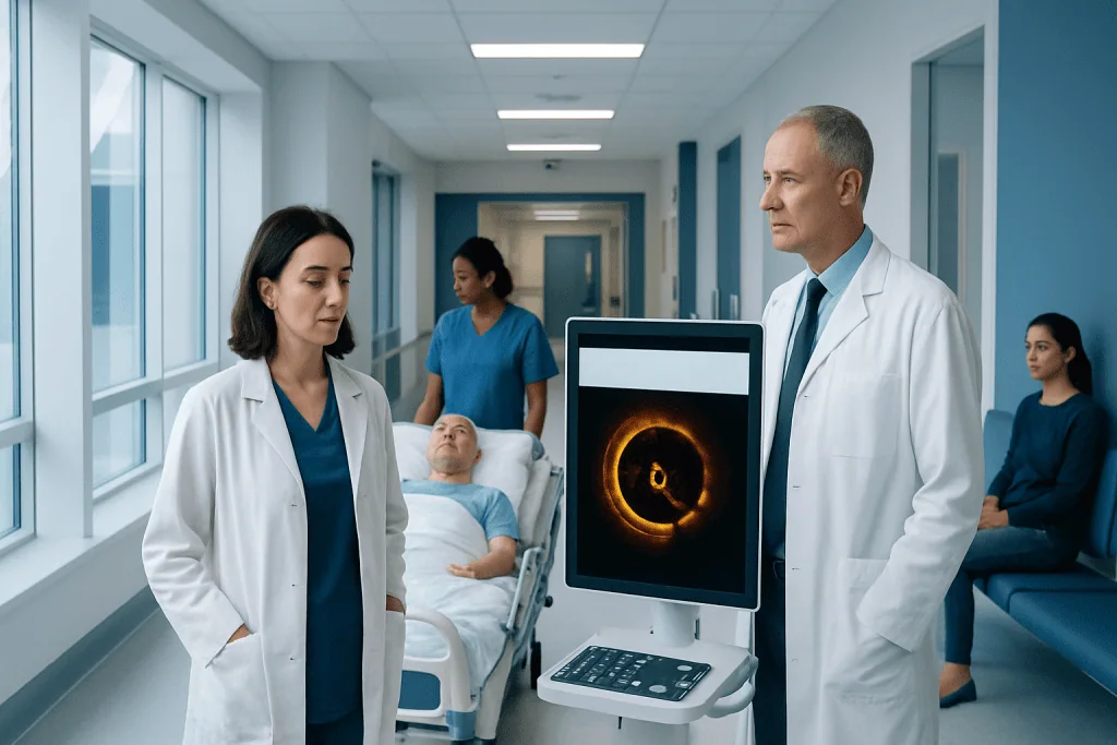
Coronary OCT uses light-based imaging to create very detailed pictures from inside the artery. It is usually performed during a cardiac catheterization or angiography procedure.
Key Takeaways
- Coronary OCT uses light-based imaging to create very detailed pictures from inside the artery.
- It is usually performed during a cardiac catheterization or angiography procedure.
- The test can help identify plaque type, blood clots, stent position, and small vessel injuries.
- OCT supports treatment decisions, especially during coronary angioplasty and stent procedures.
- It does not replace all heart tests; doctors choose it when high-resolution artery imaging is especially helpful.
Optical coherence tomography in cardiology is an advanced imaging test that lets doctors view the inside of coronary arteries in very fine detail. It is commonly used during cardiac catheterization to assess plaque, guide stent placement, and understand why a narrowed artery is causing symptoms.
Overview: What optical coherence tomography in cardiology is
Optical coherence tomography in cardiology, often called coronary OCT, is a specialized imaging technique used to look inside the coronary arteries. These arteries supply blood to the heart muscle. OCT uses near-infrared light delivered through a very thin catheter to create detailed cross-sectional images of the artery wall and any material inside the vessel, such as plaque or clot.
This test is different from standard coronary angiography. Angiography shows how dye flows through the artery and where a narrowing may be present, but it does not show the artery wall in fine detail. OCT can reveal tiny structural features that may influence diagnosis and treatment, including plaque rupture, calcium, stent expansion, or tissue growth inside a stent.
Because OCT has very high resolution, it is especially useful when doctors need a closer view than other imaging methods can provide. It is often performed as part of an interventional cardiology procedure rather than as a separate outpatient scan. In many cases, it helps the cardiologist decide how best to treat coronary artery disease and confirm that treatment has worked as intended.
What the test shows inside the arteries
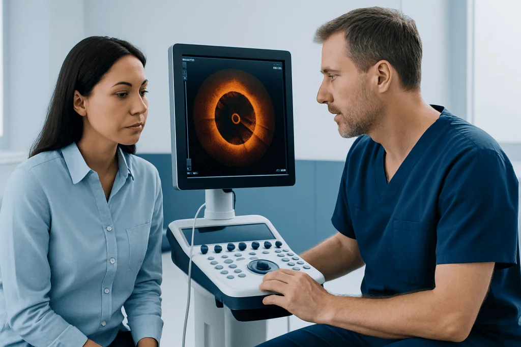
One of the main strengths of OCT is that it shows the internal structure of the coronary artery in remarkable detail. Doctors can see whether plaque is fibrous, fatty, calcified, or mixed. This matters because different plaque types behave differently and may influence which tools or techniques are used during treatment.
OCT can also identify features linked with unstable plaque, such as a thin fibrous cap, a ruptured plaque surface, or a small blood clot. In a person with chest pain or a suspected heart attack, these details may help explain why symptoms happened even when angiography alone is not fully clear. This can be valuable in selected patients with coronary artery disease or complex coronary findings.
Another important use is evaluating stents. After a stent is placed, OCT can show whether it is fully expanded, well attached to the vessel wall, and covering the narrowed segment properly. It may also reveal reasons for later problems, such as tissue overgrowth, a clot inside the stent, or a stent edge injury.
- Plaque composition and burden
- Calcium thickness and location
- Plaque rupture or erosion
- Small blood clots
- Stent expansion and apposition
- Tissue growth or restenosis inside a stent
When doctors may recommend coronary OCT

Coronary OCT is not needed for every person with heart symptoms. A cardiologist may recommend it when more detail is needed than angiography can provide. This is often the case in complex coronary artery narrowing, uncertain lesion severity, recurrent symptoms after a previous stent, or suspected problems such as stent underexpansion or thrombosis.
OCT is commonly used during procedures to open blocked or narrowed arteries. For example, it may help the doctor choose the right stent size, determine whether calcium needs special preparation, and confirm the final result after coronary angioplasty and stenting. In some patients, OCT can clarify the cause of an acute coronary syndrome when the anatomy is difficult to interpret.
It may also be considered in selected cases of chest pain with unclear findings, follow-up of previous coronary interventions, or research-based evaluation of plaque characteristics. The decision depends on the patient’s symptoms, the location of the lesion, kidney function, the amount of contrast dye likely to be used, and the overall goals of the procedure.
How the procedure is performed
OCT is usually done in a cardiac catheterization laboratory. The patient lies on an examination table while the cardiology team monitors heart rhythm, blood pressure, and oxygen levels. A catheter is inserted through an artery, commonly in the wrist or groin, and guided to the coronary arteries, similar to the process used for angiography.
Once the OCT catheter is positioned, contrast dye is briefly injected to clear blood from the section of artery being imaged. This is necessary because blood interferes with the light signal. The OCT catheter then performs a rapid pullback, collecting detailed images from inside the artery in just a few seconds.
Patients are usually awake but receive local anesthesia at the access site and often medication to help them relax. The procedure itself is typically brief, especially when added to an already planned angiogram or intervention. If OCT is being used as part of a broader treatment plan, the cardiologist may combine it with coronary angiography or other catheter-based therapies during the same session.
After the imaging is complete, the doctor reviews the pictures carefully to decide whether further treatment is needed. In experienced centers, OCT findings can immediately guide the next step, such as balloon dilation, calcium modification, stent placement, or optimization of an existing stent.
Benefits, limits, and possible risks
The main benefit of OCT is image clarity. It provides much higher resolution than many other intravascular imaging methods, allowing the cardiologist to see tiny details that may affect both diagnosis and treatment. This precision can be especially helpful in complex lesions, in evaluating stent results, and in understanding why a previous treatment may not be working as expected.
However, OCT also has limitations. Because it uses light, it cannot penetrate deeply through tissue as well as some ultrasound-based methods. It also requires contrast injection to clear blood from the artery during image acquisition. For some patients, especially those with reduced kidney function or certain unstable conditions, this may limit its use.
As with other catheter-based heart procedures, OCT carries some procedural risk, although serious complications are uncommon in appropriate settings. Risks may include bleeding or bruising at the catheter site, artery injury, irregular heart rhythm, contrast-related problems, or, rarely, damage to the coronary artery. The cardiologist weighs these risks against the potential value of the information gained.
Patients often ask whether OCT is painful. In general, the access site is numbed, and the imaging itself is not usually felt. Some people notice a brief sensation when contrast is injected, but the team closely monitors comfort and safety throughout the procedure.
How OCT compares with other heart imaging tests
OCT is one of several tools used to assess the coronary arteries. Standard angiography remains the usual first-line test during cardiac catheterization because it shows blood flow and the outline of the artery. Intravascular ultrasound, or IVUS, is another catheter-based technique that uses sound waves rather than light. Compared with IVUS, OCT generally offers sharper surface detail, while IVUS may visualize deeper vessel layers more effectively.
Noninvasive tests such as CT coronary angiography, stress testing, echocardiography, or nuclear imaging answer different questions. These tests can help detect reduced blood flow, estimate risk, or visualize heart structure and function, but they do not provide the same microscopic-like intravascular view as OCT. For that reason, OCT is usually reserved for situations in which direct internal artery imaging can change management.
In some cases, the cardiologist may combine OCT findings with pressure-based measurements or other techniques to decide whether a narrowing is truly significant. If disease is found, further treatment may include medicines, lifestyle changes, or procedures linked to broader heart disease care. Choosing among these tests depends on the clinical question, the patient’s condition, and the expertise available.
Preparation, recovery, and questions to ask
Preparation for coronary OCT is usually similar to preparation for angiography or cardiac catheterization. The care team may ask the patient not to eat or drink for several hours before the procedure. Blood tests may be needed to check kidney function, blood counts, and clotting status. The team also reviews medications, allergies, and any previous reaction to contrast dye.
After the procedure, patients are observed for a period of time while the access site is checked and vital signs are monitored. Recovery depends on whether OCT was purely diagnostic or part of a treatment such as stent placement. Some patients go home the same day, while others stay longer if they had a heart event or a more complex intervention.
It is helpful for patients to ask why OCT is being recommended, what information it may add, whether other tests are possible, and how the results could affect treatment. Questions about contrast dye, kidney health, blood-thinning medicines, and recovery time are also appropriate. A clear discussion can help patients feel informed and prepared.
Near the end of the care pathway, some patients may seek evaluation at specialized centers. Acibadem International’s multidisciplinary heart specialists and JCI-accredited hospitals diagnose and treat coronary conditions for international patients, including advanced catheter-based imaging when appropriate.
When to speak with a cardiologist
A person should speak with a cardiologist if they have chest pain, shortness of breath with exertion, unexplained fatigue, or symptoms that could suggest reduced blood flow to the heart. Medical review is also important after a previous coronary procedure if symptoms return or suddenly worsen. OCT is not a screening test for healthy people, but it may be useful when a doctor needs a closer look at a known or suspected coronary problem.
People who have already been told they need a coronary intervention may also ask whether intravascular imaging could help guide the procedure. This is especially relevant in complex blockages, heavily calcified lesions, or repeat treatment of a previously stented artery. In selected cases, OCT may support planning for other advanced therapies such as atherectomy when calcium or plaque characteristics need careful assessment.
Urgent medical attention is needed for possible heart attack symptoms, including severe chest pressure, pain spreading to the arm or jaw, fainting, or sudden shortness of breath. In those situations, emergency evaluation comes first, and OCT may be considered later if it can safely help clarify the coronary findings or optimize treatment.
Frequently asked questions
Is optical coherence tomography in cardiology the same as a regular angiogram?
No. A regular angiogram shows the outline of the artery using X-ray dye, while OCT shows detailed images from inside the artery using light. The two tests are often used together because they provide different but complementary information.
Why would a doctor choose OCT during a heart procedure?
A doctor may choose OCT when very high-resolution imaging is needed to understand plaque, guide stent placement, or assess a stent problem. It can help clarify findings that are not fully explained by angiography alone. This can make treatment planning more precise in selected patients.
Does an OCT heart test hurt?
Most patients do not find the test painful. The access site is numbed with local anesthesia, and the imaging catheter is used during a catheterization procedure that is routinely monitored for comfort and safety. Some people feel a brief warm or unusual sensation during contrast injection.
How long does coronary OCT take?
The imaging part itself usually takes only a short time, often just seconds for each scan. However, total procedure time depends on whether OCT is being added to angiography alone or combined with treatment such as balloon angioplasty or stent placement.
Are there any risks from coronary OCT?
OCT shares the general risks of cardiac catheterization, such as bleeding at the access site, artery injury, irregular heartbeat, or contrast-related issues. Serious complications are uncommon when the test is performed for appropriate reasons by experienced teams. The cardiologist considers overall health, kidney function, and the expected benefit before recommending it.
Can OCT detect blocked arteries by itself?
OCT can show the inside of an artery in great detail, including plaque and narrowing, but it is not usually used alone as a first diagnostic test. It is most often performed during catheterization after a coronary artery has already been reached. Other tests may be used first to determine whether invasive evaluation is needed.
References
- American Heart Association
- European Society of Cardiology
- Society for Cardiovascular Angiography and Interventions
- National Heart, Lung, and Blood Institute
- Mayo Clinic
This article is for general information only and is not a substitute for professional medical advice. Please consult a qualified doctor about your individual situation.
Cardiology in Turkey — costs, top hospitals & a free quote
JCI-accredited · board-certified surgeons · reply within 24h
Add us as a Preferred Source to see more of our trusted health content across Google Search, AI Overviews and Discover.
Check your numbers in seconds
BMI, calories, due date, blood pressure and 30+ more clinical calculators — free, instant, doctor-reviewed ranges.

Cardiology Department
Diagnosis and treatment of heart and vascular conditions, from prevention to advanced interventional procedures.
90 specialists in this unit