

Basal Cell Carcinoma Removal Step by Step: Local Anesthesia, Excision and Closure
Key Takeaways
- Basal cell carcinoma accounts for about 80 percent of skin cancers and grows slowly, which is why excisions are routinely scheduled weeks after biopsy without harming the outcome.
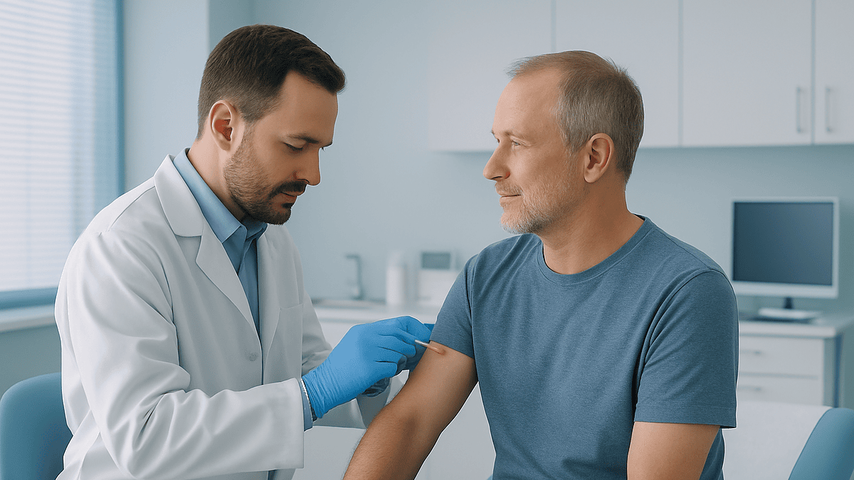
- Nearly all BCC removals are done under local anesthetic while you are awake; the injection's brief sting is usually the most uncomfortable part of the day.
- Surgeons excise an ellipse roughly three times longer than it is wide so the wound closes flat without puckered ends, aligning it with natural skin creases.
- Mohs surgery checks margins under the microscope during the visit and can take several hours, whereas standard excision takes under an hour but pathology results arrive days later.
- Surface stitches come out anywhere from three to twenty-one days after surgery, with facial wounds at the shorter end because good blood supply speeds healing.
- Having one basal cell carcinoma raises the risk of developing another, so ongoing skin examinations and lifelong sun protection are part of the treatment plan, not an afterthought.
Basal cell carcinoma removal is usually a same-day outpatient procedure done under local anesthetic. The clinician numbs the skin with an injection, cuts out the tumor with a narrow margin of healthy skin, sends the tissue to a laboratory, and closes the wound with stitches or, in some cases, a flap or graft. Most people go home within hours; stitches typically come out over the following one to three weeks.
The biopsy result arrives as a two-line message in a patient portal: basal cell carcinoma, nodular type, excision recommended. The spot in question is a pearly bump beside the nose, no bigger than a lentil, that has bled twice while shaving and healed and reopened again. Now it has a name, and a small surgery attached to it.
If you are the person reading that message, the questions come quickly. How soon does this need to happen? Will it hurt? What will my face look like afterward? Searching basal cell carcinoma removal what to expect returns pages that describe the steps but rarely the texture of them: the sting of the numbing needle, the odd tugging sensation during stitching, the wait for the pathology report.
This explainer walks through the whole sequence, from the moment you sit down in the procedure chair to the day the last stitch comes out, grounded in what mainstream medical evidence actually says rather than what a waiting-room conversation might suggest.
Basal cell carcinoma removal: what to expect starts with knowing what this cancer does
Basal cell carcinoma (BCC) is a cancer that begins in the basal cells, the small round cells in the deepest layer of the epidermis that constantly produce new skin. According to the Cleveland Clinic, it accounts for roughly 80 percent of all skin cancers, which makes it the most common cancer of any kind in the United States, a point the CDC also underlines in its skin cancer overview.
The behavior of this tumor shapes everything about how it is removed. BCC grows slowly, often over months or years, and it very rarely spreads to lymph nodes or distant organs. The Mayo Clinic describes it as a cancer that, left untreated, tends to grow wider and deeper into nearby tissue, skin, cartilage and occasionally bone, rather than one that travels through the bloodstream. That local behavior is why the treatment is local too: cut out the tumor completely, confirm under the microscope that no cancer cells remain at the edges, and the problem is usually resolved at that site.
Two features make this more complicated than it sounds. First, BCC often extends beyond what the eye can see, sending thin strands of tumor cells under normal-looking skin. Second, the places it favors, the face, ears, scalp and neck, are precisely where skin is precious and tight. Surgeons therefore balance two goals that sometimes pull against each other: remove enough to be confident the cancer is gone, and preserve enough to keep function and appearance.
Every step described below, from the choice between standard excision and Mohs surgery to the type of closure, follows from that balance. Understanding it turns a confusing menu of options into a logical set of trade-offs your treating team will weigh with you.
How urgent is BCC removal? Slow-growing does not mean optional
The honest answer is that this is a cancer with a calendar measured in months, not days. Because BCC grows slowly and almost never metastasizes, the NHS and the Mayo Clinic both classify it as a non-melanoma skin cancer that is highly treatable, and clinics routinely schedule excisions weeks after a biopsy without compromising the outcome. Nobody needs to rush from a biopsy result to an operating room the same afternoon.
Slow, however, is not the same as harmless. The Mayo Clinic notes that untreated BCC can invade surrounding structures, and the further it spreads under the skin, the larger the eventual wound and the more complex the reconstruction. A tumor that could have been removed with a simple line of stitches can, after years of neglect, require a skin flap or graft, or involve the eyelid margin, the cartilage of the nose, or the ear canal. Delay rarely changes whether the cancer can be treated; it often changes how much skin the treatment costs.
Certain features push clinicians to move faster. Aggressive subtypes, such as infiltrative or morpheaform BCC, tend to have wider hidden extension. Tumors near the eye, nose, lips or ears sit in what dermatologists call high-risk zones, where even small growth matters. Very large lesions, recurrent tumors, and people whose immune systems are suppressed by medication or illness also warrant a shorter timeline.
What this means in practice: if your team suggests a date several weeks away, that is normal and evidence-based. If a scheduling delay stretches well beyond that and the lesion is visibly changing, bleeding more, or growing, call and ask whether the plan should be revisited. The decision about timing belongs to your treating team, but you are entitled to understand the reasoning behind it.
What actually happens: the local anesthesia step
Nearly all BCC removals happen while you are fully awake, sitting or lying in a procedure room rather than an operating theater. General anesthesia, the medicine-induced sleep used for major surgery, is uncommon for this procedure and generally reserved for very large or complex reconstructions.
The process begins with the clinician marking the visible tumor with a skin pen, often drawing a second line a few millimeters outside it to indicate the planned margin. Photographs are common, both for the record and because after numbing and swelling the edges can be harder to see. The skin is then cleaned with an antiseptic.
Next comes the part most people dread: the injection. A local anesthetic, a medicine from the class that includes lidocaine, is injected into and around the lesion. These medicines work by blocking sodium channels in nerve fibers, which stops the electrical signal that would otherwise carry pain to the brain. Many clinicians combine the anesthetic with a small amount of epinephrine, a hormone that narrows blood vessels; it reduces bleeding during the surgery and keeps the anesthetic in place longer. Which agents are used, and how much, is entirely the clinician’s decision.
The Mayo Clinic describes the injection as producing a brief stinging or burning sensation that fades within seconds. Some clinicians buffer the solution or inject slowly to soften this. After a short wait, the skin is tested with a gentle touch or pinch. You should feel pressure and movement but no sharpness. If you do feel a sharp sensation at any point, say so; more anesthetic can be added at once.
From here on, the experience for most people is strange rather than painful: a sense of tugging, the smell of the cautery device used to seal small blood vessels, the murmur of the surgical team.
The excision itself: margins, depth and where the tissue goes
Standard surgical excision is the removal of the tumor together with a surrounding rim of normal-looking skin, called the margin, in a single piece. The margin exists because BCC cells can extend invisibly beyond the visible edge; taking healthy-appearing skin around the tumor improves the odds that the whole cancer comes out at once. The width of that margin depends on the tumor’s size, subtype and location, and is chosen by the surgeon according to established guidelines.
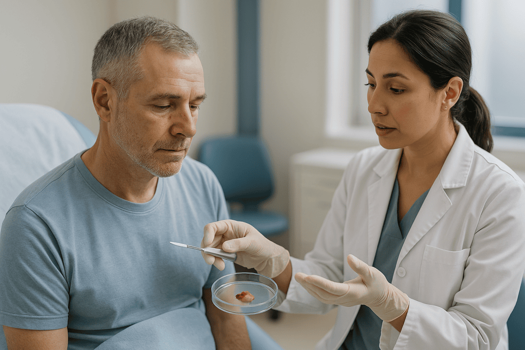
The surgeon typically draws an ellipse, an elongated oval shape, around the lesion. That shape is not arbitrary. A circular wound closed with stitches puckers at each end into small cones of skin sometimes called dog-ears; an ellipse roughly three times as long as it is wide closes flat. The long axis is usually aligned with natural skin creases so the eventual scar hides in a line the face already has.
Using a scalpel, the surgeon cuts along the marked ellipse and down through the full thickness of the skin into the fatty layer beneath, then lifts the specimen away. BCC arises in the epidermis and can push into the dermis, the thicker middle layer, so the excision must include both. A small stitch or ink mark is often placed on the specimen to orient it, so the pathologist can report exactly which edge, if any, still contains tumor.
Bleeding is controlled with a handheld electrocautery device, which uses heat to seal small vessels. The specimen is placed in preservative and sent to the laboratory, where it is processed into thin slices and examined under a microscope. According to MedlinePlus, this pathology review confirms both the diagnosis and whether the margins are clear. Results generally take days, which is why standard excision involves a wait after the wound is already closed.
Mohs surgery vs excision: how the two paths differ
Mohs micrographic surgery is a form of excision in which the surgeon removes the tumor in thin layers and examines each layer under the microscope while you wait, continuing only where cancer cells remain. The Mayo Clinic and Johns Hopkins describe it as a technique designed to remove all of the cancer while sparing as much healthy tissue as possible, which is why it is often chosen for the face, ears, nose, eyelids, and for recurrent or aggressive tumors.
The patient experience differs mainly in rhythm. After each layer is removed, the wound is bandaged and you return to a waiting area, sometimes for an hour or more, while the tissue is frozen, sliced and stained. If the edges are clear, the surgeon closes the wound. If not, more anesthetic is added and another layer is taken from the precise spot where cancer was seen. The Mayo Clinic notes that the whole process can take several hours, and it is not possible to predict in advance how many stages will be needed.
| Feature | Standard excision | Mohs surgery |
|---|---|---|
| Margin check | Pathology days later | During the visit, layer by layer |
| Time in clinic | Usually under an hour | Several hours, per Mayo Clinic |
| Tissue removed | Preset margin around tumor | Only where cancer is found |
| Typical use | Trunk, limbs, low-risk tumors | Face, ears, recurrent or aggressive tumors |
| If margins positive | Second procedure needed | Handled the same day |
Neither approach is universally better. Guidelines match the technique to the tumor and its location, and your treating team will explain which fits your case. Other options, including curettage and electrodesiccation (scraping followed by heat), cryosurgery (freezing), radiation therapy, and prescription topical medicines for certain superficial tumors, are listed by the Mayo Clinic and NHS as alternatives chosen for specific circumstances rather than substitutes for surgery in general.
Closure: stitches, flaps, grafts, and sometimes nothing at all
Once the cancer is out and the margins are clear, or the surgeon is confident enough to proceed while awaiting pathology, the wound must be closed. Four broad options exist, and the choice depends on the size and depth of the wound, the tightness of the surrounding skin, and how the site moves.
Primary closure, the simplest, brings the two edges of the ellipse together and stitches them in one line. Surgeons often use two layers: deeper absorbable stitches that dissolve on their own and hold the wound’s strength, and finer surface stitches that shape the skin edge and are removed later. On the face these surface stitches are very fine to minimize track marks.
A local flap is used when the edges cannot meet without pulling. The surgeon loosens a nearby piece of skin, still attached by its own blood supply, and rotates or slides it into the defect. Flaps preserve the color and texture of the original area because the skin comes from next door, at the cost of a longer, sometimes geometric scar.
A skin graft, by contrast, is a piece of skin removed entirely from another site, often behind the ear or the collarbone, and stitched into the wound, where it grows new blood vessels over several days. Grafts suit large or awkward defects but can heal paler or slightly sunken.
Finally, some wounds are simply left to heal on their own, a process called second-intention healing. The Mayo Clinic notes this is sometimes chosen for concave sites like the inner ear or the temple, where new skin fills in from the edges and the result can be surprisingly good. Whichever method is chosen, a pressure dressing is applied and you are given written wound-care instructions before leaving.
Is basal cell carcinoma removal painful? What patients actually report
Most people describe the injection of local anesthetic as the single most uncomfortable moment of the entire day, and even that lasts only a few seconds. Once the skin is numb, the surgery itself should not hurt. You will feel pressure, tugging, and the vibration of instruments, and you may hear or smell the cautery, but sharp pain means the anesthetic needs topping up, not that pain is an expected part of the process.
There are a few honest caveats. The nose, lips, fingertips and ears are densely supplied with nerve endings, and injections there sting more than on the back or arm. Very anxious patients sometimes experience the tugging sensation as more intense than it is; telling the team you are nervous allows them to talk you through each step, which many people find helps. Long Mohs procedures may require re-numbing between stages as the anesthetic wears off, and each top-up brings a smaller version of the initial sting.
After the anesthetic fades, usually within a few hours, the wound typically feels sore, tight and tender to touch. MedlinePlus and the Mayo Clinic describe this as mild to moderate discomfort that most people manage with over-the-counter pain relief, cold compresses, and keeping the head elevated for facial wounds to limit swelling. Which pain reliever suits you, if any, is a question for your team, particularly if you take blood thinners or have kidney, liver or stomach conditions.
Throbbing pain that worsens after the second day, rather than easing, is not typical and is one of the red flags covered later in this article. The pattern the evidence describes is simple: a sting at the start, numbness in the middle, and a sore, manageable evening.
Who is usually offered excision, and who is asked to wait or consider another route
Surgical excision, whether standard or Mohs, is the mainstay of BCC treatment for most adults. The Mayo Clinic and NHS list it as the first-line approach for tumors that can be removed with an acceptable functional and cosmetic result, which describes the great majority of cases. If you are generally healthy, the lesion is a manageable size, and you can tolerate lying still under local anesthetic, you will most likely be offered surgery.
Certain situations prompt a pause or a different plan. People taking anticoagulants, the class of medicines that thin the blood, are not usually told to stop them; guidelines increasingly favor continuing them because the risk of a clot outweighs the risk of manageable bleeding. The decision, however, rests with the prescribing clinician and surgeon together, never with the patient alone. Anyone with a pacemaker or implanted defibrillator should mention it, since electrocautery settings may be adjusted.
Active infection at or near the site usually delays surgery until it clears. A recent heart attack, stroke or major surgery may lead the team to wait until recovery is stable. Very frail patients, or those with many low-risk superficial tumors, may be offered non-surgical options such as curettage, cryosurgery, radiation, or topical prescription treatments, which the Mayo Clinic describes as appropriate for specific tumor types and patient circumstances.
Radiation therapy, which uses focused high-energy beams to destroy cancer cells over a course of sessions, is often considered for people who cannot undergo surgery or for tumors where surgery would be disfiguring. For the rare BCC that has grown too large or deep for surgery or radiation, the Mayo Clinic lists targeted medicines called hedgehog pathway inhibitors, which block a signaling route the tumor depends on. These are specialist decisions and fall well outside routine excision.
BCC excision recovery time: what the first two weeks usually look like
Recovery from a basal cell carcinoma excision is quicker than most people fear, but it follows a recognizable arc. The day of surgery is mostly about pressure and protection: keeping the initial dressing dry and in place, usually for the first day or two, and resting with the wound elevated. A small amount of blood on the dressing is normal; steady bleeding that soaks through is not.
Days two through four are peak swelling and bruising, especially around the eyes and forehead, where fluid drifts downward under gravity. A wound on the temple can produce a black eye that alarms coworkers more than it bothers you. Soreness generally eases over these days rather than intensifying.
Wound care during the first one to two weeks typically involves gently cleansing the site, applying a thin layer of plain petroleum-based ointment to keep it moist, and covering it with a fresh non-stick dressing. MedlinePlus describes this moist-wound approach as supporting faster healing and less crusting than letting the wound dry out. Your team will tell you when showering is permitted and whether the site may get wet.
Surface stitches are removed at a follow-up visit. MedlinePlus notes that stitches generally stay in for anywhere from three to twenty-one days depending on the location, with facial wounds at the shorter end because good blood supply speeds healing and early removal reduces track marks, and wounds on the back or legs at the longer end.
Activity restrictions are short but real. Bending, lifting and strenuous exercise raise blood pressure at the wound and can cause bleeding or stretch the closure; most teams advise avoiding them for one to two weeks, longer for flaps and grafts. Desk work and gentle walking are usually fine within a day or two.
Scars, sun protection and the months that follow
Every excision leaves a scar. What changes with time and care is how visible it is. In the early weeks the line appears pink or red and slightly raised, because the body has flooded the area with new blood vessels and collagen to knit it together. This is normal healing, not a sign the scar will stay that way.
Over the following months the scar remodels. Redness fades toward skin tone, the ridge flattens, and the collagen reorganizes into a more orderly pattern. Scars on the face generally mature faster than those on the trunk or limbs, again because of blood supply. The final appearance is not the appearance at stitch removal; judging a scar at two weeks is like judging a house at the framing stage.
Sun protection matters more than any cream. Ultraviolet light darkens healing scars, sometimes permanently, and the same UV exposure caused the cancer in the first place. The CDC and Mayo Clinic recommend broad-spectrum sunscreen, protective clothing, hats, and seeking shade during peak hours for everyone who has had a skin cancer. On a fresh scar, physical coverage is preferable until the wound is fully closed, after which sunscreen can be applied directly.
Silicone gel sheets or gels are commonly suggested once the wound has closed; the evidence for them is moderate rather than definitive, and your team can advise whether they suit your scar. Gentle massage of a matured scar may soften it, though it should never be started before the surgeon clears it.
Occasionally a scar heals thick, itchy and raised, a pattern called a hypertrophic scar, or spreads beyond the original wound, a keloid. Both are more common on the chest, shoulders and in people with darker skin tones, and both are treatable with options your dermatologist can discuss. Mention any thickening early rather than waiting.
Risks of BCC removal and what the evidence says about recurrence
Basal cell carcinoma removal is a low-risk procedure, but no surgery is risk-free, and a realistic picture helps you recognize problems early. The complications described by MedlinePlus and the Mayo Clinic fall into a few categories.
Bleeding is the most common early issue, usually as oozing that responds to firm pressure for ten to fifteen minutes. A hematoma, a collection of blood under the closed skin, is less common and may need to be drained. Infection affects a small minority of clean skin wounds and shows up as spreading redness, warmth, increasing pain and sometimes pus, generally after the first two days.
Nerve injury is site-specific. Small sensory nerves are always cut during excision, so some numbness around the scar is expected and often improves over months. Rarely, deeper surgery on the temple, jaw or neck can affect motor nerves controlling facial movement; surgeons discuss this beforehand when the location warrants it. Wound-edge separation, called dehiscence, can happen if stitches are stressed too early. Flap or graft failure, where the moved tissue does not survive, is uncommon and more likely in smokers and people with diabetes.
Then there is recurrence. The Mayo Clinic notes that BCC can come back at the same site, particularly if margins were involved or the tumor had an aggressive pattern, and that people who have had one BCC have a higher risk of developing new ones elsewhere. This is why follow-up skin examinations are recommended after treatment; the interval is set by your team based on your history. Reported recurrence figures vary widely by technique, tumor type and follow-up length, and any specific percentage should come from your surgeon with reference to your own pathology rather than from a general article.
What people often get wrong about basal cell carcinoma removal
Misconceptions travel fast in waiting rooms and comment sections. A few deserve correcting because they change how people behave.
“It’s just a skin thing, not real cancer.” BCC is a genuine malignancy, and the Mayo Clinic is explicit that untreated tumors invade surrounding tissue. Its low tendency to spread does not make it benign; it makes it treatable.
“If the biopsy removed the bump, the cancer is gone.” A biopsy samples tissue to establish a diagnosis. Even when the visible lesion seems to vanish afterward, tumor cells commonly remain in the deeper skin, which is why excision follows biopsy in most cases.
“Mohs is always the best option.” Mohs offers precise margin control and tissue sparing, which is valuable on the face. For a small low-risk tumor on the back, standard excision achieves the goal in a fraction of the time, and guidelines match the technique to the tumor rather than ranking one above the other.
“Clear margins mean I’m done with dermatology.” The Mayo Clinic notes an elevated risk of new BCCs after a first one. Ongoing skin checks are part of the plan, not an optional extra.
“I should stop my blood thinner before surgery.” Never change a prescribed medicine on your own. Many surgeons prefer patients to continue anticoagulants for minor skin surgery; the decision belongs to the prescribing clinician and the surgeon.
“Creams can replace surgery.” Prescription topical treatments are listed by the Mayo Clinic for certain superficial BCCs only, under close supervision. They are not interchangeable with excision for nodular or deeper tumors.
“Once it’s healed, I can skip the sunscreen.” UV exposure drove the first tumor and raises the risk of the next. Protection is lifelong.
Are you a cancer survivor if you had basal cell carcinoma?
The question comes up more often than clinicians expect, sometimes with a note of embarrassment. People who have had a small BCC removed in an afternoon may feel it would be presumptuous to use a word associated with chemotherapy and hospital stays. Others feel dismissed when relatives shrug off their diagnosis.
By strict definition, yes. BCC is a cancer, classified as such by the CDC, the NHS and every major medical body, and a person who has been diagnosed with cancer and treated for it fits the widely used definition of a survivor from the day of diagnosis onward. Whether the label feels right is personal, and there is no wrong answer.
What matters more than the word is what the diagnosis tells you. It confirms that your skin has accumulated enough ultraviolet damage to produce a malignancy, and the Mayo Clinic notes that a first BCC raises the likelihood of others. That knowledge should change behavior in concrete ways: regular skin checks by a clinician at intervals your team recommends, monthly self-examination for new or changing spots, and consistent sun protection.
Some practicalities also follow. Life and travel insurance applications typically ask about cancer history, and BCC usually needs to be declared even though insurers often treat it differently from other cancers. Registries in many regions count non-melanoma skin cancers separately or not at all, which is one reason public awareness lags behind how common the disease is.
A BCC diagnosis is not a reason for alarm, and it is not a reason for dismissal either. It sits in the space between: a real cancer, highly treatable, that asks for lifelong attention to the skin rather than fear.
Questions to ask your care team before basal cell carcinoma removal
A ten-minute consultation moves quickly, and the questions you meant to ask often surface on the drive home. Writing them down beforehand helps, and a companion who takes notes helps more. The list below covers what patients most often wish they had asked.
- What subtype of BCC did the biopsy show, and does that affect which technique you recommend?
- Are you proposing standard excision or Mohs surgery, and why for this location?
- How large do you expect the final wound to be, and what closure do you anticipate: a straight line, a flap, or a graft?
- Roughly how long will I be in the clinic, and should I bring someone to drive me home?
- Should I continue all my regular medicines, including blood thinners and aspirin, and who makes that call?
- What wound care will I need to do at home, and for how long?
- When will stitches come out, and when can I shower, exercise, and return to work?
- What will the pathology report tell us, when will I hear, and what happens if margins are not clear?
- Where will the scar sit, and what can I realistically expect it to look like at a year?
- Which nerves or structures near this site could be affected?
- How often should I have skin checks afterward, and who will do them?
- What symptoms should prompt me to call before my scheduled follow-up?
You are also entitled to ask about alternatives, including non-surgical options, and about what would happen if you chose to delay. A good team welcomes these questions; they indicate a patient who will follow wound-care instructions and attend follow-up, which are the two things most within your control.
When to call your doctor after basal cell carcinoma removal
Most wounds heal uneventfully, and most questions can wait for the scheduled stitch-removal visit. A small set of signs, however, should prompt a same-day call to your surgical team or, outside clinic hours, a visit to urgent or emergency care. These are drawn from the post-procedure guidance published by MedlinePlus and the Mayo Clinic.
Call promptly if you notice:
- Bleeding that continues despite fifteen minutes of firm, uninterrupted pressure, or that soaks through the dressing repeatedly.
- A rapidly enlarging, tense, painful swelling under the wound, which may indicate a hematoma.
- Redness spreading outward from the wound edges, increasing warmth, worsening pain after the second day, or pus draining from the site.
- Fever or chills, which may signal infection beyond the skin.
- Wound edges pulling apart, or a flap or graft turning dark purple, gray or black.
- New weakness in facial movement, such as difficulty closing the eye or an uneven smile, that was not present immediately after surgery.
- Numbness or tingling that spreads rather than staying confined to the scar area.
- Any allergic reaction, including hives, swelling of the lips or tongue, or difficulty breathing, which requires emergency care.
Beyond the immediate recovery period, contact your dermatologist if the scar develops a new bump, ulcer, pearly area or bleeds without injury, or if you find a new suspicious spot anywhere on your skin. These may be nothing, but they are the pattern by which recurrence and new tumors announce themselves, and early assessment keeps any further treatment small.
The threshold for calling should be low. Surgical teams would far rather reassure ten patients over the phone than miss one developing infection or bleed. Every decision about what to do next remains with the clinicians who know your case; your job is simply to report what you see.
Frequently asked questions
How urgent is it to have basal cell carcinoma removed?
It is important but rarely an emergency. BCC grows slowly over months and almost never spreads to other organs, so scheduling excision several weeks after a biopsy is standard and does not compromise treatment. Delay matters because untreated tumors keep growing into surrounding tissue, enlarging the eventual wound. Aggressive subtypes, tumors near the eyes, nose or ears, and people with weakened immunity are usually moved up the schedule by their team.
Is basal cell carcinoma removal painful?
The injection of local anesthetic stings for a few seconds, and after that the procedure itself should not hurt. You feel pressure and tugging, not sharpness; if you do feel sharp pain, the clinician adds more anesthetic. Once numbness fades, the wound is sore and tight for a few days, which most people manage with simple measures their team suggests. Pain that worsens after day two rather than easing warrants a call.
How long does it take to recover from basal cell carcinoma removal?
Most people return to desk work within a day or two and to full activity within one to two weeks, longer for flaps and grafts. Stitches are removed within three to twenty-one days depending on location, per MedlinePlus. Swelling and bruising peak around days two to four. Scars keep fading and flattening for many months, so the appearance at stitch removal is not the final result.
Are you a cancer survivor if you had basal cell carcinoma?
Yes, by the widely used definition. BCC is a genuine cancer, and anyone diagnosed and treated for cancer qualifies as a survivor from diagnosis onward. Whether the label feels right is personal. What matters practically is that a first BCC raises your risk of future skin cancers, so regular skin checks and consistent sun protection become lifelong habits, and insurance forms usually require the diagnosis to be declared.
What is the difference between Mohs surgery vs excision for BCC?
Standard excision removes the tumor with a preset margin in one step and sends it to a laboratory, with results days later. Mohs surgery removes thin layers and examines each under the microscope during the visit, continuing only where cancer remains, which spares healthy tissue but takes several hours. Mohs is often chosen for the face and for recurrent or aggressive tumors; excision suits many low-risk tumors on the trunk and limbs.
Will I be awake during basal cell carcinoma removal?
Almost certainly yes. Nearly all BCC excisions, including Mohs surgery, are done under local anesthetic in an outpatient procedure room. You lie or sit comfortably, the area is numbed by injection, and you can talk with the team throughout. General anesthesia is reserved for very large or complex reconstructions. Many people find that hearing what is happening step by step makes the experience less unsettling.
What does BCC excision recovery time look like for a wound on the face?
Facial wounds often heal faster than those elsewhere because of rich blood supply. Expect swelling and possible bruising, sometimes drifting toward the eye, for the first several days. Fine surface stitches typically come out toward the shorter end of the three-to-twenty-one-day range MedlinePlus describes. Keeping the head elevated, avoiding bending and heavy lifting, and following moist wound care help. Redness in the scar fades over months.
Should I stop blood thinners before basal cell carcinoma removal?
Do not change any prescribed medicine on your own. For minor skin surgery, many surgeons prefer that patients continue anticoagulants because the risk of a clot generally outweighs the risk of manageable bleeding. The decision belongs jointly to your surgeon and the clinician who prescribes the medicine. Tell the team about every medicine and supplement you take, including aspirin and fish oil, during the pre-procedure consultation.
What happens if the margins are not clear after excision?
It means the pathologist found cancer cells at one or more edges of the removed tissue, so tumor may remain in the skin. Your surgeon will usually recommend a further procedure, often a re-excision of the affected edge or Mohs surgery, to remove what is left. Occasionally observation or another treatment is considered based on the tumor type and location. Positive margins are a known, manageable outcome, not a failure.
Can basal cell carcinoma come back after it is removed?
It can, at the same site or as a new tumor elsewhere. The Mayo Clinic notes that recurrence is more likely when margins were involved or the tumor had an aggressive pattern, and that people who have had one BCC face a higher risk of developing others. This is why follow-up skin examinations, at intervals set by your team, and monthly self-checks are recommended after treatment.
References
- NHS: Non-melanoma skin cancer: Treatment
- MedlinePlus: Basal cell skin cancer
- MedlinePlus: Surgical wound care – open
This article is for general information only and is not a substitute for professional medical advice. Please consult a qualified doctor about your individual situation.

More from the Blog

Which Tests Classify Acute Lymphocytic Leukemia and Set the Risk Group Before Treatment?
Acute lymphocytic leukemia is classified with a complete blood count and blood smear, a bone marrow aspirate and biopsy, flow cytometry to identify B-cell…

How Myelogenous Leukemia Treatment Differs for Chronic and Acute Forms
Chronic myelogenous leukemia (CML) and acute myeloid leukemia (AML) are treated very differently because they behave differently. CML usually grows slowly and is driven…

When Hormone Therapy Joins Prostate Cancer Treatment: With Radiation, Before Surgery or Alone
Hormone therapy for prostate cancer is used in three main situations: alongside radiation for intermediate- and high-risk disease, where it shrinks the gland and…

How Long Targeted Therapy Continues: Cycles, Scans and What Decides When It Stops
Targeted therapy has no single length. Courses given after surgery to lower recurrence risk usually have a set end point, commonly around a year…
Compression Garments and Cancer Rehabilitation: Managing a Swollen Arm or Leg Day to Day
Compression garments after cancer treatment are fitted elastic sleeves, gauntlets, stockings or adjustable wraps that apply graded pressure to a limb with lymphedema, helping…
Colon Cancer Surgery Step by Step: Removing the Segment, Rejoining the Bowel, Recovery
Colon cancer surgery, usually called a colectomy or bowel resection, removes the section of colon containing the tumor together with a margin of healthy…