

Cartilage Grafts in Revision Rhinoplasty: Why Rib or Ear Cartilage Is Sometimes Needed

Key Takeaways
- The septum is the first-choice graft source, and it is usually depleted or scarred after a first rhinoplasty, which is the main reason revision surgery turns to the ear or rib.
- Ear cartilage is curved and flexible, suited to tip shaping and small contour fixes; rib cartilage is thick and strong, suited to rebuilding a whole bridge or supporting a collapsed tip.
- Only a segment of the cartilaginous front of one rib is removed, leaving the bony rib intact, so the chest wall remains stable and the ribcage does not change shape.
- Warping is a mechanical property of carved cartilage, reduced by central carving, internal reinforcement or dicing, but not eliminated by any technique.
- Mayo Clinic describes external splint removal at about one week and continuing change in nasal shape for up to a year, so early appearance is swelling rather than result.
- Rare but serious chest complications, including pneumothorax, are why sudden breathlessness or sharp worsening chest pain after rib harvest needs emergency assessment rather than watchful waiting.
Revision rhinoplasty often needs extra cartilage because the nasal septum, the usual source, was used or weakened during the first operation. Surgeons then borrow from the ear when small, flexible pieces are enough, or from a rib when the nose needs strong, straight structural support. Rib cartilage is plentiful but involves a chest incision, more early soreness and a tendency to warp, so the choice rests with the treating team.
The consultation usually starts with a mirror rather than a scan. Someone tilts their head, points to a bridge that dips where it once was straight, or a tip that has slowly pinched inward, and asks the question they have been rehearsing for months: “Can this be fixed, and why does everyone keep talking about my ribs?”
It is a fair question. A first rhinoplasty is mostly about shaping what is already there. A second one is often about rebuilding what is missing. When surgeons discuss revision rhinoplasty rib cartilage, they are not proposing something exotic; they are acknowledging that the nose may no longer contain enough of its own building material to hold a new shape.
What follows is an honest walk through that reasoning: where graft cartilage comes from, why the ear sometimes suffices and the rib sometimes does not, what the chest incision genuinely feels like, and which trade-offs a careful care team weighs before choosing either.
Why does a revision rhinoplasty need a cartilage graft at all?
Picture the nose as a small tent. The skin is the canvas; the cartilage underneath is the set of poles. A primary rhinoplasty reshapes those poles, often by trimming them. Take away too much, or let scar tissue tighten the canvas over weakened supports, and the tent begins to sag in predictable ways: a scooped bridge, a drooping or pinched tip, nostrils that collapse on a deep breath.
A cartilage graft is a piece of the patient’s own cartilage moved from one place to another to restore missing support or shape. In revision surgery it serves three broad purposes. It can rebuild structure, such as a strut that holds the tip up. It can add volume where a bridge was over-reduced. And it can reinforce the side walls of the nose so they no longer fold inward when air rushes past, a problem doctors call nasal valve collapse.
The first choice of material is almost always the septum, the cartilage wall dividing the two nasal passages, because it is straight, firm and already inside the nose. The catch with revision cases is that the septum was frequently harvested during the first operation. What remains is often thin, scarred or needed to keep the nose standing. That shortage, described in patient guidance from Mayo Clinic and Cleveland Clinic, is the single most common reason a surgeon looks elsewhere.
Elsewhere means the ear or the rib. Neither is a downgrade in principle; each is a different material with different behavior, and the decision depends on what the nose needs rather than on what sounds more dramatic. Revision rhinoplasty cartilage graft planning is really an inventory exercise: how much is missing, how strong must the replacement be, and how much of the patient’s anatomy can reasonably be borrowed.
Septum, ear or rib: where does graft cartilage come from?
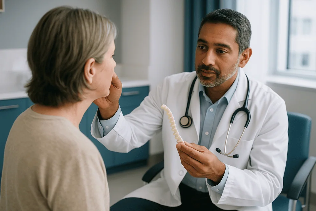
Three donor sites cover the vast majority of revision work, and each has a personality. The septum is straight and rigid, ideal for struts and spreader grafts that open the breathing passage, but supply is limited after a first surgery. Ear (auricular) cartilage, taken from the bowl of the ear, is naturally curved and springy, good for reshaping a tip or camouflaging a small dent, less good for bearing weight. Rib (costal) cartilage is the bulk supplier: long, thick, carvable into almost any shape, and stiff enough to rebuild an entire bridge.
| Donor site | Typical amount available | Character of the cartilage | Commonly used for | Main trade-offs |
|---|---|---|---|---|
| Septum | Small to moderate; often depleted after a first rhinoplasty | Straight, firm, thin | Spreader grafts, tip struts, minor bridge work | Over-harvesting can weaken nasal support |
| Ear | Small | Curved, flexible, thin | Tip refinement, side-wall support, filling small depressions | Curvature limits use for straight struts; usually a subtle change to ear contour |
| Rib | Large | Thick, strong, carvable | Full bridge reconstruction, major tip support, severe collapse | Chest incision, early soreness, tendency to warp, rare chest complications |
Two things stand out from that table. Rib cartilage is chosen for quantity and strength, not because it is intrinsically superior. And the ear is chosen when the problem is shape rather than structure. Many revisions use more than one source in the same operation, such as a rib strut for support with ear cartilage layered over the tip for softness.
The choice is made by the operating surgeon after examining the nose inside and out, often with the earlier operative notes in hand. No table replaces that assessment; it only explains the vocabulary.
Revision rhinoplasty rib cartilage: what actually happens in the operating room
The operation runs in two halves that often overlap. The first is harvest. Under general anesthesia, the surgeon makes a short incision on the lower chest, usually along a fold beneath the breast or just below it, where clothing tends to cover the scar. The muscle is parted rather than cut, exposing the cartilaginous front portion of a rib. A segment of that cartilage is removed, leaving the outer covering, called the perichondrium, and the bony rib intact. The wound is checked carefully to confirm the lining around the lung has not been breached, then closed in layers.
The second half is the nose itself. Most revisions that involve rib are performed through an open approach, meaning a small incision across the strip of skin between the nostrils lets the surgeon lift the skin and see the entire framework. Scar tissue from the first operation is released. Whatever cartilage remains is assessed and, where possible, preserved.
Then the carving begins. Rib cartilage arrives as a curved block. The surgeon slices it into the pieces the nose needs: a long strip to rebuild the bridge, a straight strut to prop the tip, thin sheets to reinforce collapsing side walls. Many surgeons cut from the center of the block rather than the edges, and some leave the carved pieces sitting in saline for a while to see how they bend before placing them. Both habits exist to anticipate warping, which has its own section below.
Grafts are secured with fine sutures, the skin is redraped, the incisions are closed, and an external splint is applied. A revision using rib generally takes longer than a first-time rhinoplasty because two operative sites are involved and scar tissue slows every step. Length is a planning detail the anesthesia team factors in, not a measure of difficulty for the patient to worry about.
Ear cartilage rhinoplasty: when a smaller donor site is enough
Not every revision needs a rib. A tip that lost definition, a small visible dent on the bridge, a side wall that folds on inhalation but is otherwise intact: these are problems of contour and modest reinforcement, and the ear supplies exactly that kind of cartilage.
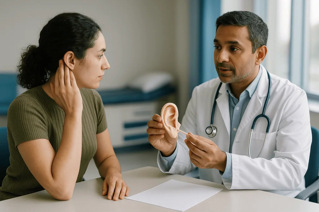
The graft comes from the concha, the cup-shaped hollow of the outer ear just outside the ear canal. The surgeon reaches it through an incision either behind the ear or inside the bowl, lifts the skin, removes a piece of cartilage while leaving the ear’s outer rim and structural folds untouched, and closes the wound. Because the skin on both sides of the ear is preserved, the ear usually keeps its overall shape, though some people notice a subtle change in the depth of the bowl when they look closely.
Inside the nose, ear cartilage behaves differently from rib. Its natural curve makes it awkward for a straight strut but useful for wrapping around a tip or bolstering the arch of a nostril. It is softer, so it blends into thin skin without the sharp edges that thicker material can telegraph. Surgeons often stack two pieces back-to-back, curve against curve, when they need something straighter.
Recovery at the donor site is typically gentler than at the chest. Soreness is localized, and there is no muscle or rib involved. Bruising behind the ear and a feeling of numbness over the bowl are common early on and generally settle as the small nerves recover.
The limitation is honest and simple: there is not much of it. When a nose needs a whole new bridge or the tip has no remaining support, ear cartilage alone is rarely sufficient. That is the moment the conversation turns toward the rib, and a good surgeon will explain that shift in terms of quantity and load rather than as a reflection on how “bad” the nose is.
Who is usually offered rib cartilage, and who is asked to wait
Rib grafting tends to be proposed when the deficit is structural and large. Typical scenarios include a bridge that was over-reduced and now dips into a saddle shape, a tip that has lost its supporting cartilage and droops or collapses, side walls that fold inward badly enough to obstruct breathing, and cases where two or more prior operations have used up the septum and ear. Some noses damaged by injury or infection rather than surgery fall into the same category.
Timing matters as much as anatomy. Mayo Clinic notes that a nose continues to change for up to a year after rhinoplasty as swelling resolves and tissues settle, so surgeons commonly ask patients to wait roughly that long before committing to a revision. Operating earlier risks correcting a problem that would have softened on its own, and it means working through tissue that is still inflamed and unpredictable.
A care team may also counsel waiting or reconsidering when the main concern is very subtle, when skin is unusually thin and would show every edge of a thick graft, or when expectations point toward a level of perfection that no reconstruction can reliably deliver. People with conditions affecting wound healing, those who smoke, and those with uncontrolled medical problems are typically asked to address those factors first; nicotine narrows small blood vessels and is linked to poorer healing after nasal surgery in guidance from the NHS and Mayo Clinic.
Older adults are sometimes told that rib cartilage calcifies with age, becoming harder to carve. That is true as a tendency, not a rule, and imaging of the chest can help a surgeon judge whether a particular rib is usable. None of these considerations is a verdict. They are the questions a thoughtful team works through before recommending revision rhinoplasty rib cartilage, or steering toward a smaller procedure, or advising that the wiser course is no further surgery at all.
How painful is a rib cartilage graft? An honest answer
Most people who have had the operation describe the chest as the part they notice, not the nose. The rib site aches in a way that is deep rather than sharp, and it flares with anything that moves the chest wall: a deep breath, a laugh, a cough, rolling over in bed. The first two or three days are usually the peak, after which the ache typically settles into a bruised feeling that eases over the following weeks. That pattern is consistent with the general recovery arc described for rhinoplasty by Mayo Clinic, with the added donor site layered on top.
Why does it hurt? The incision passes through skin and separates muscle fibers to reach the rib, and those muscles are part of every breath. The cartilage itself has no nerve supply of consequence, but the perichondrium wrapped around it and the surrounding tissue do. Local anesthetic infiltrated at the end of the operation, and in some centers a longer-acting regional technique, can blunt the earliest hours; the prescribing clinician decides what pain relief is appropriate afterward and for how long. This article does not discuss doses or specific drugs, and any plan should come from the treating team.
The nose, by comparison, is more about pressure and congestion than pain. A splint sits across the bridge, the inside is swollen, and breathing through the mouth for a few days leaves the throat dry. Many people rate nasal discomfort as mild.
Ear harvest is generally the least troublesome of the three sites, producing localized soreness and some numbness rather than the chest’s deep ache.
Two practical realities are worth knowing. Splinting the chest with a pillow when coughing or sitting up genuinely helps. And taking slow, full breaths despite the discomfort matters, because shallow breathing after chest wall surgery lets the lower lungs under-inflate; nursing teams usually coach this before discharge.
Rib cartilage warping: the trade-off every plan has to account for
Warping is the tendency of a carved piece of rib cartilage to bend gradually after it is placed, sometimes over weeks and sometimes over many months. It is the most discussed drawback of rib grafting, and it is worth understanding rather than fearing.
The mechanism is mechanical. Cartilage is a living tissue under internal tension, a little like a bent bow held in equilibrium by its outer layers. Cut a straight piece from a curved rib, and the released stresses on each side are no longer balanced. The piece slowly curls toward its former shape. Grafts carved from the outer edge of the block warp more; pieces taken from the central core, where the forces are more symmetrical, warp less. That is the reason surgeons carve centrally, cut balanced slices, and sometimes let pieces rest in saline before choosing which to use.
Other strategies exist. A very fine wire or a splint of the patient’s own septal cartilage can be placed through the middle of a graft to resist bending. Some surgeons dice rib cartilage into tiny fragments and wrap them in a thin sheet of the patient’s own connective tissue, creating a moldable graft that cannot warp as a single unit; this diced-cartilage approach trades rigidity for predictability and suits bridge augmentation better than tip support. Each method has advocates, and the published surgical literature compares them without a single agreed winner, which is a fair reflection of the evidence.
For the patient, warping usually shows up as a slight curve or deviation of the bridge that was not there at the first check-up. Minor degrees may be unnoticeable to anyone else. Larger degrees can require a small touch-up procedure. A frank surgeon will raise this possibility beforehand rather than afterward, and asking about their warping strategy is a legitimate consultation question.
Rib graft rhinoplasty recovery: what the first days and weeks usually look like
The first night is often spent in the hospital or an overnight facility so the chest can be watched and pain managed. Breathing is checked, the dressing over the rib site is inspected, and staff encourage early gentle walking, which lowers the risk of blood clots after any general anesthetic.
Days one to three are about congestion, chest soreness and fatigue. Bruising under the eyes is common with any rhinoplasty, according to Mayo Clinic and MedlinePlus, and it may be more pronounced in revision work because scarred tissue bleeds more readily. Sleeping with the head elevated, avoiding bending and lifting, and not blowing the nose are standard instructions.
Around the one-week mark, the external splint is typically removed, a timeline described by both Mayo Clinic and MedlinePlus. The nose that appears underneath is swollen and does not yet look like the final result. Rib site stitches may be dissolvable or removed at this visit. Many people manage a return to desk-based work in the following week or two, though the chest continues to remind them not to twist or reach quickly.
Weeks two to six are the gradual phase. Mayo Clinic advises avoiding strenuous exercise, swimming and anything that risks a knock to the nose for several weeks; the chest incision adds a reason to hold off on heavy upper-body lifting until the surgeon clears it. Glasses often need to be taped or propped away from the bridge during this window.
Then comes the long tail. Mayo Clinic is clear that the nose keeps changing for up to a year as swelling resolves, and tip swelling in particular lingers. The rib graft itself integrates gradually with surrounding tissue over months. It is not “healed” at six weeks in any final sense; it is stable enough for ordinary life while the shape continues to refine. Patience, and photographs taken at each follow-up, are the tools that make that year tolerable.
Will a rib graft make my nose bigger?
This worry surfaces in almost every consultation, and it deserves a straight answer: a rib graft adds material, so in the narrow sense it does add volume. Whether the nose ends up looking bigger is an entirely different matter, and usually the opposite is true.
Consider what the graft is replacing. A saddle-shaped bridge looks short and wide because the profile has collapsed and the skin has spread. Rebuilding the bridge restores height, which visually narrows and lengthens the nose. A drooping tip lifted by a strut projects further forward, yet a supported tip reads as more refined than a collapsed one. Side walls braced against collapse may add a millimeter of width and still make the nose appear more balanced because the pinched, over-narrowed look has gone.
The realistic caveats matter too. Rib cartilage is thick, and under thin skin the edges of a graft can be visible or palpable unless the surgeon bevels them and sometimes adds a soft layer of the patient’s own connective tissue on top. Early swelling exaggerates any added volume for months; the bridge often looks heavier at week six than it will at month twelve, consistent with the year-long settling that Mayo Clinic describes. And a graft that was placed to correct one deficit cannot simultaneously shrink another feature; if the goal is a smaller nose overall, revision with rib is usually a structural rescue first and a cosmetic refinement second.
Surgeons often use computer imaging to show the planned profile, and it is reasonable to ask for this. It is a communication tool rather than a guarantee, since living tissue does not follow software. The question to put to the care team is not “Will it be bigger?” but “Where exactly will material be added, and what will that do to the overall proportions?”
Risks of revision rhinoplasty rib cartilage: chest, scar and breathing
Every revision carries the general risks of rhinoplasty listed by Mayo Clinic, Cleveland Clinic and the NHS: bleeding, infection, an adverse reaction to anesthesia, numbness of the nasal skin, persistent asymmetry, difficulty breathing through the nose, a small hole in the septum, and the possibility of needing further surgery. Revision cases add scar tissue that makes bleeding and unpredictable healing somewhat more likely than in a first operation.
Rib harvest brings its own list. The most serious, and rare, is pneumothorax, a puncture of the lining around the lung that lets air leak into the chest cavity. Surgeons check for it during the operation by filling the wound with fluid and asking the anesthesia team to inflate the lungs; if bubbles appear, the leak is repaired on the spot. Chest pain that worsens rather than eases, or breathlessness in the days after surgery, is the reason to seek urgent assessment. Other donor-site issues include a visible scar, prolonged soreness, a small contour dip where the cartilage was taken, fluid collection under the wound, and infection.
Graft-specific problems are warping, described earlier, and resorption, the slow reabsorption of some graft volume by the body over years. Rib cartilage is considered relatively resistant to resorption compared with some alternatives, though no graft is immune, and a mild loss of height years later is possible. Visible or palpable edges under thin skin, and a bridge that feels unusually firm to the touch, are also reported.
Breathing can improve, stay the same or, less commonly, worsen. Grafts placed to open the internal valve generally aim to help airflow, but swelling can obstruct for months, and rarely a graft shifts.
Balanced against all this is the reason the operation is considered at all: a nose that cannot be rebuilt without adequate material. Weighing that balance is the treating team’s task, in conversation with the patient.
Alternatives: keeping an existing graft, donor rib and synthetic implants
A revision plan does not always mean starting over. If a previous operation already placed a rib or ear graft that is straight, stable and well positioned, many surgeons will keep it and work around it, adding smaller pieces where needed rather than removing sound structure. The decision rests on what is found at surgery; a graft that has warped, shifted or is causing an edge to show is more likely to be reshaped or replaced.
When a patient’s own rib is unsuitable, perhaps because of calcification, a previous chest operation or a wish to avoid the donor site, some surgeons use cartilage from a tissue bank. This irradiated homologous costal cartilage comes from a screened deceased donor and is treated to reduce infection risk. It avoids a chest incision and its soreness. The published evidence on how it behaves over the long term is mixed: some series report resorption and warping rates comparable to a patient’s own rib, others report greater loss of volume over years. Neutral counseling would describe it as an option with a different and less certain long-term profile, not as equivalent or inferior across the board.
Synthetic implants made of medical-grade polymers are another route to bridge augmentation without any harvest. Their appeal is availability and a predictable shape. Their drawback is that foreign material carries a lifelong, if small, risk of infection and extrusion, meaning the implant works its way toward the skin surface, and infected implants generally have to be removed. Many surgeons prefer the patient’s own tissue in revision cases for that reason, particularly where skin is thin or scarred.
Injectable fillers are sometimes discussed for tiny contour dips, but they add no structural support and carry their own vascular risks in a scarred nose. Each alternative is a legitimate conversation to have; none should be presented as the one right answer.
What people often get wrong about rib and ear cartilage grafts
“Rib cartilage means the surgery failed badly.” It means the nose needs more material than it contains. Plenty of revisions after technically sound first operations end up requiring rib simply because the septum was appropriately used the first time and there is nothing left to borrow.
“They remove a rib.” Only a segment of the cartilage at the front of one rib is taken. The bony rib stays. Chest wall stability is not meaningfully affected, and the ribcage does not look different.
“The rib pain lasts forever.” Deep soreness is real and peaks in the first days. It ordinarily fades over weeks. Persistent or worsening chest pain is not the expected course and should be reported, as the next sections explain.
“Ear cartilage will deform my ear.” The graft comes from the bowl, not the rim or the folds that give the ear its outline. A subtle change in the depth of the bowl is possible; a visibly different ear is not the usual outcome.
“Warping means the surgeon made a mistake.” Warping is a property of cartilage, not of technique alone. Good carving and reinforcement reduce it and cannot abolish it. A surgeon who says it is impossible is overstating what the evidence supports.
“The result at splint removal is the result.” Mayo Clinic describes a year of continuing change. Judging a revision at one week, or even three months, is judging swelling.
“Rib always makes the nose bigger.” It adds material where material is missing; rebuilding a collapsed bridge typically makes the nose look narrower and more proportionate, not larger.
“Donor rib from a tissue bank is identical to my own.” It avoids the chest incision but has a different and less certain long-term record. It is an alternative with trade-offs, not a free upgrade.
Questions to ask your care team before agreeing to a rib or ear graft
A revision consultation works best as an interview in both directions. Bring your previous operative notes if you can obtain them from the earlier surgeon; they tell the new team what was removed and what was placed.
- What exactly is missing or weakened in my nose, and which grafts would you use to address it?
- Why rib rather than ear, or ear rather than rib, in my particular case? Could a combination be used?
- Is there any usable septal cartilage left, and how will you know before the operation?
- Where will the chest incision sit, and what will the scar look like at a year?
- What is your approach to warping: central carving, reinforcement, dicing? What would we do if the bridge curved later?
- How thick is my skin, and does that change the risk of visible graft edges?
- What is the realistic effect on my breathing, and how will that be measured afterward?
- If my existing graft from the last surgery is sound, would you keep it?
- What would you recommend if I chose not to have rib harvested: tissue-bank cartilage, an implant, or no surgery?
- How will pain at the chest be managed, and who do I call at night if it changes?
- How long should I plan away from work, and when can I lift, exercise and wear glasses?
- At which follow-up points will you photograph the nose, and when do you consider the result settled?
- What would make you advise waiting longer or not operating at all?
The answers should be specific to your anatomy rather than generic reassurance. A team comfortable with revision work will welcome the warping question and the “what if it goes wrong” question in particular, because they have a plan for both. The decision to proceed, and with which material, belongs to you and that team together.
When to call your doctor
Most of what follows surgery is expected: congestion, bruising, a dull chest ache, fatigue, a nose that looks swollen and unfamiliar. A short list of signs falls outside that expected pattern and should prompt a same-day call to the surgical team or, where marked, emergency care.
Seek emergency help for sudden or worsening shortness of breath, sharp chest pain that intensifies with breathing, a racing heartbeat with lightheadedness, or coughing up blood. These can signal a pneumothorax, a clot in the lung or another chest complication, and they are not symptoms to monitor at home. The same urgency applies to heavy nasal bleeding that does not slow with gentle pressure and head elevation, and to a fever accompanied by confusion or a rapidly spreading area of hot, red skin.
Call the surgical team the same day for a temperature above the level your discharge instructions specify, increasing rather than easing pain at the nose or chest after the first few days, redness, warmth, swelling or discharge at either incision, a rib wound that is gaping or leaking fluid, sudden new swelling or asymmetry of the nose after a knock, a foul smell or thick colored drainage from the nostrils, or vision changes and severe headache.
Mention at the next appointment anything slower to develop: a bridge that seems to be curving weeks or months in, an edge you can feel under the skin, breathing that has become more blocked after initially improving, persistent numbness of the tip beyond the expected months, or a chest scar that is thickening or itching intensely.
The general red flags for nasal surgery listed by Mayo Clinic and the NHS apply throughout, and rib harvest adds the chest to the list. When in doubt, the correct default is to call; surgical teams would far rather hear about a false alarm than miss a true one. Every decision about what to do next belongs to the clinicians who know your operation.
Frequently asked questions
Is rib cartilage good for rhinoplasty?
Rib cartilage is a well-established graft material when the nose needs substantial, strong support, because it is plentiful and can be carved into long, rigid pieces. It is not automatically better than septal or ear cartilage; it is chosen when those are insufficient. Its drawbacks are a chest incision, early soreness and a tendency to warp, which the treating team weighs against the reconstruction the nose requires.
How painful is a rib cartilage graft?
The chest is usually the sorer site. People describe a deep ache that flares with deep breaths, coughing and turning in bed, peaking in the first few days and settling into a bruised feeling over the following weeks. The nose itself tends to feel congested and pressured rather than painful. Pain relief is planned by the prescribing clinician, and worsening chest pain is a reason to call rather than wait.
How long does it take to recover from a revision rhinoplasty with a rib graft?
Typical patterns, based on general rhinoplasty guidance from Mayo Clinic and MedlinePlus, are splint removal at about a week, a return to desk work in the following week or two, and avoidance of strenuous exercise for several weeks. The chest adds a reason to hold off on heavy lifting until cleared. Final nasal shape continues to settle for up to a year, so the timeline is measured in months, not weeks.
How long does rib cartilage take to heal after rhinoplasty?
The rib graft integrates with surrounding nasal tissue gradually over months, and it is stable enough for ordinary life once the surgeon clears normal activity after the early weeks. The donor site on the chest usually stops aching within weeks, though the scar keeps maturing for a year. Mayo Clinic notes the nose itself changes for up to a year, and any warping tends to declare itself within that window.
What is a revision rhinoplasty cartilage graft, in plain terms?
A cartilage graft is a piece of your own cartilage moved from one place to another to rebuild support or shape in the nose. In revision surgery the graft may come from the septum, the ear or a rib, depending on how much material is needed and how strong it must be. Grafts are carved to size, sutured into position and gradually integrate with the surrounding tissue.
Does ear cartilage rhinoplasty change the shape of my ear?
Usually not in a way others notice. The cartilage is taken from the bowl of the ear, leaving the outer rim and the folds that define its outline untouched, and the skin on both sides is preserved. Some people see a slightly deeper bowl on close inspection. Soreness and temporary numbness over the harvest area are common early on and typically fade as small nerves recover.
What causes rib cartilage warping and can it be prevented?
Warping happens because cartilage holds internal tension; when a straight piece is cut from a curved rib, released stresses bend it slowly over weeks or months. Surgeons reduce the risk by carving from the central core, cutting balanced slices, reinforcing grafts with fine wire or septal cartilage, or dicing the cartilage and wrapping it. These methods lower the chance of warping but cannot remove it entirely.
Will a rib graft make my nose look bigger?
It adds material, but rebuilding a collapsed bridge or supporting a drooping tip generally makes the nose look narrower and more proportionate rather than larger. Early swelling exaggerates volume for months, so the bridge often looks heavier at six weeks than at a year. Under thin skin, thick grafts can be visible at their edges unless carefully beveled, which is worth discussing with the surgeon.
Can a surgeon keep an existing rib graft during a revision rhinoplasty?
Often, yes. If a graft placed previously is straight, stable and well positioned, many surgeons preserve it and add smaller pieces around it rather than removing sound structure. A graft that has warped, shifted or is showing through the skin is more likely to be reshaped or replaced. The decision is made at surgery based on what is actually found.
What are the alternatives to using my own rib cartilage?
Options include cartilage from a screened tissue-bank donor, which avoids a chest incision but has a mixed long-term record for resorption and warping, and synthetic implants, which are readily available but carry a lifelong small risk of infection and extrusion. For small deficits, ear cartilage or remaining septal cartilage may suffice. Each carries trade-offs, and the treating team should explain why they favor one in your case.
References
- MedlinePlus Medical Encyclopedia – Rhinoplasty
- NHS – Nose reshaping (rhinoplasty)
- Cleveland Clinic – Rhinoplasty (Nose Job): Procedure details, risks and recovery
This article is for general information only and is not a substitute for professional medical advice. Please consult a qualified doctor about your individual situation.

More from the Blog

Does the Compression Garment After Neck Contouring Matter? Wear Time, Fit and Sleep
Yes, most surgeons consider a compression garment after neck contouring a meaningful part of early recovery. It applies gentle, even pressure that limits swelling,…

Neck Contouring Options Compared: Liposuction, Muscle Tightening and Skin Removal
Neck contouring options fall into three surgical approaches. Liposuction removes fat beneath the chin through tiny incisions. Platysmaplasty tightens the platysma, the thin sheet…

Who Is a Candidate for a Forehead Lift? Brow Position, Hairline Height and Skin Quality
A forehead lift candidate is usually a healthy adult whose eyebrows have dropped low enough to crowd the upper eyelids or create a tired…

Zygoma Reduction Recovery: Soft Diet, Swelling Stages and When the New Contour Appears
Zygoma reduction recovery usually unfolds in stages: swelling and bruising peak in the first two to three days, a soft or liquid diet is…

Facial Augmentation Recovery: Swelling, Head Elevation and When Contours Start to Show
Facial augmentation recovery usually means visible swelling that peaks around the second or third day, eases noticeably over two to three weeks, and settles…

Facial Aesthetic Surgery Preparation: Photographs, Blood Tests and the Smoking Question
Preparing for facial aesthetic surgery usually means three things in the weeks before the operation: standardized photographs so the surgeon can plan and later…