

How 3D Planning Models Shape Joint Surface Reconstruction Before the First Incision

Key Takeaways
- 3D planning for jaw joint reconstruction rebuilds a patient's own CT scan into a digital model that can be virtually cut, fitted with an implant and checked for bite alignment before surgery is scheduled.
- Temporomandibular disorders affect an estimated 5 to 12 percent of people according to the National Institute of Dental and Craniofacial Research, yet only a small minority ever need joint surgery of any kind.
- Printed cutting guides fit onto the exposed bone in only one position, so a snug fit in the operating room confirms that the anatomy still matches the scan the plan was built from.
- A printed anatomical model is a reference object made of resin; the implant that enters the body is manufactured separately from titanium alloy under medical device regulation.
- Stock and patient-specific TMJ implants are both established options, and current comparative evidence is too limited to declare one superior for every patient.
- Bone models contain no soft tissue, so the facial nerve, the disc and previous scar remain matters of surgical judgment that no amount of planning removes.
3D planning for joint surface reconstruction uses a patient's own CT or cone-beam scan to build a digital and often printed replica of the jaw joint, so the surgical team can rehearse bone cuts, check how the new joint surfaces will meet, and shape guides or implants before the operation begins. It supports the surgeon's judgment rather than replacing it, and the treating team decides whether it suits an individual case.
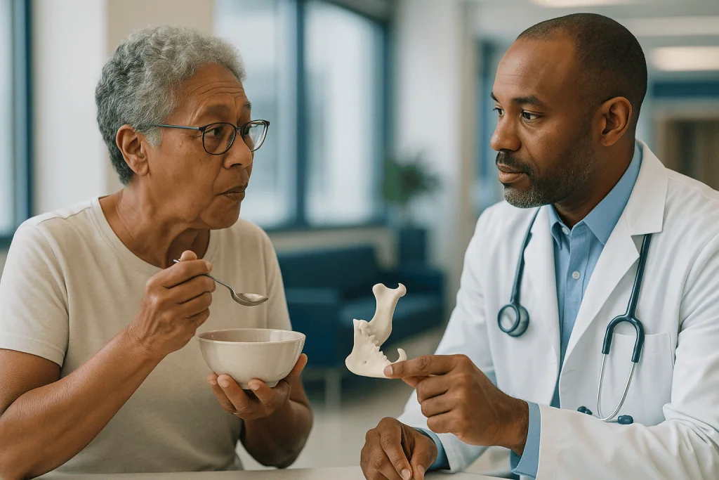
The model sits on the clinic desk between them: a pale resin copy of one person’s lower jaw, small enough to hold in two hands. She turns it over and finds the worn spot she has been feeling for years, the rounded knob at the back where the jaw meets the skull, now flattened and pitted. The surgeon points with a pen. This is where the bone will be trimmed. This is where the new surface will sit.
For someone facing 3D planning joint surface reconstruction of the temporomandibular joint, that moment can be the first time the problem looks like something concrete rather than a pain that flares when chewing bread crust. The technology behind it sounds futuristic. In practice it is closer to a tailor cutting a pattern before touching the cloth.
What follows explains what the models are, what they change, what they cannot promise, and the questions worth bringing to your own care team.
What is 3D planning in joint surface reconstruction?
Joint surface reconstruction means rebuilding the two surfaces that glide against each other inside a joint. In the face, that joint is almost always the temporomandibular joint, or TMJ: the hinge just in front of each ear where the lower jaw (the mandible) meets the skull. Its moving parts are the condyle, a rounded knob of bone on top of the jaw, and the glenoid fossa, the shallow socket in the skull that receives it. A disc of cartilage sits between them like a washer.
When arthritis, injury, previous surgery, tumor removal or a growth disorder destroys those surfaces, the options range from reshaping the bone to replacing the whole joint with an artificial one. Whatever the operation, the surgeon needs to know the exact shape of what remains, which is where 3D planning enters.
The process starts with a computed tomography (CT) scan, an X-ray technique that takes many thin cross-sectional images and stacks them into a three-dimensional picture. Software then separates bone from soft tissue and rebuilds the jaw, skull and joint as a digital object that can be rotated, measured and virtually cut. That digital model may be printed as a physical replica, used to design guides that steer the saw or drill, or sent to manufacture an implant contoured to that one person’s anatomy.
The phrase ‘before the first incision’ matters. Much of the decision making that once happened in the operating room, with the surgeon judging angles by eye and by feel, now happens days or weeks earlier at a workstation. The surgery itself becomes the execution of a plan rather than its discovery.
None of this makes an operation automatic. The TMJ disorders that lead people here affect an estimated 5 to 12 percent of the population according to the National Institute of Dental and Craniofacial Research, and only a small minority ever need surgery of any kind. Planning models serve the cases where reconstruction is already the considered choice.
Why the jaw joint is such a hard surface to rebuild
Most joints in the body move in one or two planes. The knee bends and straightens; the elbow does much the same. The TMJ is different. It rotates like a hinge for the first part of opening the mouth, then the condyle slides forward and down along the socket so the jaw can open wide. Chewing adds side-to-side grinding. Both joints, left and right, are connected by the same bone, so neither can move without the other following.
That combined motion is demanding enough in a healthy joint. In reconstruction it creates three particular difficulties.
First, space. The condyle sits millimeters from the ear canal, the facial nerve that moves the muscles of expression, and major blood vessels. A cut placed slightly too deep, or an implant seated at the wrong angle, has little room for error. Second, symmetry. Because the two joints share one bone, a new surface on one side changes how the teeth meet on both. Even a small change in the height of the condyle can leave the back teeth touching on one side and not the other, which patients notice immediately. Third, the socket is thin. The roof of the glenoid fossa is a sliver of bone separating the joint from the brain’s protective lining, so fixing anything into it requires precise knowledge of thickness.
Traditional two-dimensional X-rays flatten all of this into overlapping shadows. A panoramic dental film shows the condyle, but not how far it sits from the nerve behind it or how thin the bone above it has become. Three-dimensional imaging, and the models built from it, restore depth. The surgeon can see that this person’s socket is shallower on the right, that the condyle has drifted forward, or that scar from an earlier operation has fused bone where a gap should be.
The Mayo Clinic notes that imaging is a standard part of assessing TMJ problems that have not responded to simpler measures. For reconstruction specifically, three-dimensional detail moves from helpful to close to essential.
How virtual surgical planning for TMJ actually works, step by step
Virtual surgical planning, often shortened to VSP, is the process of performing an operation in software before performing it on a person. For the jaw joint it usually runs through a recognizable sequence.
It begins with imaging. A fine-slice CT of the skull and jaw is the standard source. A cone-beam CT, a lower-radiation scanner common in dental settings, is sometimes used, particularly for bone detail alone. MedlinePlus describes CT as fast and painless; the person lies still on a table that moves through a ring-shaped scanner. A dental impression or an intraoral digital scan of the teeth is often added, because CT shows teeth poorly around metal fillings and the bite must be planned to the tooth surfaces.
Next comes segmentation, meaning the software identifies which pixels belong to bone, which to teeth and which to soft tissue, and lifts the bone out as a separate object. The result is a digital skull and mandible that match the patient to within a fraction of a millimeter.
Then the surgeon, usually with a biomedical engineer, plans. They mark the line where diseased condyle will be removed. They check the distance from that line to the nerve canal running inside the jaw. They position a virtual implant, or a virtual bone graft, and watch how the teeth come together as the jaw is moved through opening and closing on screen. If the bite is off, they adjust and check again.
Once the plan is settled, the software produces outputs: cutting guides that clip onto the patient’s own bone and have slots showing exactly where to saw, drill guides for screw positions, a printed anatomical model for reference, and if an implant is being custom made, the manufacturing file for it.
Finally, a review. The surgeon approves the design, and any custom parts are made and sterilized. Only then is the operation scheduled.
Each step adds days to the calendar. That trade, time before surgery in exchange for certainty during it, is the whole bargain of virtual planning.
From scan to 3D printed TMJ model: what gets made before surgery
Not every 3D-planned operation produces something you can hold. When it does, the physical objects fall into a few categories, each with a distinct job.
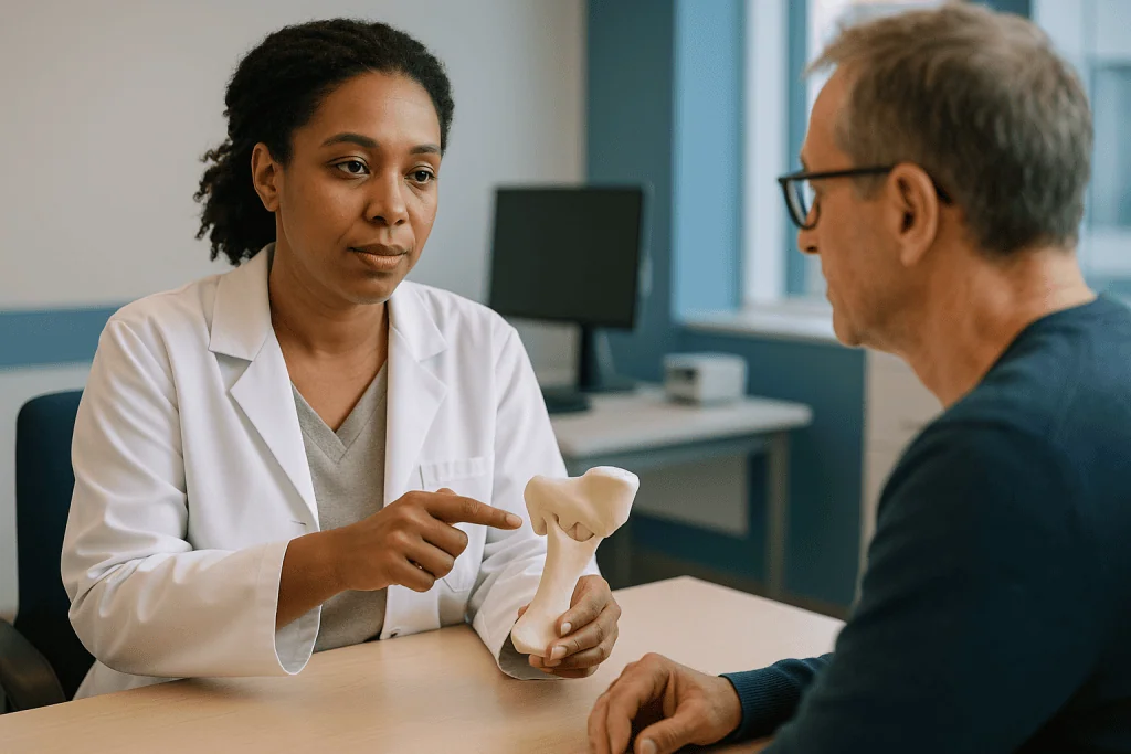
The anatomical model is a replica of the patient’s jaw and joint, printed in resin or nylon from the segmented scan. It is used for two things: helping the surgeon rehearse and helping the patient understand. Surgeons can practice the bone cut on the model and pre-bend fixation plates against it, so metal that would otherwise be shaped by hand in the operating room already fits. Patients, meanwhile, often find that a model makes a conversation about ‘condylar resorption’ suddenly intelligible.
Cutting and drilling guides are the workhorses. These are small templates that fit onto the exposed bone in only one position, like a key in a lock, with slots or tubes that direct the saw or drill along the planned path. They translate the on-screen plan into physical constraint. Guides are typically single-use and made from a medical-grade polymer that can be sterilized.
Positioning guides or splints hold the jaw at the planned bite while the new joint is fixed. Because the teeth are the most accurate reference points on the head, a splint made to fit the teeth in the planned position tells the surgeon that the jaw is where the software said it should be.
The implant itself, when custom made, is the most involved output. A patient-specific joint prosthesis consists of a fossa component that lines the socket and a condylar component that replaces the head and part of the jaw. Both are manufactured to match the individual’s scanned bone, usually in titanium alloy with a polymer bearing surface.
What a 3D printed TMJ model cannot show is soft tissue. Muscles, the disc, scar and nerve are absent from a bone print. The surgeon carries that knowledge separately, from the scan’s soft-tissue windows and from experience. The model is a map of the terrain, not the weather.
Patient-specific TMJ implants versus stock implants: what the difference means
When the plan is total joint replacement, the surgeon chooses between a stock implant, made in a range of standard sizes, and a patient-specific implant designed from the individual’s scan. Both are established options and both rely on 3D imaging; the distinction lies in how much the implant is adapted to the person versus how much the person’s bone is adapted to the implant.
| Feature | Stock implant | Patient-specific implant |
|---|---|---|
| How it is sized | Selected from preset sizes based on the scan | Designed to the patient’s own bone contours |
| Bone shaping needed | Bone may be trimmed so the standard part sits flat | Minimal, since the part follows existing bone |
| Lead time before surgery | Shorter; parts are kept in stock | Longer; design, approval and manufacture come first |
| Suits unusual anatomy | Less well when bone is badly distorted or missing | Designed for exactly these situations |
| Screw placement | Determined at surgery | Planned in software to avoid nerve and thin bone |
| Role of 3D planning | Sizing, rehearsal, guide design | Central to the entire process |
A stock device can be an entirely appropriate choice when the socket and jaw are reasonably normal in shape, when surgery is needed sooner than a custom timeline allows, or when the team’s judgment favors it. A patient-specific device is often considered when previous surgery has left the anatomy distorted, when bone has been lost to tumor or trauma, or when the joint must be built alongside other jaw repositioning.
The temptation is to assume ‘custom’ means ‘better’. The honest position is that the comparative evidence is limited, mostly observational, and does not permit a blanket statement that one type produces superior outcomes for everyone. What is fair to say is that patient-specific design shifts decision making earlier and reduces improvisation on the day. Whether that matters enough in a given case is a judgment your treating team makes with you.
Who is usually offered 3D planning joint surface reconstruction, and who is asked to wait
Joint reconstruction is not a treatment for ordinary jaw pain. The NHS describes temporomandibular disorder as a condition that usually settles with self-care, and most people never see a surgeon. The path toward reconstruction typically involves years, a documented structural problem, and a series of less invasive treatments that have not helped.
People who are commonly considered include those with end-stage arthritis of the joint, where imaging shows the joint surfaces have collapsed or fused; those with ankylosis, meaning the joint has become locked by bone or dense scar; those whose condyle has been destroyed by a tumor, by infection, or by injury; those with conditions in which the condyle dissolves over time; and those who have had several previous joint operations that have failed. In children and teenagers with growth-related jaw asymmetry, reconstruction using the person’s own rib cartilage may be planned, since a growing joint is generally not a candidate for a fixed artificial implant.
Being asked to wait is common and is not a dismissal. A surgeon may want to see whether pain and function improve with a period of conservative management, since the Cleveland Clinic and other centers note that many TMJ symptoms fluctuate and improve without surgery. Someone whose main complaint is muscle pain rather than a damaged joint surface will usually be steered toward physical therapy and bite management, because rebuilding the bone does not address a problem that lives in the muscle. Active infection, uncontrolled diabetes, heavy smoking, and certain bone diseases may also lead a team to postpone.
Age alone rarely rules anyone in or out. Overall health, the state of the bone, what the person needs the jaw to do, and realistic expectations carry more weight.
The point of naming these groups is to show that eligibility is a clinical judgment involving imaging, history and often a multidisciplinary discussion. The 3D model is a tool within that judgment, not a ticket to surgery.
What happens on the day and how the plan travels into the operating room
Reconstruction of the jaw joint is performed under general anesthesia, meaning the person is fully asleep. A breathing tube is usually placed through the nose rather than the mouth so the surgeon can bring the teeth together during the operation to check the bite.
The approach is through one or two incisions: one just in front of the ear, following a natural skin crease, to reach the joint itself, and often a second beneath the angle of the jaw to reach the lower part where a condylar component is fixed. The facial nerve runs through this territory, so the surgeon works in the layer beneath it, and many teams use a nerve monitor that sounds if the nerve is touched.
Here the planning becomes visible. Once bone is exposed, the printed cutting guide is seated. Because it was designed to fit only one way on that person’s bone, a snug fit confirms that reality matches the scan. The surgeon cuts through the guide’s slot, removes the diseased condyle, and checks the result against the model on a nearby stand. If a socket component is planned, the fossa is prepared and the component placed, with drill guides showing screw positions chosen in software to avoid the thin roof of the socket.
The positioning splint is then wired to the teeth, setting the jaw exactly where the plan specified. The condylar component is brought in, seated against the fossa component, and fixed to the jaw with screws through pre-planned holes. The jaw is put through a range of motion while the surgeon watches the new surfaces glide. The splint is released, the bite is checked, and the wounds are closed in layers.
Most people ask how long this takes. Duration varies widely with the complexity of the case, and your team is the right source for a realistic estimate. Some centers keep patients in hospital for a short period afterward for pain control, swelling and the start of jaw movement exercises.
What the first days and weeks after jaw joint reconstruction usually look like
The pattern of recovery is broadly predictable even though its pace varies. Timelines below are typical shapes of the journey, not promises; your surgeon’s instructions take precedence.
In the first days, swelling in front of the ear and along the jawline is the dominant feature, often peaking a couple of days after surgery before receding. Numbness of the skin near the incisions is common as small sensory nerves recover. Some people notice weakness of the eyebrow or the corner of the mouth on the operated side; this usually reflects bruising of the facial nerve rather than injury, and most such weakness resolves over weeks to months, although permanent weakness is a recognized risk and one your surgeon will have discussed.
Eating starts with liquids and progresses to soft foods that need little chewing. The jaw is not wired shut in most modern reconstructions, and early gentle movement is encouraged because a joint left still tends to stiffen. Many teams begin opening exercises within days, sometimes with a simple device that gradually stretches the mouth open. The goal is to keep the new surfaces gliding while the soft tissues heal around them.
Over the following weeks, swelling fades, the incisions settle into pale lines within skin creases, and the range of mouth opening improves with regular exercise. The bite may feel unfamiliar as muscles adapt to a joint that sits at a slightly different height, and a dentist or orthodontist may be involved if adjustments are needed. Return to work and normal activity depends on the job and the individual; heavy chewing, contact sports and anything that risks a blow to the jaw are usually deferred longer than desk work.
Follow-up imaging, often a plain X-ray or CT, confirms that implants remain in position. For people with artificial joints, long-term review continues because, like any prosthesis, the components can wear over years. The Johns Hopkins Medicine overview of TMJ disorders describes surgery as an option reserved for severe cases, and lifelong attention to the joint reflects that seriousness.
Does 3D planning change how the face looks afterward?
Because this topic sits within facial aesthetics, it is worth being direct about what joint reconstruction does and does not do to appearance.
The condyle sets the vertical height of the back of the jaw on its side. When a condyle has collapsed, the chin often swings toward that side, the jawline looks shorter there, and the bite tilts. Rebuilding the joint surface to its original height can restore symmetry, and 3D planning is the tool that lets the surgeon decide precisely how much height to restore. On screen, the mandible can be rotated back to level, the chin midline checked against the midline of the face, and the profile viewed from every angle before anything is committed.
That is a functional correction with an aesthetic consequence, not a cosmetic procedure. The changes people see are generally a straighter chin, a more even jawline and a bite that closes evenly, not a different face. Surgeons planning a reconstruction alongside orthognathic surgery, meaning repositioning of the jaws to correct a bite, can plan both in the same virtual session, which is one of the situations where 3D planning has changed practice most.
Two cautions belong here. Soft tissue does not follow bone exactly. Software can predict the new skin surface, but the predictions are estimates, and swelling obscures the result for weeks. Anyone hoping to judge the outcome in the first month will be looking at swelling, not at the final shape.
Second, the incisions leave scars. Placed in the crease in front of the ear and under the jaw, they generally fade to fine lines, but they are permanent, and the way an individual’s skin heals is not something a model can predict.
The realistic hope is a face that looks more like itself before the joint failed. Anyone whose primary goal is a change in appearance rather than function should say so early, because it changes the conversation about whether this operation is the right one.
Risks and limits of 3D planning: what models cannot tell you
A plan that looks flawless on screen inherits every limitation of the data it was built from. Understanding those limits protects against overconfidence, in the surgeon and in the patient.
The scan is a snapshot. If months pass between imaging and surgery, bone that is actively dissolving or a joint that is progressively fusing may no longer match the model. Teams often repeat imaging if the delay is long, and a guide that does not seat snugly on the day is a warning that anatomy has changed.
Metal in the mouth scatters the X-ray beam, producing streaks that can blur the teeth and nearby bone. This is why dental impressions or optical scans are merged with the CT; the fusion step itself introduces small errors that must be checked.
Soft tissue is largely absent. The model shows bone, not the muscles that will pull on the new joint, the nerve that must be protected, or the scar from previous surgery that can make dissection unpredictable. The disc, which is often the very structure that has failed, does not appear on a bone print at all.
The surgical risks themselves are not removed by planning. They include facial nerve weakness, temporary or permanent; numbness of the ear or cheek; infection, which around an implant may require its removal; bleeding; a bite that does not meet as intended; limited mouth opening from scar; heterotopic bone, meaning new bone forming where it should not and restricting movement; and, with artificial joints, loosening or wear over years. Allergy to implant metals is uncommon but is screened for. Planning can reduce the chance of some of these, particularly screw placement into vulnerable structures, but the evidence that it lowers overall complication rates is drawn mainly from case series rather than randomized trials, so it should be stated cautiously.
Radiation from CT is small and, as MedlinePlus explains, is weighed against the benefit of the information gained. Cone-beam scanners deliver less. For an operation this precise, most teams judge the trade worthwhile, but it is a fair question to raise.
What are the alternatives to joint surface reconstruction?
Reconstruction sits at the far end of a long ladder. Understanding the rungs below helps explain why a surgeon may propose something less, and what ‘trying conservative measures first’ actually means.
The NHS and Mayo Clinic both describe self-care as the foundation: a softer diet during flares, avoiding wide yawning and chewing gum, warmth or cold to the joint, and gentle stretching. Physical therapy addresses the muscles that clench and guard around a painful joint. A bite splint worn at night can reduce grinding load. Medicines used include anti-inflammatory and muscle-relaxant classes; how they are used, and for how long, is a decision for the prescribing clinician, and this article does not describe doses.
Minimally invasive procedures follow. Arthrocentesis flushes the joint with fluid through fine needles to wash out inflammatory debris and free a stuck disc. Arthroscopy inserts a slim camera to inspect and treat the joint through small punctures. Both can relieve pain and improve opening in selected cases without altering the bone.
Open joint surgery without full replacement includes reshaping a rough condyle, repositioning or repairing the disc, and removing bony bridges in ankylosis while placing a soft tissue spacer to prevent them re-forming. These preserve the person’s own joint.
Reconstruction with the person’s own tissue, most often rib cartilage attached to a segment of rib, rebuilds the condyle biologically. It is the usual choice in growing children and an option in adults who prefer to avoid a permanent implant, though grafts can overgrow or resorb unpredictably.
Total joint replacement with an artificial prosthesis, stock or patient-specific, is the most definitive option and the one where 3D planning has the largest role.
Each step up brings more certainty about the structure and more irreversibility. The right rung depends on what is actually causing the problem, which is why imaging and careful diagnosis precede any talk of models and implants. Your team will explain which alternatives remain realistic in your case and which have already been tried or excluded.
What people often get wrong about 3D planning models
Enthusiasm for technology breeds its own myths. A few deserve correcting.
The first is that a 3D-planned operation is a guaranteed-precision operation. The plan is precise. The surgery is performed by hands on living tissue, with bleeding, muscle pull and scar that no model contains. Planning narrows the range of surprise; it does not eliminate it. Surgeons still make judgments in the room, and sometimes those judgments override the plan for good reason.
The second is that a printed implant is the same as a printed model. The resin replica handed across a desk is a reference object. The implant that goes into the body is manufactured separately under medical device regulation, from titanium alloy or similar materials, and often by machining rather than printing. Conflating the two leads people to imagine plastic parts holding up their jaw.
The third is that custom always beats stock. As the comparison above sets out, the evidence does not support a universal ranking. Stock devices have long track records, and a well-sized stock implant in suitable anatomy is not a compromise.
The fourth is that if imaging shows a damaged joint, surgery is needed. Imaging findings and symptoms often disagree. The National Institute of Dental and Craniofacial Research notes that many people with TMJ disorders improve without invasive treatment, and worn-looking joints can be painless while normal-looking ones hurt. The decision to operate rests on function and symptoms, with imaging as one input.
The fifth is that the model shows how the face will look. It shows bone. Predicted soft-tissue overlays exist, but they are estimates and should be presented as such.
A sixth, quieter misconception is that more technology means the surgeon matters less. The reverse is true. Someone must decide where to place the cut, how much height to restore and what to do when the guide does not fit. The workstation makes those decisions visible and reviewable. It does not make them.
Questions to ask your care team
A consultation about joint reconstruction is dense with new vocabulary, and the presence of a model on the desk can make the plan feel more settled than it is. Written questions help. The following are ones patients commonly wish they had asked.
- What exactly is damaged in my joint, and how do you know the joint surface rather than the muscles or the disc is the main source of my symptoms?
- Which less invasive options have already been tried or ruled out, and why?
- Are you proposing reshaping, a graft of my own tissue, a stock implant or a patient-specific implant, and what makes that the right choice for my anatomy?
- Will my scan be repeated before surgery if there is a delay, and how old is the imaging the plan is built on?
- How will the plan be checked in the operating room, and what happens if the guide does not fit as expected?
- Which structures are closest to the planned cuts and screws, and what are the specific risks to the facial nerve, hearing and the ear canal in my case?
- How is the new height of the joint chosen, and will my bite change; will a dentist or orthodontist be involved afterward?
- What will the first days and weeks realistically involve for eating, speaking and jaw exercises?
- If an artificial joint is used, what long-term follow-up is needed, and what happens if a component loosens or wears years from now?
- How many such reconstructions does the team perform, and is the planning done in-house or with an external engineering partner?
- Can I see and, if possible, keep the model, and can the plan be explained to me on screen?
- What would you advise if I decided to wait?
Ask for answers to be written down or for a summary letter. A plan you understand is a plan you can consent to properly, and a good team will welcome the questions.
When to call your doctor
Before surgery, the signals that warrant prompt contact with a clinician rather than waiting for a scheduled review are those suggesting something beyond a worn joint. The NHS advises seeking medical advice for jaw pain that stops you eating or drinking, that is severe or that keeps returning, and for a jaw that locks open or closed. Sudden facial weakness, numbness of the face, persistent swelling in front of the ear, hearing change, or a hard lump that grows should be assessed without delay, since these can point to nerve involvement, infection or a mass rather than to routine joint wear.
After surgery, your team will give specific instructions, and those come first. In general, call the surgical team the same day, or go to an emergency department if you cannot reach them, for any of the following:
- Fever, chills, or spreading redness, heat and increasing pain around an incision, which may signal infection near an implant
- Pus or cloudy fluid draining from a wound, or a wound that opens
- Bleeding that soaks a dressing and does not stop with firm pressure
- Swelling that increases rapidly rather than slowly settling, or that makes swallowing or breathing feel difficult
- New or worsening weakness of the face, particularly if the eye on the operated side will not close fully, because an unprotected eye can be damaged
- A sudden change in the bite, a grinding or clunking sensation in the joint, or an inability to bring the teeth together as they did in the first days after surgery
- Clear fluid running from the nose or ear, or a severe headache with neck stiffness
- Pain that escalates despite the prescribed plan, or calf pain, chest pain or breathlessness, which need urgent assessment for a clot
Seek emergency care immediately for difficulty breathing, heavy bleeding, or signs of a severe allergic reaction such as facial or throat swelling with hives.
None of these signs mean the operation has failed. They mean something needs a clinician’s eyes sooner than planned, and early attention is what keeps small problems small. Every decision about what happens next belongs with your treating team.
Frequently asked questions
What is virtual surgical planning for TMJ surgery?
Virtual surgical planning is the rehearsal of a jaw joint operation in software using the patient’s own CT scan before the real procedure. The surgeon marks bone cuts, positions a graft or implant, and checks how the teeth meet as the jaw moves on screen. The approved plan is then turned into printed guides, splints or a custom implant so the operation follows a predetermined path.
How is a 3D printed TMJ model made from my scan?
Software separates bone from soft tissue in your CT images, a step called segmentation, and rebuilds the jaw and skull as a digital object. That file is sent to a 3D printer, which builds a resin or nylon replica layer by layer. The model is used for rehearsal, for pre-shaping plates and for explaining the plan to you; it is not the implant itself.
What is a patient-specific TMJ implant?
It is an artificial jaw joint designed from an individual’s scan so the socket and condylar components follow that person’s bone contours exactly. It is usually considered when anatomy is distorted by previous surgery, tumor, trauma or bone loss. Stock implants in standard sizes remain an appropriate alternative for many people, and the choice is a clinical judgment made by the treating team.
How long does TMJ replacement surgery planning take before the operation?
Planning adds time between the decision to operate and the surgery date, because imaging, virtual design, surgeon review and manufacture of any custom parts all happen first. Stock implants shorten this because parts are already available. Your team is the right source for a realistic estimate in your case, since it depends on complexity and whether custom components are being made.
Does 3D planning make jaw joint surgery safer?
It reduces certain specific risks, particularly placing screws into the thin roof of the socket or near the nerve canal, because those positions are chosen with full three-dimensional information. Evidence that it lowers overall complication rates comes mainly from case series rather than randomized trials, so the honest position is that it supports precision without removing the recognized risks of the operation.
Will I be able to see the 3D model of my jaw?
In most centers, yes. Surgeons often use the printed model or the on-screen plan to explain what is damaged and what will change, and many patients find this the clearest part of the consultation. It is reasonable to ask to see the plan and to have the proposed cuts and implant positions pointed out on the model before you consent.
Does a worn-looking joint on a scan mean I need reconstruction?
No. Imaging findings and symptoms often disagree; joints that look badly worn can be painless, and normal-looking joints can hurt. Reconstruction is considered when structural damage clearly explains disabling symptoms and less invasive treatments have not helped. The National Institute of Dental and Craniofacial Research notes that many people with TMJ disorders improve without invasive treatment.
What are the alternatives to jaw joint reconstruction?
Options below reconstruction include self-care and a softer diet during flares, physical therapy, night splints, medicines prescribed by a clinician, arthrocentesis to flush the joint, arthroscopy through small punctures, and open surgery that reshapes bone or repairs the disc while keeping the natural joint. Reconstruction with the person’s own rib cartilage is another alternative to an artificial implant, especially in growing patients.
Will joint reconstruction change how my face looks?
It can restore symmetry when a collapsed condyle has shortened one side of the jaw and pulled the chin off center, because 3D planning lets the surgeon set the new joint height precisely. The effect is a face that looks more like it did before the joint failed rather than a different face. Swelling hides the result for weeks and incisions leave fine permanent scars.
What are the red-flag signs after jaw joint surgery?
Contact your surgical team the same day for fever, spreading redness or pus at an incision, bleeding that does not stop with pressure, rapidly increasing swelling, new facial weakness especially if the eye will not close, a sudden change in the bite, or clear fluid from the nose or ear. Seek emergency care for breathing difficulty, heavy bleeding, chest pain or calf pain.
References
- TMD (Temporomandibular Disorders), National Institute of Dental and Craniofacial Research, NIH
- Temporomandibular disorder (TMD), NHS
- Temporomandibular Disorders (TMD), Cleveland Clinic
- CT Scans, MedlinePlus
This article is for general information only and is not a substitute for professional medical advice. Please consult a qualified doctor about your individual situation.

More from the Blog

Microsurgery Risks Explained: Why Blood Flow Is Watched Hourly in the First Days
Microsurgery complications most often involve the tiny reconnected blood vessels that feed transplanted tissue, called a flap. A clot in the artery or vein…

Scar Treatment Results: What Realistic Improvement Looks Like and Why Sessions Repeat
Realistic scar treatment results mean a scar that is flatter, softer, paler and less noticeable, not a scar that vanishes. Scar tissue is permanent,…

Neck Lift Recovery Time: Healing Stages, Scars and Results That Settle
Most people look socially presentable about two to three weeks after a neck lift, once visible bruising fades and swelling starts to settle. Full…

How to Prepare for Microneedling: Sun, Retinoids and the Two Weeks Before Your Session
Preparing for microneedling usually means keeping the skin out of strong sun and free of sunburn or tan for the two weeks beforehand, pausing…
IPL Photorejuvenation Results: Why Spots Darken Before They Fade and How Sessions Build
IPL photorejuvenation results usually appear gradually. Treated brown spots often turn darker and coffee-ground-like for several days before flaking away over one to two…

Surface Reconstruction Recovery: Early Supervised Walking, Physical Therapy and Milestones
Surface reconstruction recovery after ocular surface surgery usually unfolds in stages: the corneal surface layer re-covers over days, comfort and light sensitivity settle over…