Surface Reconstruction
Surface reconstruction is an orthopedic surgical procedure that restores damaged joint surfaces, often helping selected patients with arthritis reduce pain and preserve mobility.
Quick answer
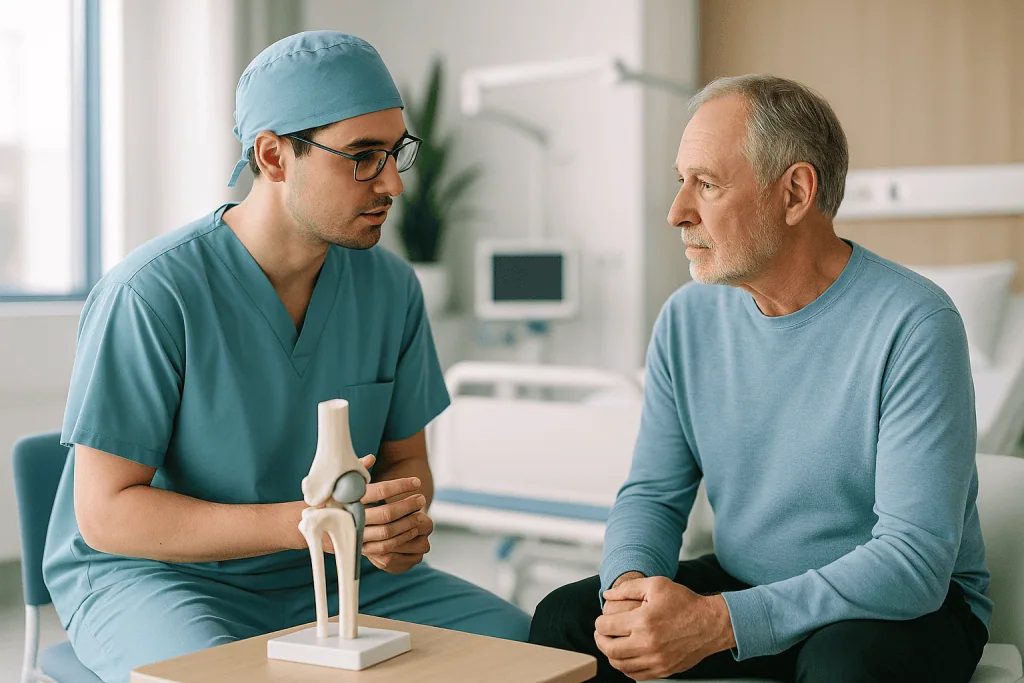
Surface reconstruction is a group of orthopaedic procedures that repair or resurface the damaged bearing surface of a joint: the area where cartilage-covered bones meet and move. Depending on the joint and the defect, it can involve cartilage repair, osteochondral grafting, bone correction or partial resurfacing. It is considered when a targeted, joint-preserving operation may relieve pain without replacing the whole joint.
What Is Surface Reconstruction?
Surface reconstruction is an orthopaedic surgical approach that repairs, reshapes or resurfaces the damaged bearing surface of a joint: the area where bones meet and glide against each other, normally covered by smooth articular cartilage. It is considered for carefully selected patients whose pain comes from a worn or injured joint surface, when anatomy, age, activity level and the pattern of arthritis suggest that a joint-preserving operation may serve them better than replacing the joint entirely. The goal is practical: make movement smoother, address the mechanical source of pain, and keep as much of your own bone and joint structure as possible.
One point of clarity before anything else. The same phrase is also used in engineering for computer techniques that turn scan data into three-dimensional surface models: the kind of model surgeons study when planning complex joint operations. Because the two meanings are easily confused, this page covers both: the surgery itself in detail, and the role that 3D surface modelling plays in preparing for it.
To understand what the operation addresses, it helps to picture what goes wrong inside the joint. Healthy articular cartilage lets bones move against each other with very little friction. When that cartilage becomes worn, cracked or lost, through arthritis, injury, deformity or previous surgery, the underlying bone can become exposed. Bone moving against bone creates friction, inflammation, stiffness, swelling and pain. The joint no longer moves the way it was designed to, and the body often starts compensating in ways that create problems elsewhere.
The term covers several distinct techniques, and which one applies depends on the joint involved and the type of damage. In some patients it means reshaping a worn area and resurfacing it with a carefully positioned implant. In others it means cartilage restoration, osteochondral grafting, correction of the underlying bone, or partial joint resurfacing. The principle stays constant across all of them: the surgeon concentrates on the damaged surface rather than replacing the whole joint, wherever a more targeted approach is genuinely suitable.
Surface reconstruction is most often discussed for the hip, knee, shoulder and ankle, and for certain small joints, although the techniques differ greatly between them. A patient with a localised cartilage defect in the knee needs a very different operation from a patient with early arthritis in the hip. That is why individualised planning matters so much. The procedure is chosen according to the joint, the size and location of the defect, bone quality, limb alignment, ligament stability, and your age, weight, activity level and medical history.
Is surface reconstruction the same as joint replacement?
No, and the distinction matters when you compare your options. Surface reconstruction preserves and restores the existing joint surface; replacement removes the worn surfaces and substitutes them with prosthetic components. For advanced arthritis affecting the entire joint, severe deformity, major bone loss or marked stiffness, total joint replacement usually offers more predictable pain relief, and it would not serve you to pretend otherwise. Programmes in adult reconstruction cover that end of the spectrum. For appropriately selected patients, however, surface reconstruction addresses the mechanical source of pain while preserving more of the natural joint, which is precisely why the selection process described below is taken so seriously before any decision is made.
How joint surface damage changes daily life
Living with arthritis or a damaged joint surface is rarely just a physical problem. Patients describe organising their whole day around pain: choosing lifts over stairs, limiting travel, giving up sport, avoiding long walks because they cannot predict how the joint will feel tomorrow. For international patients weighing up treatment abroad, the decision carries extra layers, comparing surgical options, wondering whether the joint can still be preserved, and estimating how much recovery time is needed before travelling home safely. The sections below are written to make each of those judgements easier to think through, including where the answer may be that a different treatment suits you better.
Who May Need Surface Reconstruction?
Patients considered for surface reconstruction usually have joint pain that has become persistent, function-limiting and clearly linked to damage of the joint surface itself. Symptoms often begin gradually, although they can also follow a specific injury. Pain may appear during walking, climbing stairs, kneeling, squatting, standing up from a chair, lifting the arm or turning the hip. Some patients notice swelling, grinding, catching, stiffness after rest, or simply a sense that the joint no longer moves smoothly. None of these symptoms, on its own, proves that surgery is the answer, but together with imaging they help define whether the surface is the true source of the problem.
A typical candidate has already tried nonsurgical care: physical therapy, anti-inflammatory medication, activity modification, weight management, bracing or injections. These treatments may still have a place, and surgery does not cancel them out. But when symptoms keep interfering with daily life despite well-directed conservative care, an operation enters the conversation. The key question is always the same: is the joint damage localised, or structurally correctable, enough for a surface-focused procedure to be worthwhile?
How is a joint assessed for surface reconstruction?
Assessment starts with a careful clinical examination, not a scan. The orthopaedic surgeon evaluates range of motion, areas of tenderness, joint stability, limb alignment, walking pattern, muscle strength and signs of inflammation. Your history carries equal weight: when the symptoms began, what worsens them, what relieves them, whether there was a past fracture or ligament injury, and, critically, what activities you hope to return to after treatment. A plan built for comfortable daily walking looks different from a plan built for recreational sport.
Imaging then fills in the structural picture. X-rays assess joint space, bone shape, alignment, osteophytes, deformity and the stage of arthritis. Magnetic resonance imaging evaluates cartilage, meniscus, labrum, ligaments, tendons, bone marrow changes and localised cartilage defects. Computed tomography helps when detailed bone mapping is needed, for complex deformity, previous surgery or implant planning, and CT data is also what feeds the three-dimensional surface models described later on this page. In selected cases, laboratory tests are requested to exclude inflammatory arthritis, infection or metabolic bone problems, because those conditions change the treatment logic entirely.
Surface reconstruction may be discussed for patients who have:
- Persistent joint pain caused by localised cartilage loss or early-to-moderate arthritis.
- Mechanical symptoms such as catching, grinding or painful restriction related to surface damage.
- Damage after trauma, osteochondral injury, avascular necrosis or previous joint instability.
- A joint that remains reasonably mobile and has enough bone quality to support reconstruction.
- Symptoms that have not improved sufficiently with well-directed nonsurgical treatment.
- A wish to preserve mobility and, where possible, delay or avoid a more extensive joint replacement.
Patient selection is central to everything that follows. A younger, active patient with localised damage is generally a stronger candidate than a patient with advanced diffuse arthritis, severe deformity or inflammatory disease affecting the whole joint. Even so, age alone does not decide suitability. Surgeons weigh the entire clinical picture, including medical conditions such as diabetes, osteoporosis, vascular disease and autoimmune disorders, as well as smoking, all of which influence healing capacity and surgical risk. Being told you are not a candidate is not a dismissal; it is the selection process protecting you from an operation unlikely to help.
Conditions Surface Reconstruction May Address
Surface reconstruction is used across several orthopaedic conditions in which the smooth cartilage layer, or the bone directly beneath it, has been damaged. The indication depends on the joint involved, the extent of disease, and whether the remaining joint structures can support a durable reconstruction.
Osteoarthritis
Osteoarthritis is one of the most common reasons patients ask about this procedure. In early or moderate osteoarthritis, cartilage wear is sometimes concentrated in one region of the joint rather than spread throughout it. If alignment, stability and bone quality are favourable, a surface-focused procedure may reduce pain and improve function in that region. In advanced osteoarthritis, widespread cartilage loss with significant deformity, total joint replacement is usually the more appropriate recommendation, and a candid surgeon will say so.
Focal cartilage defects
Focal cartilage defects can occur after sports injuries, falls or repetitive overload. They may be small yet disproportionately painful, particularly when they sit in a weight-bearing area of the joint. Depending on the size and depth of the defect, reconstruction may involve cartilage repair techniques, grafting or localised resurfacing. Treating a defect while it is still contained is one of the situations where surface-focused surgery has the clearest rationale.
Osteochondral lesions
Osteochondral lesions involve both the cartilage and the bone underneath it, and they occur in the knee, ankle, elbow and other joints. When the damaged area fails to heal properly, patients develop swelling, pain and mechanical symptoms such as catching. Here the reconstruction has two jobs at once: restoring the bony support and rebuilding the cartilage surface above it, because a smooth surface over collapsing bone does not last.
Avascular necrosis (osteonecrosis)
Avascular necrosis, also called osteonecrosis, develops when the blood supply to part of a bone is reduced, which in some patients leads to collapse of the joint surface. Early diagnosis makes a genuine difference here. Depending on the stage, surface reconstruction or other joint-preserving procedures may be considered before collapse becomes advanced, after which the range of realistic options narrows considerably.
Post-traumatic arthritis
Post-traumatic arthritis can develop after fractures that extend into a joint, ligament injuries that alter joint mechanics, or previous dislocations. In these cases the surgeon must evaluate not only the damaged surface but also alignment, stability, scar tissue and any previous implants or hardware, which is why this work often overlaps with broader reconstruction after trauma. A surface repaired without correcting the mechanics that damaged it tends to fail the same way the original surface did.
Congenital and developmental joint problems
Congenital or developmental joint problems can load a joint surface abnormally for years before symptoms appear. In selected patients, surface reconstruction is combined with corrective procedures that improve the mechanics and reduce overload on the reconstructed area, treating the cause and the consequence in the same plan.
Because these conditions vary so widely, surface reconstruction is not one standardised operation. It is better understood as a family of joint-preserving and surface-restoring strategies, and the right member of that family emerges only after the diagnosis is matched to your anatomy and your functional goals.
How Surface Reconstruction Is Performed
Before surgery: diagnosis, planning and preparation
The process begins well before the day of surgery. A detailed consultation confirms the diagnosis, reviews previous imaging, establishes your goals and sets realistic expectations, including the honest possibility that a different treatment is more appropriate. For patients coming from abroad, planning usually begins with existing X-rays, MRI or CT scans and medical reports, reviewed before any travel is arranged, so that the initial assessment happens on paper rather than at an airport.
Preoperative preparation may include updated imaging, blood tests, an anaesthesia assessment, and cardiac or internal medicine evaluation where needed. The surgical and anaesthesia teams review every medication and supplement you take, blood thinners, diabetes medication and immune-modifying drugs receive particular attention, and any adjustment before surgery is decided by your treating doctors, not by a checklist. If infection is suspected, or if the same joint has been operated on before, additional tests may be required before a date is set.
The type of anaesthesia depends on the joint, the procedure and your general health. Some operations are performed under general anaesthesia; others use regional anaesthesia or a combination of the two. The anaesthesiology team discusses the safest plan with you beforehand and monitors you throughout the procedure.
What happens during the operation?
The surgeon reaches the affected joint through arthroscopic instruments, a limited incision or an open approach. Arthroscopy uses a small camera and specialised instruments inserted through small incisions, allowing certain types of damage to be inspected and treated with less soft-tissue disruption. Open surgery is chosen when the reconstruction demands direct visualisation, precise implant positioning, graft placement or correction of bone deformity. Neither approach is inherently superior; each suits different problems. Whichever route is taken, the operation follows a recognisable sequence:
- Step 1: Access and inspection. The joint is exposed or visualised arthroscopically, and the surgeon confirms that the damage seen on imaging matches what is actually there.
- Step 2: Surface preparation. Unstable cartilage fragments are removed, irregular edges are smoothed, the underlying bone may be reshaped, impingement is corrected, and a stable bed is prepared for any graft or implant.
- Step 3: Reconstruction. If resurfacing is performed, the implant or reconstructive material is positioned to match the joint’s natural contour as closely as possible. If a graft is used, it is fitted securely to restore structural support and create a congruent surface.
- Step 4: Verification. Alignment, positioning and the accuracy of the reconstruction are checked, with intraoperative imaging where appropriate, and the joint is moved through its range to confirm smooth tracking.
- Step 5: Closure and protection. The tissues are closed in layers and the joint is protected according to the demands of the specific reconstruction performed.
How long does the operation take?
It varies, and the variation is normal. A smaller arthroscopic cartilage procedure may be relatively short, while a complex reconstruction involving bone correction, grafting or partial resurfacing takes considerably longer. Surgical time on its own is a poor measure of quality. What matters is careful planning, precise execution, protection of the healthy tissue around the defect, and a rehabilitation programme matched to what was actually done inside the joint.
What happens immediately after surgery?
You are moved to a recovery area where pain control, circulation, breathing and nerve function are monitored. Some patients go home the same day; others stay in hospital for one or more nights, particularly after larger joint procedures or when medical monitoring is needed. International patients receive practical guidance covering accommodation, wound care, mobility support and the safe timing of the flight home: a detail that deserves as much planning as the operation itself.
When does rehabilitation begin?
Early, but always tailored to the procedure. For some patients, gentle movement starts within the first day to reduce stiffness and support circulation. Weight-bearing may be restricted for a period if the reconstructed surface needs protection; in other cases, partial or full weight-bearing is allowed sooner. Physical therapy focuses first on swelling control, safe movement and muscle activation, then progresses to strength, balance, gait training and activity-specific conditioning. Recovery is not only about the incision healing: the deeper tissues, the reconstructed surface, the bone and the surrounding muscles all need their own time. You are given clear instructions on wound care, warning signs, exercises and follow-up appointments, and for patients returning to another country, the care plan should include communication between the surgical team and your local physician or physiotherapist wherever possible.
3D Surface Modelling: The Technology Behind Surgical Planning
3D surface modelling is a computational technique, not an operation, but it earns its place on this page because modelling of exactly this kind sits behind modern surgical planning. The short explanation below describes what those 3D joint models your surgeon may show you actually are, and how far to trust them.
What is 3D surface modelling?
Surface modelling algorithms convert a cloud of measured points, each with a position and an orientation, into a smooth, closed three-dimensional surface, smoothing measurement noise so that the result is watertight and reliable. These techniques were developed for computer graphics and engineering, and the same family of algorithms now runs inside the software used to build anatomical models from medical scans. The research field remains active, but that debate belongs to engineers. As a patient, you do not need to evaluate algorithms; you only need to know that 3D planning rests on established, verifiable mathematics.
How is a point cloud converted into a surface?
Converting a point cloud to a surface follows a broadly consistent pipeline in medical planning. First, a CT or MRI scan captures the joint as a stack of cross-sectional images. Second, software or a technician segments the bone and cartilage outlines on each slice, producing thousands of points, each defined by its coordinates in space. Third, the modelling algorithm fits a continuous surface through those points, closing gaps and smoothing measurement noise. Finally, the resulting mesh is checked against the original scan images before anyone plans surgery on it.
Why does 3D modelling matter for joint surgery?
Because a surface reconstruction operation succeeds or fails on millimetres. A faithful 3D model lets the surgical team measure the size, depth and location of a cartilage or bone defect before the joint is ever opened, analyse limb alignment, plan the position of an implant or graft, and in selected cases prepare patient-specific planning tools. Two honest limits apply. The model is only as good as the scan and the segmentation behind it, errors in either propagate straight into the mesh. And no surgeon operates on a model alone: the decision to reconstruct a surface rests on whether the imaging findings match your symptoms and examination, never on the geometry by itself. Technology sharpens judgement; it does not replace it.
Why Acting Early Matters
Joint surface damage tends to progress when the underlying mechanical problem continues. A small cartilage defect may enlarge. A joint with abnormal alignment concentrates increasing pressure on one area. Instability after a ligament injury accelerates wear. Pain leads to reduced activity, which causes muscle weakness and stiffness, which further disturbs joint mechanics: a loop that rarely interrupts itself.
Early evaluation does not mean early surgery. For many patients, the first recommendation is targeted physical therapy, weight reduction where appropriate, medication, injections or bracing. What timely assessment does is establish whether the problem is still at a stage where joint-preserving, surface-focused options remain on the table. Waiting until the joint is severely damaged can quietly remove those options.
The risks of delay are concrete: worsening cartilage loss, progressive bone changes, loss of motion, deformity, chronic inflammation and declining muscle strength. Many patients also develop compensatory pain, in the back, the opposite hip or knee, or other joints, because they have changed how they walk and move. In advanced cases, a less invasive reconstruction may no longer be feasible and a larger operation becomes the realistic path, sometimes requiring the kind of staged planning found in complex joint reconstruction.
For international patients there is a further, practical reason to start early: time. Collecting medical records, obtaining imaging in the correct format, addressing medical conditions before travel, and understanding the recovery period all take weeks, not days, particularly when work leave, family support, visa arrangements and rehabilitation at home must line up with a surgical date.
Potential Benefits of Surface Reconstruction
When the procedure genuinely fits the diagnosis and forms part of a structured treatment plan, patients may experience several meaningful benefits. Note the conditional language: these are the aims of a well-selected operation, not promises attached to every case.
| Benefit | What It Means for You |
|---|---|
| Pain reduction | By addressing the damaged joint surface that creates friction and inflammation, the procedure may reduce pain during daily activities such as walking, climbing stairs or standing. |
| Preservation of joint structure | In selected patients, reconstruction may preserve more of the natural joint and bone compared with more extensive replacement procedures. |
| Improved mobility | A smoother, more stable joint surface can make movement feel less restricted and may support better range of motion during rehabilitation. |
| Delay of larger surgery | For some patients, surface reconstruction may help postpone the need for total joint replacement, though future surgery may still be necessary if arthritis progresses. |
| Activity-specific recovery goals | The treatment plan can be aligned with your lifestyle, whether the priority is comfortable walking, travel, recreational sport or maintaining independence. |
| Personalised surgical planning | Imaging and functional evaluation help the surgeon choose a technique matched to the location, size and cause of the surface damage. |
Recovery Timeline After Surface Reconstruction
Recovery varies with the joint treated, the surgical technique, the extent of reconstruction and your overall health. The timeline below is a general orientation, not a schedule: your surgeon’s instructions for your specific procedure always take precedence over any table.
| Time Period | What Patients Can Expect |
|---|---|
| Day 1 | Pain control, monitoring, early movement, and instructions for protecting the reconstructed joint. Some patients begin walking with assistance or start gentle range-of-motion exercises. |
| First Week | Swelling and discomfort gradually improve. The focus is wound care, safe mobility, prescribed exercises, and following weight-bearing restrictions where they apply. |
| First Month | Physical therapy becomes more structured: restoring motion, activating muscles, improving walking mechanics, and protecting the reconstructed surface as healing continues. |
| Two to Three Months | Many patients build strength and tolerance for daily activity. Depending on the procedure, some restrictions ease, while higher-impact activity is usually still limited. |
| Longer Term | Functional recovery continues over several months. Return to sport, heavy labour or demanding travel depends on healing, strength, imaging findings where needed, and surgeon approval. |
Factors That Influence Outcomes
The result of surface reconstruction depends on more than the operation itself. A good outcome begins with matching the right patient to the right procedure. The pattern of joint damage is one of the most important variables: localised defects, stable joints, correctable alignment and good bone quality create a far more favourable setting than diffuse arthritis, severe deformity or major instability.
The stage of arthritis matters just as much. Surface reconstruction works best when the joint still has enough healthy cartilage, motion and structural support around the treated area. If the joint is already severely narrowed, stiff or deformed, reconstructing one surface cannot adequately address the overall disease process, and a surgeon who explains that plainly is protecting your outcome, not withholding treatment.
Alignment and stability are critical to durability. A reconstructed surface can be overloaded if the leg, hip, knee, ankle or shoulder keeps moving in a mechanically unfavourable way. For that reason, surgeons sometimes recommend additional procedures alongside the reconstruction: correcting alignment, repairing ligaments: an unstable knee may need ACL reconstruction, for instance, addressing meniscal deficiency, or treating impingement. These additions are not scope creep; they exist to protect the reconstruction and improve long-term function.
Patient-related factors influence healing in ways that are easy to underestimate. Smoking impairs tissue and bone recovery. Poorly controlled diabetes increases infection risk. Excess body weight raises the load on weight-bearing joints. Osteoporosis, inflammatory arthritis, vascular disease and immune-suppressing medication can all affect the surgical plan and the pace of recovery. None of these automatically rules out surgery, but each must be managed deliberately, and how they are managed is a decision for your treating doctors.
Rehabilitation is the other half of the operation. Patients who follow weight-bearing instructions, attend physiotherapy, do the home exercises and resist returning to high-impact activity too early protect the reconstructed surface most effectively. The failure modes run in both directions: doing too much too soon jeopardises healing, while doing too little leads to stiffness and weakness.
Finally, expectations should be realistic, because that is where satisfaction is won or lost. Surface reconstruction aims to reduce pain, improve function and preserve mobility in well-selected patients. It may not make an arthritic joint feel exactly like a healthy one, and it cannot stop all future degeneration. Some patients will eventually need further surgery. A frank discussion before treatment, covering the likely recovery path, its limits, and the signs that should prompt medical review, is not pessimism; it is the foundation of a decision you will not regret.
How Acibadem Approaches Surface Reconstruction
Patients travelling for orthopaedic care need more than a well-executed operation. They need a reliable diagnostic process, clear communication, coordinated scheduling and a treatment plan that stays practical after they return home. Acibadem’s approach to surface reconstruction is organised around those needs.
Complex joint problems are evaluated by experienced orthopaedic physicians, and where the case demands it, input is sought from radiology, physical medicine and rehabilitation, rheumatology, anaesthesia, internal medicine, cardiology, endocrinology or infectious disease specialists. This multidisciplinary review matters most for patients with inflammatory arthritis, previous surgery on the same joint, significant medical comorbidities, complex imaging findings, or genuine uncertainty about whether surface reconstruction or joint replacement is the better path.
Diagnostic pathways follow evidence-informed orthopaedic practice. Advanced imaging defines the cartilage defects, bone anatomy, alignment and soft-tissue condition, but the findings are always read together with the clinical examination, never in isolation. A scan may show cartilage wear; the decision to operate depends on whether that finding matches your symptoms and functional limitations. Plenty of worn-looking joints do not hurt, and treating images instead of patients is a known way to disappoint both.
Technology supports accuracy at each stage. Depending on the case, this may include high-resolution MRI, CT-based bone assessment, digital radiographic alignment analysis, arthroscopic visualisation, intraoperative imaging, and the 3D planning tools described earlier: the practical, clinical application of the surface-modelling techniques explained above. The purpose is unglamorous and important: identify the true source of pain, select the correct procedure, position implants or grafts accurately where they are used, and reduce avoidable variability.
For international patients, Acibadem International coordinates the practical side of care before, during and after travel: appointment planning, medical record transfer, translation and interpretation, hospital admission guidance, accommodation coordination and communication with the clinical team. In orthopaedic surgery this coordination carries real clinical weight, because timing, mobility needs, rehabilitation planning and return-flight recommendations all interact with the surgery itself.
Treatment planning stays personalised throughout, which sometimes means the plan is not surgery at all. Some patients who ask about surface reconstruction are advised that nonsurgical care remains the right course for now. Others learn that a partial or total joint replacement would give a more predictable result for their stage of disease. For those who are genuine candidates, the surgical technique, anaesthesia plan, hospital stay, rehabilitation protocol and follow-up schedule are adapted to both the condition and the travel circumstances.
Deciding Between Surface Reconstruction and Other Treatments
If joint pain is changing how you walk, work, sleep or travel, the useful next step is understanding, precisely, which option fits your joint. The right recommendation always depends on the details: the joint involved, the stage of arthritis, the quality of bone and cartilage, alignment, stability, previous treatments, and what you personally want your joint to do again. Surface reconstruction suits selected patients with surface-level damage who value preserving natural joint structure; joint replacement suits advanced, whole-joint disease; continued nonsurgical care suits joints that have not yet exhausted it.
Many international patients arrive at this question holding conflicting advice: one physician suggests waiting, another recommends replacement, a third proposes arthroscopy. That is not unusual, and it does not mean anyone was careless: it reflects genuine judgement calls at the boundaries between treatments. A structured second opinion, built on your imaging and a fresh examination, can clarify the diagnosis, the true stage of the joint damage, the realistic benefits and limits of surface reconstruction, and the consequences of delay. That clarity, more than any single technique, is what lets you make a decision about surgery abroad with confidence.
Preparation
- Before surface reconstruction, orthopedic specialists review imaging, medical history, medications, and overall fitness for surgery. Blood tests, anesthesia assessment, and joint-specific planning are usually required. Patients may be asked to stop certain blood thinners and avoid eating or drinking before surgery.
Aftercare
- After surgery, pain control, wound care, and early supervised mobilization are important. Physical therapy helps restore strength, range of motion, and walking ability. Follow-up visits monitor healing, implant or joint surface position, and return to daily activities.
Frequently Asked Questions
What affects the cost of surface reconstruction?
The final cost depends on the joint involved, the severity of damage, the chosen technique, implant or graft requirements, surgeon and hospital fees, anesthesia, imaging, hospital stay, rehabilitation, and travel-related services. A free consultation can help determine which items apply to your case.
How can I get a personalised quote?
You can share recent medical reports, imaging, previous treatment history, and a description of your symptoms with the international patient team. An orthopedic specialist can review your case and provide a personalised quote after assessing the likely treatment plan.
Are travel, hotel, and translation services included?
Some international patient packages may include coordination support such as appointment planning, translation, airport transfers, or hotel guidance. Inclusions vary, so they should be confirmed in writing before travel.
Does the quote include rehabilitation?
Rehabilitation may be included, partially included, or planned separately depending on the hospital package and the procedure. Physiotherapy is often an important part of recovery, so patients should ask what is covered before confirming treatment.
Is surface reconstruction suitable for every patient with arthritis?
No. Suitability depends on the pattern of joint damage, bone quality, alignment, age, activity level, general health, and expectations. A specialist examination and imaging review are needed to decide whether surface reconstruction or another option is more appropriate.
Medically reviewed by the Acıbadem International Medical Board September 13, 2026
See our medical review board →
Update history
- PublishedJune 8, 2026
- Medical review approvedSeptember 13, 2026
- Last content updateSeptember 12, 2026
Trusted care for international patients
From the Acibadem Blog

When Arthritis Damage Calls for Surface Reconstruction Rather Than Joint Replacement
Surface reconstruction is usually considered when arthritis damage is confined to a limited patch of cartilage, the rest of the joint is healthy, the…

How 3D Planning Models Shape Joint Surface Reconstruction Before the First Incision
3D planning for joint surface reconstruction uses a patient's own CT or cone-beam scan to build a digital and often printed replica of the…