

How Laser Treatment for Diabetic Retinopathy Is Done and Why It Sometimes Takes Sessions

Key Takeaways
- Scatter laser protects central vision by deliberately treating oxygen-starved peripheral retina so it stops releasing VEGF, the protein that drives abnormal new vessel growth.
- The NHS describes a laser session as taking around 20 to 40 minutes under numbing drops, with blurred vision and aching typically settling within hours.
- Scatter laser is usually delivered in two or more sessions because a single heavy session raises discomfort and can temporarily worsen macular swelling.
- Abnormal vessels regress over the weeks after treatment, so a review at that point decides whether fill-in laser is needed; needing more spots is expected, not a failure.
- Focal laser cannot be placed on the fovea, which is why anti-VEGF injections now lead for swelling that involves the very center of the macula.
- Permanent reductions in side vision, night vision and color perception are recognized trade-offs of scatter laser, weighed against preventing hemorrhage and retinal detachment.
Diabetic retinopathy laser treatment uses a focused beam to seal leaking retinal vessels or to treat oxygen-starved peripheral retina so abnormal new vessels shrink. It is an outpatient procedure done with numbing drops, usually taking about 20 to 40 minutes. Scatter laser is often split into two or more sessions to limit discomfort and swelling, and follow-up visits check whether extra spots are needed.
The letter from the eye clinic says two words that make most people reread the paragraph: laser treatment. Someone who has managed diabetes for years, kept up with the annual retinal photographs, and never noticed a thing wrong with their sight now has an appointment for a procedure they can picture only from science fiction. The first question is usually not “will it work?” It is “what exactly are they going to do to my eye?”
The second question follows a week later, when the specialist mentions that one visit may not finish the job. That is the moment people start searching for information about diabetic retinopathy laser treatment at midnight, and much of what they find is either a two-line definition or an animation of a retina turning red.
This explainer takes the opposite approach. It walks through what the laser does at the level of individual blood vessels, why the retina’s own biology sets the pace, and why a second or third session is a design feature rather than a sign that something went wrong.
What diabetic retinopathy laser treatment actually does inside the eye
The retina is the light-sensing lining at the back of the eye, thinner than a sheet of paper and packed with the highest oxygen demand of any tissue in the body. Diabetes damages the tiny vessels that feed it. Over years, walls weaken and leak, some capillaries close off entirely, and patches of retina begin to starve. According to the National Eye Institute, this progression is what clinicians call diabetic retinopathy, and it can advance for a long time without any change a person would notice.
A starving retina does something that sounds helpful but is not. It releases signaling proteins, chiefly vascular endothelial growth factor (VEGF), a molecule that tells the body to grow new blood vessels. The vessels that answer the call are fragile and grow in the wrong place, on the retinal surface and into the vitreous, the clear gel filling the eye. They bleed easily and pull scar tissue behind them.
Laser photocoagulation, which simply means using light energy to create a controlled, tiny burn, addresses this problem from two directions. When aimed at scattered points across the peripheral retina, it converts oxygen-hungry, poorly perfused tissue into small inactive scars. Less hungry retina produces less VEGF, and the abnormal vessels lose their chemical instruction to keep growing. The Mayo Clinic describes this as shrinking the vessels and making them less likely to bleed.
When aimed instead at specific leaking spots near the center of vision, the same energy seals the leak, much as heat closes a pinhole in a plastic bag. Fluid that had been pooling in the retina can then drain away. The two approaches use similar equipment and very different maps, which is why the next section matters more than most patients expect.
Scatter laser versus focal laser for macular edema: two procedures, one machine
Most confusion about laser for diabetic eye disease comes from one word covering two treatments. The NHS and Mayo Clinic both describe them separately, and it helps to hold the distinction in mind for every later section.
Panretinal photocoagulation, often shortened to PRP or called scatter laser, treats proliferative diabetic retinopathy. “Proliferative” means the abnormal new vessels have already started growing. The ophthalmologist places a broad pattern of small laser spots across the peripheral retina, deliberately avoiding the center. The goal is to reduce the retina’s overall demand for oxygen and switch off the VEGF signal driving the new vessels. Because the area to be covered is large, this is the treatment that most often requires more than one session.
Focal or grid laser treats diabetic macular edema. The macula is the small central zone of the retina responsible for reading, faces and fine detail. Edema means swelling from leaked fluid. Here the surgeon targets individual leaking microaneurysms (tiny balloon-like bulges in capillary walls) or lays a light grid over a zone of diffuse leakage. Far fewer spots are used, and the energy is gentler, because the tissue being treated is the tissue you read with.
The two can be needed in the same eye at different times, and the order matters. Heavy scatter treatment can temporarily worsen macular swelling, so a specialist who finds both problems may treat the macula first, or combine laser with injections, before completing the peripheral pattern. Cleveland Clinic notes that treatment plans are built around which stage and which complications are present, rather than a single fixed sequence.
A newer generation of pattern-scanning lasers delivers many spots in a rapid grid within a fraction of a second. The physics is the same; the pace is faster and often more comfortable. Whether a clinic uses a single-spot or pattern system does not change the underlying plan.
Who is usually offered laser, and who is usually asked to wait
Laser is not a treatment for having diabetic retinopathy. It is a treatment for specific complications of it, and the boundaries are drawn fairly clearly in mainstream guidance.
Scatter laser is typically offered when the retinopathy has become proliferative, meaning new vessels are visible on the retina or the optic nerve, or when it is severe enough that the risk of progression in the near term is judged high. The Mayo Clinic lists it as a standard option at this stage. Focal laser is considered when macular edema is affecting or threatening central vision, especially when leakage is localized enough to target.
People who are commonly asked to wait fall into several groups:
- Mild or moderate non-proliferative retinopathy without macular swelling. The NHS states that treatment is not usually needed at this stage. The plan is closer monitoring and tighter control of blood glucose, blood pressure and cholesterol.
- Macular edema involving the very center of the macula. Laser cannot safely be placed on the fovea, the pinpoint center of sharpest vision. Anti-VEGF injections, a class of medicines that block the growth signal, are more often used first here, with laser reserved for surrounding leakage.
- Eyes with blood clouding the view. Laser needs a clear path. If a hemorrhage into the vitreous blocks the retina, the team may wait for it to clear or move toward surgery.
- Pregnancy. Retinopathy can accelerate during pregnancy, so monitoring is more frequent and timing of any treatment is individualized.
None of these are rigid rules. A person with early changes but poor access to follow-up, or a rapidly worsening picture in the other eye, may be treated sooner. The threshold is set by the treating ophthalmologist who can see the actual retina, and the decision belongs with that team.
What happens on the day of diabetic retinopathy laser treatment
The appointment is longer than the laser itself. The NHS describes the procedure as taking around 20 to 40 minutes, but the preparation and settling-down time before and after can stretch the visit to a couple of hours.

It begins with dilating drops, which widen the pupil so the surgeon can see and reach the retina. Vision blurs and light feels harsh; this is expected and passes over several hours. Numbing drops are added so the surface of the eye does not feel the contact lens that comes next. For most scatter treatments, a special lens is placed against the front of the eye. It holds the lids open, steadies the eye and focuses the laser. The sensation is pressure, not pain.
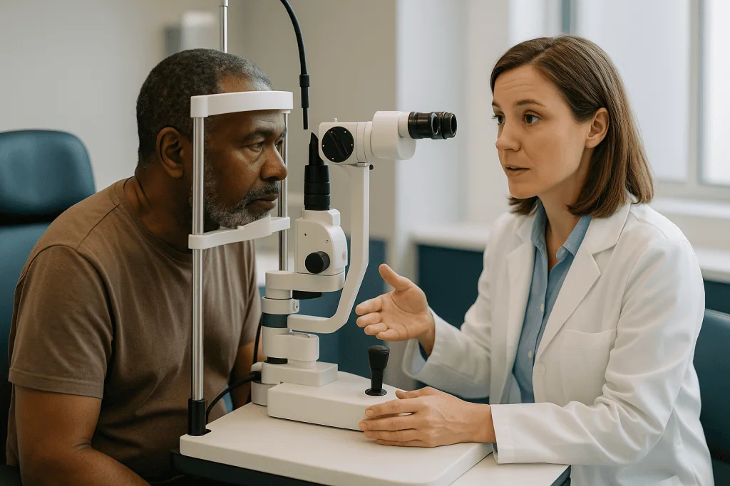
You then sit at a slit lamp, the familiar chin-rest microscope from every eye examination, while the ophthalmologist aims through the lens. A steady fixation target helps you keep the eye still. Each laser pulse arrives as a bright flash, sometimes with a sound like a soft click. With a pattern laser, a whole cluster of spots lands at once.
Occasionally the treatment is delivered differently. If the view through the pupil is poor, or laser is being done during a surgical operation, an indirect delivery system with a headset can be used instead. Some specialists also offer an injection of anesthetic around the eye for people who find scatter laser too uncomfortable with drops alone, a decision made case by case.
When the session ends, the lens is removed, the eye may be rinsed, and you sit for a while as the glare settles. Because your pupil stays dilated and the treated eye is blurry, the NHS advises against driving afterward. Arrange a ride or a companion, wear sunglasses, and plan nothing visually demanding for the rest of the day.
Is laser eye treatment for diabetic retinopathy painful?
The honest answer is that it depends on which laser, how much of it, and where in the retina it lands. Focal laser for macular edema uses fewer, gentler spots and is usually described as uncomfortable at most, with the bright flashes being more bothersome than any sensation.
Scatter laser is a different experience. The peripheral retina sits close to nerves that run forward to the eye’s focusing muscle, and heavier spots in certain zones, particularly the far edge and the area toward the nose, can produce a deep ache that people compare to a dull toothache behind the eye. The NHS lists discomfort during the procedure and aching afterward among the recognized effects, and both typically settle within hours.
Several factors nudge the experience one way or the other:
- Session size. Placing a very large pattern in a single sitting raises the cumulative discomfort. This is one of the main reasons scatter laser is often divided into two or more sessions, as the Mayo Clinic notes.
- Spot duration. Pattern-scanning systems use much shorter pulses, and shorter pulses generally hurt less because less heat spreads into surrounding tissue.
- Previous treatment. Retina that has already been treated has fewer active nerve endings, so fill-in sessions are often easier than the first.
- Anxiety and fatigue. Tight jaw, held breath and a long wait beforehand all amplify pain. Speaking up early and asking for a pause makes a measurable difference.
If drops alone are not enough, ophthalmologists can use a local anesthetic injection beside the eye to numb the nerves. Ask before the session begins if you have had a difficult experience previously. Pain that continues to build for hours after you get home, rather than fading, is not a normal part of the procedure and should be reported the same day.
Why diabetic retinopathy laser treatment often takes more than one session
This is the question that brought most readers here, and the answer has four parts, each grounded in how the retina responds rather than in scheduling convenience.
Coverage. Scatter laser has to treat a wide band of peripheral retina to bring the eye’s oxygen demand down enough to switch off the growth signal. Delivering that entire area at once means a long, uncomfortable session and a heavy dose of inflammation landing on the eye in a single afternoon. The Mayo Clinic notes the treatment is usually completed in two or more sessions for this reason.
Swelling risk. Every laser spot produces a small inflammatory response. Heavy treatment in one sitting has been associated with temporary worsening of macular edema and, less commonly, fluid gathering under the retina or behind the iris that can transiently raise eye pressure. Spacing the sessions lets each round settle before the next.
Biology sets the timetable. The abnormal vessels do not vanish when the laser stops. They regress over the following weeks as VEGF levels fall. Only after that interval can the ophthalmologist tell whether the pattern was sufficient. If new vessels persist or fresh ones appear, additional spots are placed in untreated gaps, a step sometimes called fill-in laser.
Two eyes, two plans. When both eyes need treatment, they are almost always done on separate days so you are never left with two blurry, aching eyes at once and so the response in the first eye can guide the second.
There is one more reason that has nothing to do with the laser. Blood in the vitreous, cataract or a poorly dilating pupil can block the view partway through, forcing a stop. The plan resumes when the view clears. In every case, needing another session reflects the size of the task and the retina’s pace, not a failure of the first visit.
How many laser sessions for diabetic retinopathy is typical?
There is no fixed count, and any source that offers one is guessing about your retina. What can be said with confidence is how the number is decided.
For scatter laser, mainstream references describe an initial course delivered over two or more visits, usually spaced a week or a few weeks apart, followed by a review. The NHS advises that more than one visit to a laser clinic may be needed. At the review, the ophthalmologist examines the retina, often with photographs and a scan, and asks a single question: are the new vessels regressing? If yes, the plan shifts to surveillance. If not, fill-in treatment is added. Eyes with very active disease, or where the first course was deliberately light to protect a swollen macula, are more likely to need additional rounds.
For focal laser, a single session commonly treats the identified leaks, but the retina takes months to absorb the fluid, and the effect of laser continues to build during that time. Retreatment is considered only after that interval and only if leakage persists in areas that can be safely targeted.
Several things stretch the timeline:
- Poor glucose or blood pressure control, which keeps driving new leakage.
- Combination plans where injections are given between laser sessions.
- Disease in both eyes, which doubles the number of visits even when each eye needs the same amount.
- Cataract or hemorrhage that limits the view and requires pauses.
Once regression is achieved, scatter laser is often durable, and many people need no further laser for years, though monitoring never stops because diabetes does not. The CDC continues to recommend a dilated eye examination at least yearly for adults with diabetes, and more often when retinopathy is present. Ask your specialist to describe the plan in stages rather than sessions; it is a more useful way to understand where you are.
Laser, injections or surgery: how the main options compare
Laser now shares the stage with two other treatments, and most eye units use them in combination rather than as rivals. The table summarizes what each does and where it fits, drawing on descriptions from the Mayo Clinic, the NHS and the National Eye Institute. It is a map, not a menu; the route is chosen by the treating team.
| Approach | What it does | Typically used for | Pace and pattern | Main trade-offs |
|---|---|---|---|---|
| Scatter laser (panretinal photocoagulation) | Treats peripheral retina to reduce oxygen demand and VEGF release | Proliferative retinopathy | Two or more sessions, then review over weeks | Some loss of side and night vision; durable once vessels regress |
| Focal or grid laser | Seals leaking spots near the macula | Macular edema outside the very center | Often one session; effect builds over months | Small scars in treated spots; limited use at the fovea |
| Anti-VEGF injections | Medicine blocks the growth signal directly | Center-involving macular edema; also proliferative disease | Repeated injections over months, schedule set by response | Needs frequent visits; effect fades if stopped; small infection risk per injection |
| Corticosteroid implants | Reduce inflammation and leakage | Macular edema not responding to other options | Longer-acting; fewer visits | Can raise eye pressure and accelerate cataract |
| Vitrectomy surgery | Removes vitreous gel, blood and scar tissue | Persistent hemorrhage, traction, retinal detachment | Single operation, weeks of recovery | Surgical risks; often combined with laser during the operation |
Two patterns stand out. Injections have taken over much of the treatment of center-involving macular edema because laser cannot be placed on the fovea. Laser, meanwhile, remains valuable for proliferative disease partly because its effect does not depend on a person returning every few weeks for years, a genuine advantage for anyone whose life makes frequent appointments hard. Neither fact makes one option better in the abstract; each fits a different problem and a different life.
The first days after laser: what recovery usually looks like
The rest of treatment day belongs to the sofa. Dilation keeps vision blurry and light-sensitive for several hours, and the treated eye may ache in a way that feels like eye strain after a long drive. The NHS lists blurred vision lasting a few hours and aching afterward as common; the Mayo Clinic describes blurry vision for about a day. Simple over-the-counter pain relief is usually enough, but check with the clinic or pharmacist about what suits your other medicines.
By the next morning most people are back to ordinary activities. A few practical points from mainstream guidance:
- You may notice new floaters, small drifting shapes in your vision, or a faint gray patch. These often reflect the laser spots settling and tend to fade over days to weeks.
- Reading and screens are safe when comfortable; they cannot damage the treated retina. Stop if the glare is unpleasant.
- There are usually no eye drops to take after laser unless the ophthalmologist prescribes them for a specific reason, which sets it apart from injections and surgery.
- Return to work depends on the job. Desk work is generally fine the next day; roles requiring sharp peripheral vision or working at heights may need a longer pause and a conversation with the clinic.
Driving is the most common question. The dilation must fully wear off, and you must meet your region’s legal vision standard in both eyes together. After a single scatter session most people are able to drive within a day or two once the blur clears, but after an extensive course of scatter laser in both eyes, peripheral vision may be reduced enough to require a formal visual field test before driving is permitted. Ask directly rather than assuming.
What recovery does not include is a dramatic change in vision for the better. Laser prevents future loss; it rarely sharpens sight in the days after. Knowing this in advance saves a great deal of unnecessary worry.
Weeks and months later: how the retina responds and what follow-up checks
The real work of laser happens after you leave. Each treated spot heals into a small, pale scar over the following two to three weeks. As the peripheral retina’s oxygen demand falls, VEGF production drops, and the abnormal vessels begin to shrink and close. The Mayo Clinic describes this regression unfolding over weeks rather than days, which is why the review appointment is set with that gap deliberately in mind.
At the review, the specialist looks at three things:
- The new vessels. Are they thinner, fewer or gone? Partial regression may mean fill-in laser in untreated gaps.
- The macula. A scan called optical coherence tomography (OCT), which builds a cross-section image of the retina using light, shows whether swelling has increased, stayed flat or improved.
- The vitreous. Any fresh bleeding, or signs that the regressing vessels are pulling on the retina and creating traction.
For focal laser, the timeline stretches further. Leaked fluid is absorbed slowly, and the sealing effect strengthens over months. Judging the result too early leads to unnecessary retreatment, so specialists wait before deciding on a second round.
Once the retina is stable, follow-up intervals lengthen but never disappear. The CDC recommends at least yearly dilated examinations for everyone with diabetes, and the interval after treated proliferative disease is usually shorter, set by the ophthalmologist. Keep every one of these appointments even when vision feels fine. Proliferative retinopathy can reactivate silently, especially if glucose or blood pressure control slips, and a small fill-in session caught early is a far smaller intervention than treating a hemorrhage.
People who also receive injections will have a parallel schedule of visits, and the two plans are coordinated by the same team. Ask for a written summary after each review that states what was found and what the next step is; it turns a confusing series of appointments into a plan you can follow.
Panretinal photocoagulation side effects and the trade-off nobody should hide
Scatter laser works by sacrificing a portion of the peripheral retina to protect the center. That is a deliberate trade, and patients deserve to hear it stated plainly rather than discovered later.
The NHS lists the recognized effects of laser treatment as blurred vision for a few hours, increased sensitivity to light, aching or discomfort, seeing floaters, reduced side vision, and difficulty seeing in dim light. The Mayo Clinic adds that scatter treatment can cause some loss of peripheral vision, night vision and color vision. Most of the short-term effects fade within hours to days. The peripheral, night and color changes can be permanent, though for many people they are subtle and noticed mainly when driving at dusk or walking into a dark room.
Less common effects include:
- Temporary worsening of macular swelling, one reason sessions are spaced and sometimes combined with injections.
- A short-term rise in eye pressure or a shallow front chamber from fluid shifts after heavy treatment, which usually settles but is monitored.
- Small blind spots if a laser spot lands closer to the center than intended, or if scars enlarge over years, a phenomenon called scar creep.
- Rarely, a laser burn to the fovea, which is why the central zone is treated with great caution or not at all.
Set against these is the alternative the laser is preventing. Untreated proliferative retinopathy leads to vitreous hemorrhage, tractional retinal detachment and neovascular glaucoma, a painful form of glaucoma driven by abnormal vessels growing on the drainage tissue. The National Eye Institute describes these as the pathways to severe, often irreversible vision loss. Losing some twilight vision to keep reading vision is the calculation behind every scatter pattern, and a good consultation makes that calculation explicit so you can weigh it with your own eyes in mind.
How blood sugar, blood pressure and cholesterol shape what the laser can do
Laser treats the consequences of vessel damage. It does not touch the cause, and the cause continues every day between sessions. This is why the ophthalmologist keeps asking about glucose readings, and why the answer genuinely changes the plan.
Three measures matter most, according to the CDC and the National Eye Institute:
- Glucose control, tracked by HbA1c. HbA1c is a blood test reflecting average glucose over roughly three months. Persistently high values keep damaging capillaries, feeding new leakage after focal laser and new VEGF release after scatter laser, so treated eyes reactivate sooner.
- Blood pressure. High pressure strains already weakened vessel walls and drives macular edema. The NHS lists blood pressure control alongside glucose as a core part of slowing retinopathy.
- Cholesterol and triglycerides. Lipid deposits, visible as yellow flecks called hard exudates, accumulate where vessels leak and are more prominent when blood fats run high.
One counterintuitive point deserves airtime. Rapidly tightening glucose control after years of high readings can cause a temporary worsening of retinopathy in the first months before the long-term benefit appears. This is well recognized and is not a reason to avoid improving control; it is a reason the eye team and the diabetes team should know what each other is doing so monitoring can be adjusted. Any change to diabetes medicines belongs with the prescribing clinician, and the eye clinic will usually ask to be kept informed.
Smoking cessation, kidney function and sleep apnea also influence retinal health, and each is worth raising with your primary care team. The practical message is not moralizing. It is that laser buys time and stability, and the value of that stability is set by what happens to the vessels afterward. People who leave the laser clinic with a renewed conversation about their broader diabetes care tend to get more out of the treatment they just had.
What people often get wrong about laser for diabetic retinopathy
Some myths about eye laser are borrowed from vision-correction procedures, others from a relative’s experience decades ago. Several deserve a direct correction.
“Laser will give me back the vision I have lost.” Scatter laser is preventive. Its purpose is to stop the retina from bleeding and detaching, not to restore sight already gone. Focal laser can stabilize and sometimes modestly improve vision as swelling resolves, but neither should be expected to sharpen sight in the days after treatment. The Mayo Clinic frames laser as slowing or stopping leakage and abnormal growth.
“Needing a second session means the first failed.” As explained earlier, splitting scatter laser into stages is built into the treatment to protect the macula and manage discomfort, and fill-in treatment after review is expected in a proportion of eyes.
“It is the same as laser eye surgery for glasses.” Refractive laser reshapes the cornea at the front of the eye and aims to sharpen vision. Retinal laser treats the back of the eye and aims to protect it. They share only the word.
“If my vision is fine, I do not need treatment.” Proliferative retinopathy is frequently painless and symptom-free until a hemorrhage occurs. The National Eye Institute notes that the disease often has no early warning signs, which is the entire rationale for screening.
“Injections have replaced laser.” Injections now lead for center-involving macular edema and are an option for proliferative disease, but laser remains a core treatment for proliferative retinopathy, valued partly for its durability. Many people receive both.
“Once treated, I am finished with eye appointments.” Diabetes continues, and so does monitoring. The CDC’s recommendation of at least yearly dilated exams applies for life.
One more, gentler misunderstanding: that asking to pause during the procedure is a nuisance. It is not. Steady, unhurried treatment produces a better pattern, and every experienced laser surgeon would rather stop for thirty seconds than place a spot on a moving eye.
Questions to ask your care team before and after laser
A laser consultation moves quickly, and the questions that matter most often surface in the car on the way home. Bringing a short written list changes that. These are the ones seasoned patients tend to wish they had asked.
Before the first session
- Which laser am I having, scatter or focal, and which eye? What specifically is it treating?
- Do I also have macular swelling, and if so, will that be treated first, alongside, or afterward?
- How many stages do you anticipate for this eye, and roughly how far apart?
- What options exist for comfort if the treatment is painful?
- Are injections an alternative or an addition in my case, and what would tip the balance?
- What changes to my side vision or night vision should I expect, and how might they affect my driving or work?
After each session
- How much of the planned pattern was completed today?
- What symptoms are normal this week, and which ones should prompt a same-day call?
- When may I drive, and will I need a formal visual field test at any point?
- When is my review, and what will you be looking for?
At the review
- Are the new vessels regressing? Does the scan show the macula flat or swollen?
- Is fill-in laser needed, and where?
- How often will I be seen once things are stable?
- What glucose, blood pressure and cholesterol targets would help my eyes most, and who is coordinating that with the eye clinic?
Ask for the answers in writing or ask permission to record them. If you have a diabetes educator or primary care clinician, sharing the eye clinic’s summary closes a loop that often stays open. None of these questions challenge the specialist’s judgment; they help the plan become something you carry rather than something done to you.
When to call your doctor
Mild ache, blur, glare and a few new floaters are expected for a day or so after laser. The following are not, and each warrants a same-day call to the eye clinic or, out of hours, urgent eye care. Mainstream sources including the NHS and the Mayo Clinic list these as signs of complications that need prompt assessment.
- A sudden shower of new floaters, cobwebs or a red or dark haze across the vision, which can signal bleeding into the vitreous.
- Flashes of light, especially with a shadow or curtain moving across part of the visual field, a possible sign of retinal tear or detachment.
- A sudden drop in vision in the treated eye that does not lift as the dilation wears off.
- Severe, deep eye pain with nausea, headache or a hazy cornea, which can indicate a sharp rise in eye pressure and needs assessment within hours.
- Pain that worsens over the following days rather than fading, or a red, sticky or swollen eye, particularly if you also had an injection.
- A new fixed dark spot in the center of your vision that was not present before treatment.
Outside the immediate recovery period, the same signals apply for as long as you have diabetes. Proliferative retinopathy can reactivate without warning, and a bleed caught early is far easier to manage than one discovered weeks later.
For anything less dramatic, such as glare that lingers longer than expected, difficulty judging steps in dim light, or uncertainty about driving, contact the clinic during working hours rather than waiting for the scheduled review. The people who run laser clinics field these calls every day and would rather hear from you than have you guess. Every decision about further treatment, and about whether a symptom needs to be seen, rests with the ophthalmology team that knows your retina.
Frequently asked questions
How many laser sessions for diabetic retinopathy will I need?
There is no fixed number; the plan depends on how much retina must be treated and how the vessels respond. Scatter laser is commonly delivered over two or more visits, followed by a review a few weeks later to check whether the abnormal vessels are regressing. Fill-in treatment is added if they persist. Focal laser for macular swelling is often a single session, reassessed after months. Your ophthalmologist sets the schedule.
Is laser eye treatment for diabetic retinopathy painful?
Focal laser is usually mildly uncomfortable at most, with the bright flashes more bothersome than any sensation. Scatter laser can cause a deep ache, especially with larger sessions or spots near the far periphery. Numbing drops are standard, shorter-pulse pattern lasers are generally more comfortable, and an anesthetic injection beside the eye can be offered when drops are not enough. Tell the surgeon early if it hurts so the pace can be adjusted.
What are the main panretinal photocoagulation side effects?
Short-term effects include blurred vision for hours, light sensitivity, aching and new floaters, which usually fade within days. Longer-term effects can include some permanent loss of peripheral vision, poorer vision in dim light and subtle changes in color perception, because peripheral retina is deliberately treated. Less common effects are temporary worsening of macular swelling and a brief rise in eye pressure. These are weighed against preventing hemorrhage and retinal detachment.
What is focal laser for macular edema, and how is it different from scatter laser?
Focal laser targets individual leaking spots near the macula, the central area used for reading, to seal them and let swelling drain. It uses far fewer and gentler spots than scatter laser, which covers a wide band of peripheral retina to control abnormal vessel growth. Focal laser cannot be used on the very center of the macula, so anti-VEGF injections are often preferred when swelling involves that point.
Will laser treatment improve my eyesight?
Usually not in a noticeable way. Scatter laser is preventive; its aim is to stop bleeding and retinal detachment, not to restore vision already lost. Focal laser can stabilize vision and sometimes allow modest improvement as swelling resolves over months. Expect blur for a day and then a return to roughly your previous vision. The benefit is measured in what does not happen over the following years.
Can I drive after diabetic retinopathy laser treatment?
Not on the day. Dilating drops blur vision and increase glare for several hours, so you need someone to take you home. Most people can drive within a day or two after a single session once the blur clears. After an extensive course of scatter laser in both eyes, peripheral vision may be reduced enough to require a formal visual field test before driving. Ask your clinic for specific guidance.
Why did my ophthalmologist recommend injections instead of laser?
The most common reason is macular swelling that involves the fovea, the pinpoint center of sharpest vision, where laser cannot safely be placed. Anti-VEGF medicines, a class that blocks the growth and leakage signal, can be used there. Injections are also an option for proliferative disease and are sometimes combined with laser. The choice depends on where the problem sits and on how often you can attend follow-up.
How long does recovery take after laser for diabetic retinopathy?
Blurred vision and aching typically settle within hours to about a day, and most people return to normal activities the next morning. New floaters or a faint gray patch may take days to weeks to fade. The retina’s response continues beneath the surface for several weeks after scatter laser and for months after focal laser, which is why the review appointment is scheduled with that interval in mind.
Does laser treatment mean my diabetic retinopathy is gone?
No. Laser treats a complication of retinopathy, not the underlying vessel damage, which continues as long as diabetes does. Successfully treated eyes can reactivate, particularly if glucose or blood pressure control slips, and reactivation is often silent. The CDC recommends dilated eye examinations at least yearly for everyone with diabetes, and people with treated proliferative disease are usually seen more often.
Can I have laser if there is blood inside my eye?
It depends on how much blood is present. Laser needs a clear view of the retina, so a dense vitreous hemorrhage can block treatment. Ophthalmologists may wait for the blood to clear, use injections to reduce the risk of further bleeding, or recommend vitrectomy surgery, during which laser can be applied directly. Your treating team decides based on how quickly the view improves and how active the disease is.
References
- NHS: Diabetic retinopathy: Treatment
- National Eye Institute (NIH): Diabetic Retinopathy
- CDC: Diabetes and Vision Loss
- Cleveland Clinic: Diabetes-Related Retinopathy
This article is for general information only and is not a substitute for professional medical advice. Please consult a qualified doctor about your individual situation.

More from the Blog

Life With an Intraocular Lens: Halos, Night Driving and How the Brain Adapts
Living with an intraocular lens usually means clearer vision within days of cataract surgery, with full recovery taking around four to six weeks according…

Vitrectomy Recovery With a Gas Bubble: Face-Down Positioning, Drops and Weekly Milestones
After a vitrectomy with a gas bubble, the surgeon usually asks you to hold a specific head position, often face down, for several days…
How OCT and Fundus Examination Map Macular Disease Before Any Treatment Is Planned
Optical coherence tomography (OCT) and a dilated fundus examination together map macular disease by showing the retina in two complementary ways: the fundus exam…

Low Pressure, Leaks and Infection After Glaucoma Surgery: What Your Team Watches For
After glaucoma surgery, care teams watch most closely for eye pressure that drops too low (hypotony), a leak from the small drainage blister called…

Farsightedness Myths: Does Reading in Dim Light or Screen Time Make It Worse?
No. Reading in dim light and spending long hours on screens can tire the eyes and cause temporary blur or headache, but neither changes…

Keratoconus Treatment Ladder: Lenses, Cross-Linking, Rings and When Transplant Is Discussed
Keratoconus treatment options form a ladder rather than a single choice. Glasses and soft lenses help early; rigid gas-permeable, hybrid or scleral lenses restore…