

How Melanoma Follow-Up Is Scheduled: Skin Examinations, Imaging and Self-Checks
Key Takeaways
- UK national guidance (NICE) discharges stage IA melanoma after two to four checks in about a year, while stages IB to III are seen every three months for three years and then every six months to five years.
- Routine CT or PET imaging is not recommended for asymptomatic stage I to IIB melanoma because false alarms outnumber true recurrences; surveillance scans are considered from stage IIC and in stage III.
- There is no screening blood test for melanoma recurrence; LDH is used only in staging advanced disease, and vitamin D is the one blood level UK guidance asks clinicians to measure at diagnosis.
- Local recurrence often appears as a colourless firm lump under or beside the scar rather than a dark mole, so feeling the scar and the limb toward the nearest lymph nodes matters as much as looking.
- The UK two-week rule describes the wait to see a specialist after an urgent referral, not how fast melanoma grows, and the same route gets a discharged patient back to a specialist quickly.
- Having one melanoma permanently raises the risk of a second, so monthly skin self-checks using the ABCDE signs and strict sun protection should continue for life, long after scheduled visits end.
Melanoma follow-up is scheduled by stage. UK national guidance (NICE) suggests that stage IA patients have two to four skin checks over about a year, while stages IB to III are usually seen every three months for three years, then every six months up to five years, with imaging considered for higher stages. Each visit combines a full skin and lymph node examination with coaching for monthly self-checks. Your treating team sets the exact plan.
The stitches are out. The scar on your calf has settled from angry red to a quiet pink line, and the surgeon has said the words you were waiting for: the margins were clear. Then comes the sentence that catches many people off guard. We will see you again in three months. And after that? And after that?
A melanoma follow up schedule can feel like a second diagnosis in miniature. It stretches ahead for years, it involves people undressing in bright rooms, and it is rarely explained in one sitting. Yet the structure behind it is surprisingly logical. Every appointment, every scan that is offered and every scan that is deliberately not offered traces back to a single question: how likely is this particular melanoma to come back, and where would it show up first?
This explainer walks through that logic, stage by stage, including what actually happens in the room, when imaging earns its place, how to check your own skin without turning every freckle into a crisis, and which signs mean you should pick up the phone between visits.
What is the recommended follow-up for melanoma?
There is no single answer, and that is the point. Melanoma follow-up is tiered, so a thin melanoma removed early earns a very different plan from one that had already reached a lymph node. The clearest published framework comes from the UK’s national guideline body, NICE, whose recommendations the NHS applies across its melanoma services. US cancer organizations describe a similar shape, with intervals that stretch from every three months for higher-risk disease to once a year for the lowest risk.
Under the UK framework, someone treated for stage 0 melanoma (abnormal cells confined to the very top layer of skin, also called melanoma in situ) is usually discharged once the wound has healed, with advice on self-checks. Stage IA, the thinnest invasive melanoma, typically means two to four appointments over roughly twelve months, then discharge. Stages IB through IIIC are usually seen every three months for three years, then every six months for a further two years, and people with stage IIC or stage III disease are additionally considered for surveillance imaging. Stage IV follow-up is built individually around active treatment.
Why five years? Most melanoma recurrences that are going to happen declare themselves within that window, and the risk falls steadily year on year. Follow-up is front-loaded for the same reason: the earliest months carry the greatest chance of catching something treatable.
What matters most, in my reading of the evidence, is not the exact interval but the two things that happen inside it. A clinician examines all of your skin and the lymph node basins that drain the original site, and you leave knowing exactly what you are looking for at home. Everything else in the melanoma follow up schedule is scaffolding around those two acts.
How a melanoma follow-up appointment actually works
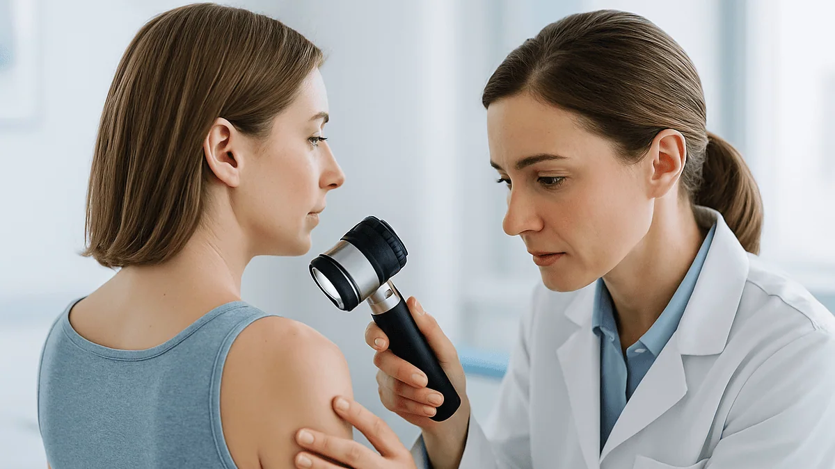
Picture a twenty-minute slot. You undress to your underwear, and the clinician, often a dermatologist, surgeon or specialist nurse, starts at the scar. They are feeling as much as looking: a recurrence at the original site, or an in-transit metastasis (a deposit of melanoma cells that has travelled a short way along lymph channels but not yet reached a node), often announces itself as a small firm lump under otherwise normal skin.

Next come the lymph node basins. For a melanoma on the leg that means the groin; for the arm, the armpit; for the scalp or face, the neck. Nodes that are enlarged, hard or fixed get attention. Then the rest of your skin is examined, because having had one melanoma raises the chance of developing a second, unrelated one. Mayo Clinic describes this head-to-toe check as the backbone of every follow-up visit.
A dermatoscope, a handheld magnifier with polarized light that lets a clinician see pigment patterns beneath the surface, is used on anything that looks even slightly out of step. Some services photograph the whole body or individual moles so changes can be compared over time rather than remembered.
The conversation is the other half of the visit. Expect to be asked about new lumps, headaches, breathlessness, bone pain, appetite or weight, not because these are expected, but because the questions are a cheap and effective screen for spread that an examination cannot reach. You should also be asked how you are coping. The NHS notes that living with the uncertainty of follow-up is one of the hardest parts of melanoma for many people, and specialist nurses exist partly for this reason.
If anything is found, the usual next step is a biopsy or an ultrasound rather than a conclusion in the room.
Why stage decides the melanoma follow up schedule
Stage is shorthand for three measurements taken from the removed melanoma and, where done, from the sentinel lymph node. Each one shifts the follow-up plan.
Breslow thickness is the depth of the melanoma in millimetres, measured under the microscope from the top of the skin to the deepest cancer cell. It remains the single strongest predictor of behaviour. Thin melanomas have simply had less opportunity to reach blood or lymph vessels.
Ulceration means the surface of the melanoma had broken down, a sign of faster growth. An ulcerated melanoma is staged one notch higher than an unulcerated one of the same thickness, and its follow-up is scheduled accordingly.
Lymph node status comes from a sentinel lymph node biopsy, a procedure in which dye and a tracer are injected around the scar so the first node receiving lymph from that area can be removed and checked for cancer cells. A negative result keeps someone in stages I or II; a positive result moves them into stage III and into a more intensive schedule that may include imaging and a discussion of adjuvant therapy (treatment given after surgery to reduce recurrence risk).
Put those three together and the pattern of the UK guidance makes sense. Stage IA carries a low enough risk that a year of checks is considered proportionate. Stage IB to IIIC carries enough risk that three-monthly visits for three years are justified. Stage IIC and III carry enough risk of internal spread that looking inside the body with scans adds something examination cannot.
Other factors nudge the plan at the edges: a mitotic rate (how many cells are dividing), a melanoma that was already a recurrence, many atypical moles, a family history, or a weakened immune system. Your team may lengthen or shorten intervals on that basis. Cleveland Clinic’s overview of melanoma notes that follow-up is individualized precisely because these risk factors vary so widely between two people with the same headline stage.
Melanoma follow-up guidelines by stage: a summary table
The table below summarizes the UK national framework (NICE, applied by the NHS) and notes where common US recommendations differ. It is a map, not a prescription: your team’s plan may legitimately differ, and local pathways vary.

| Stage at diagnosis | Typical clinical visits (UK guidance) | Imaging | Usual duration |
|---|---|---|---|
| 0 (in situ) | Discharge after wound healing, with self-check advice | None | One visit or two |
| IA | 2–4 visits over about 12 months | None routinely | About 1 year |
| IB to IIB | Every 3 months for 3 years, then every 6 months for 2 years | Not routinely, unless symptoms | 5 years |
| IIC and III | Every 3 months for 3 years, then every 6 months for 2 years | Surveillance CT considered, commonly 6-monthly for 3 years then yearly | 5 years, sometimes longer |
| IV | Individualized around treatment | Regular scans to track response | Ongoing |
Two honest caveats. First, US guidance tends to give ranges rather than fixed intervals, for example every three to twelve months for stages I and II depending on risk, and it generally leaves open whether a lower-risk patient should continue annual skin checks for life. Many dermatologists favour this, because the risk of a second primary melanoma does not expire at five years.
Second, the strength of evidence behind any particular interval is modest. Follow-up schedules were built largely on patterns of when recurrences appeared in large registries, not on randomized trials comparing three-monthly with six-monthly visits. That is why guidelines across countries agree on the shape of the schedule but quibble about the numbers. When your team departs from the table, it is usually for a reason worth asking about, not a sign of neglect.
Stage 1 melanoma follow up: who is discharged early, and who is asked to keep coming
Stage 1 is where most melanomas are now diagnosed, and it is also where the follow-up plan most often surprises people, sometimes by seeming too short.
Stage IA means a melanoma under one millimetre thick without ulceration. UK guidance treats this group as low enough risk that a year of checks is sufficient before discharge back to the family doctor, with clear instructions about self-examination and how to be seen quickly if anything changes. It can feel abrupt to be told you are done after four appointments. The reasoning is that the chance of this melanoma returning is low, the chance of a new melanoma is better managed by self-checks and prompt access than by scheduled visits, and clinic capacity is finite.
Stage IB covers slightly thicker or ulcerated melanomas, and here the plan lengthens to five years, because the risk of spread, while still modest, is meaningfully higher than for IA.
People frequently ask how often stage 1 melanoma metastasizes. The honest answer is that the risk is low and depends heavily on exact thickness, ulceration and, when performed, sentinel node result, so a single percentage would be misleading. Your pathology report and your team’s interpretation of it are more informative than any general figure. What the staging system does tell you is that follow-up intensity has been matched to that risk on purpose.
Some stage 1 patients are kept under longer surveillance regardless. Those with many unusual moles, a previous melanoma, a strong family history, fair skin with heavy past sun damage, or immune suppression after a transplant may be offered annual skin checks indefinitely. The question to ask is not just which stage you are, but which of these additional factors apply to you. A short conversation about that often explains why your plan differs from a friend’s.
Melanoma surveillance imaging: when scans help and when they do not
Scans sound reassuring, which is exactly why guidelines are cautious with them.
For stages I through IIB, routine imaging in someone without symptoms is not recommended in either UK or US guidance. The logic is statistical. When the chance of internal spread is low, a CT scan is far more likely to find an incidental shadow in the lung or liver that turns out to be nothing than to find a real recurrence. Each shadow brings weeks of anxiety, more scans and sometimes a biopsy, with radiation exposure accumulating along the way. Mayo Clinic’s treatment overview makes the same point: imaging is reserved for people whose stage or symptoms justify it.
From stage IIC and throughout stage III, the balance tips. UK guidance suggests that surveillance imaging, usually CT of the chest, abdomen and pelvis, be considered on a schedule of roughly every six months for three years and then yearly to five years, with brain imaging added in selected cases because melanoma has a known tendency to spread there. CT (computed tomography, a series of X-ray slices assembled into cross-sectional pictures) is the workhorse. PET-CT, which adds a sugar-based tracer taken up by metabolically active cells, is used when a finding needs clarifying. MRI is preferred for the brain.
Imaging is also triggered by symptoms at any stage. A persistent cough, new bone pain or an unexplained headache in someone with a melanoma history will prompt a scan regardless of what the schedule says.
What people rarely hear is that no trial has shown that routine imaging in early-stage melanoma helps people live longer. The case for scanning higher-stage disease rests on the availability of effective treatments for recurrence that work better when disease burden is small, which makes earlier detection worth the downsides. Your oncologist weighs that trade-off for your situation; the decision belongs with them.
Blood tests, lymph node ultrasound and other checks people ask about
Three tests come up repeatedly in clinic conversations, and each has a different standing in the evidence.
Blood tests. There is no reliable blood marker for early melanoma recurrence. Lactate dehydrogenase, or LDH, an enzyme released when cells break down, rises in some people with advanced melanoma and is used in staging stage IV disease, but it is neither sensitive nor specific enough to screen for recurrence in earlier stages. Routine blood tests are therefore not part of standard follow-up for stages I to III, though your team may check a full blood count or liver tests for other reasons, especially during adjuvant treatment. One exception written into UK guidance is vitamin D: NICE recommends measuring it at diagnosis, because low levels are common and may be relevant to outcomes, with any supplementation decided by the clinician.
Lymph node ultrasound. Ultrasound of the draining lymph node basin is more accurate than fingers alone at spotting small nodal recurrences. Its routine role is strongest for people who had a positive sentinel node but did not have the remaining nodes removed, a group that has grown since trials showed completion dissection does not improve survival for most. Some services use it more widely; others reserve it for suspicious findings on examination.
Genetic or molecular testing of the tumour. Tests for mutations such as BRAF are performed on the melanoma tissue itself, usually at diagnosis for stage III and IV disease, because they determine which targeted therapies might be used if treatment is needed. They do not change the follow-up schedule.
The pattern here is consistent with everything else in melanoma surveillance: tests are added when they change what a clinician would do next, and left out when they mainly add noise. Johns Hopkins’ melanoma overview summarizes follow-up as examination first, imaging where risk warrants it, and laboratory testing only in defined situations.
Melanoma recurrence signs: what returning disease can look like
This is the question underneath every other question, so it deserves a plain answer. Melanoma can return in three broad places, and each looks different.
At or near the scar. A local recurrence usually appears as a new pigmented spot within or beside the scar, or as a firm nodule under the skin that may be flesh-coloured, bluish or black. In-transit deposits appear as similar lumps further along the limb, between the scar and the nearest lymph nodes. These are often the first sign and the easiest to find during a self-check.
In lymph nodes. A swollen, firm, usually painless lump in the groin, armpit or neck on the same side as the original melanoma. Infections cause tender, mobile swellings that settle within a couple of weeks; a node that persists or grows warrants prompt review.
At distant sites. Spread to the lungs, liver, bones or brain produces symptoms that are vague on their own and matter mainly because of your history: a cough that lingers, breathlessness, pain in a bone that does not ease, loss of appetite or unintended weight loss, persistent headache, visual change, seizures or new weakness.
The NHS emphasizes that most of these symptoms, in most people, turn out to have an ordinary cause, and that is true even after melanoma. The purpose of knowing the list is not to monitor yourself for doom but to lower your threshold for mentioning things. People sometimes sit on a new lump for months because they do not want to be the anxious patient. Clinicians would far rather see ten lumps that turn out to be cysts than miss the one that is not.
How you report also matters. Note when you first noticed something, whether it has changed, and whether it is tender. That information shapes the next step.
How to do a monthly skin self-check without becoming anxious about every freckle
Self-examination is the one part of the melanoma follow up schedule you control, and the evidence suggests it matters: a substantial share of recurrences and second melanomas are first noticed by patients or partners, not at scheduled visits. Both the NHS and US cancer organizations suggest a full check about once a month.
Choose good light and a full-length mirror, with a hand mirror for the back. Work in the same order every time so it becomes automatic. Face, scalp (a comb or hairdryer parts the hair), ears, neck. Chest and torso, lifting breasts or folds of skin. Arms, including armpits, palms and between fingers. Back and buttocks using the hand mirror. Legs front and back, soles of the feet, between the toes and under toenails. Then the scar itself and the skin between the scar and the nearest lymph node basin, feeling as well as looking.
What you are looking for is change, not perfection. The ABCDE mnemonic described by MedlinePlus helps: Asymmetry, irregular Border, more than one Colour, Diameter larger than about six millimetres (roughly a pencil eraser), and Evolution, meaning any change in size, shape, colour, texture or sensation. Evolution is the one that matters most. A spot that looks different from all your others, the so-called ugly duckling, also deserves attention.
Photographs turn memory into evidence. Take a dated image of any mole you are unsure about and compare it next month. A smartphone is sufficient; dedicated mole-tracking apps have not been shown to diagnose melanoma reliably and should never replace a clinician’s view.
Keep the check to fifteen minutes and then stop. Checking daily tends to breed anxiety without adding safety, because meaningful change usually takes weeks. If a partner can help with your back, their eyes are often sharper than yours in a mirror.
What the first weeks and months after melanoma treatment usually look like
Follow-up begins before the first formal appointment. In the fortnight after wide local excision, the second operation that removes a margin of healthy skin around the biopsy site, the focus is on the wound. Dressings come off, stitches are removed or dissolve, and you are told how to watch for infection. If a sentinel node biopsy was done, the small second scar in the groin, armpit or neck heals alongside.
The pathology meeting, often around two to four weeks later, is where the final stage is confirmed and the follow-up plan is set. This is the appointment to bring a list of questions and, if possible, another pair of ears.
For stage I and II disease, the first scheduled follow-up visit typically falls about three months after surgery. Many people describe this stretch as the strangest: active treatment is over, yet it does not feel finished. The NHS acknowledges this emotionally unmoored period and points to specialist nurses and support organizations rather than expecting people to simply adjust.
For stage III, the timeline may include a discussion of adjuvant therapy with immunotherapy (drugs that release the brakes on the immune system so it attacks melanoma cells) or targeted therapy for BRAF-mutant disease. These are usually given for around a year and come with their own schedule of clinic visits, blood monitoring and scans, which generally overlaps with surveillance rather than adding to it. Whether to have adjuvant treatment, and which kind, is a decision made with your oncologist based on your stage, your health and your priorities.
Across all stages, the early months are when sun habits get rebuilt. Daily high-factor sunscreen, shade in the middle of the day and covering clothing reduce the risk of a second melanoma. Lymphedema (persistent swelling of a limb after lymph nodes are removed) can develop in the first year after node surgery; early referral to a lymphedema service makes it easier to manage.
What is the 2 week rule for melanoma?
The two-week rule has nothing to do with how quickly melanoma grows. It is the name of a referral pathway in the UK’s health service. When a family doctor sees a skin lesion that raises suspicion of melanoma, they refer under an urgent suspected cancer pathway, and the person should be offered a specialist appointment within two weeks of that referral. The NHS describes this pathway on its melanoma pages, and it exists because earlier diagnosis of melanoma is strongly associated with thinner tumours and a lower stage.
Several points often get muddled.
The rule covers the wait to be seen, not the time to diagnosis or treatment. At the specialist appointment the lesion is examined with a dermatoscope and, if concern remains, removed for biopsy. Results take a further one to two weeks, and if melanoma is confirmed, a wider excision is scheduled after that.
Being referred under the two-week rule is not a diagnosis. Most people seen on this pathway turn out to have benign moles, seborrhoeic keratoses or other harmless lesions. The threshold is deliberately low because the cost of missing a melanoma is high.
The pathway also applies to people already under follow-up who have been discharged. If you have completed your stage IA year of checks and later notice a change, your family doctor can use the same urgent route to get you back in front of a specialist quickly. Knowing this makes early discharge less unsettling: you are not being cut loose, you are moving to a different door.
Outside the UK, similar fast-track systems exist under other names, though timelines differ. If you are unsure how to be seen urgently in your own system, ask your team at your final scheduled visit. It is a far better question to ask then than when you have found something worrying.
What people often get wrong about melanoma follow-up
More scans mean safer follow-up. In early-stage disease the opposite is closer to the truth. Routine CT in someone at low risk generates false alarms far more often than it finds recurrence, and no trial has shown it improves outcomes. Guidelines limit imaging to higher stages on purpose.
Five years clear means it cannot come back. Late recurrences, beyond ten years, are uncommon but documented. More importantly, having had one melanoma permanently raises the risk of a new one. Lifelong self-checks and sun protection outlast the clinic schedule.
A blood test would pick it up. There is no screening blood test for melanoma recurrence. LDH has a role only in advanced disease.
Being discharged after stage IA means the hospital does not care. Discharge reflects a low measured risk and a rapid route back in if anything changes. It is a risk calculation, not a dismissal.
Recurrence always looks like the first melanoma. Local recurrence is often a colourless lump under the skin, not a dark mole, which is why feeling the scar matters as much as looking at it.
Sunscreen alone fixes the risk. Sunscreen is one layer. Shade, clothing and avoiding sunbeds matter at least as much, and sun exposure before diagnosis cannot be undone, which is one reason surveillance for new melanomas continues.
Skipping a visit is fine if the skin looks normal. You cannot examine your own lymph node basins well, and clinicians find things patients miss. Rescheduling is fine; quietly dropping out is not.
A negative sentinel node means zero risk. It lowers the risk substantially but does not eliminate it, which is why stage IB and II still get five years of follow-up.
Each of these myths tends to push people toward either excess worry or false reassurance. The evidence sits, as it usually does, in the calmer middle.
Questions to ask your care team about your follow-up plan
Bring these written down. Clinic time is short, and the answers shape the next five years.
- What is my exact stage, including Breslow thickness, ulceration and, if done, sentinel node result? Can I have a copy of the pathology report?
- How often will I be seen, by whom, and for how long? Will the interval change after three years?
- Will I have surveillance imaging? If so, what type, how often, and what would prompt it to stop? If not, why does my stage not warrant it?
- Which lymph node areas should I be feeling at home, and can you show me how?
- What exactly am I looking for at the scar, and what would a local recurrence feel like?
- Who do I contact between visits if I find something, and how quickly can I be seen?
- If I am discharged, how do I get back to a specialist quickly, and is my family doctor aware of the plan?
- Am I a candidate for adjuvant treatment, and how would its monitoring fit with surveillance?
- Do I have any additional risk factors, such as many atypical moles or family history, that should extend my follow-up?
- Should my close relatives have their skin checked?
- Has my vitamin D been measured, and does anything need to happen as a result?
- What support is available for the anxiety that comes with waiting between appointments?
Two further questions are worth asking even if they feel awkward. First, is there anything about my case that makes you more or less worried than the stage alone suggests? Clinicians hold nuanced impressions that rarely make it into a letter unless asked. Second, what would you do if something turned up at a visit? Hearing the pathway in advance, for example an urgent ultrasound and biopsy within a week or two, makes a future finding less frightening because it is already mapped.
The answers you receive will reflect your team’s judgement about your particular melanoma. Where they diverge from the general guidance described here, that divergence is usually the most informative part of the conversation.
When to call your doctor between melanoma follow-up appointments
Scheduled visits are only part of surveillance. The other part is you deciding to be seen early, and the threshold for that should be low. Contact your melanoma team, specialist nurse or family doctor without waiting for the next appointment if you notice any of the following.
At the scar or nearby: a new pigmented spot in or beside the scar; a firm lump under the skin at the scar or anywhere between the scar and the nearest lymph node area; a mole anywhere that is changing in size, shape or colour, bleeding, itching or crusting, or that stands out from your other moles.
In lymph node areas: a swelling in the groin, armpit or neck that persists beyond about two weeks, grows, or feels hard or fixed rather than soft and mobile.
General symptoms lasting more than a couple of weeks without an obvious cause: unintended weight loss, loss of appetite, a cough or breathlessness that does not settle, bone pain that wakes you or does not ease with rest, persistent abdominal discomfort, or unusual tiredness out of proportion to your activity.
Seek urgent same-day care, through emergency services if necessary, for a first seizure, sudden weakness or numbness on one side of the body, new confusion or difficulty speaking, a severe headache unlike any you have had before, sudden visual loss, or coughing up blood. These need assessment regardless of melanoma history, and in someone who has had melanoma they need it quickly.
If you are receiving immunotherapy or targeted therapy, your team will have given you a separate list of side effects to report, such as persistent diarrhoea, new rash, breathlessness or severe fatigue; follow that list and contact the number you were given.
Most calls end in reassurance. That is not a reason to stop making them. Your team would rather examine something harmless than learn later that you waited.
Frequently asked questions
What is the recommended follow-up for melanoma cancer?
Follow-up depends on stage. UK national guidance suggests stage IA patients have two to four skin checks over about a year, while stages IB to III are seen every three months for three years, then every six months to five years, with surveillance imaging considered from stage IIC. US guidance gives similar ranges. Every plan is individualized by the treating team.
What is the 2 week rule for melanoma?
It is the UK’s urgent suspected cancer referral pathway. When a family doctor is concerned a skin lesion could be melanoma, the patient should be offered a specialist appointment within two weeks of referral. It governs waiting time, not diagnosis, and most people seen this way do not have melanoma. The same route applies to discharged patients who notice a change.
How often does stage 1 melanoma metastasize?
The risk is low but varies with exact Breslow thickness, ulceration and sentinel node result, so a single percentage would be misleading. Stage IA carries a low enough risk that UK guidance discharges patients after about a year; stage IB carries a somewhat higher risk and earns five years of follow-up. Your pathology report and your team’s interpretation give the most accurate picture.
What are the signs that a melanoma is returning?
Melanoma recurrence signs include a new pigmented spot or firm lump at or near the scar, lumps further along the limb, a persistent swollen lymph node in the groin, armpit or neck, and general symptoms such as unexplained weight loss, lasting cough, bone pain or persistent headache. Most of these have ordinary causes, but any of them should prompt a call to your team.
Does stage 1 melanoma follow up really stop after one year?
For stage IA under UK guidance, yes: scheduled hospital visits usually end after two to four appointments in about twelve months, with discharge to self-checks and rapid re-referral if anything changes. Stage IB continues for five years. People with extra risk factors such as many atypical moles, a prior melanoma or immune suppression may be offered annual skin checks indefinitely.
Is melanoma surveillance imaging routine after treatment?
Not for most people. Routine scans are not recommended for asymptomatic stage I to IIB melanoma because they mostly find harmless incidental shadows. UK guidance suggests surveillance CT be considered for stage IIC and stage III, commonly every six months for three years then yearly to five years, with brain imaging in selected cases. Scans are also triggered by symptoms at any stage.
Do I need blood tests during melanoma follow-up?
Routine blood tests are not part of standard follow-up for stages I to III, because no blood marker reliably detects early recurrence. LDH is used in staging stage IV disease only. UK guidance does recommend measuring vitamin D at diagnosis. People on adjuvant immunotherapy or targeted therapy have blood monitoring for side effects, which is a separate purpose.
How often should I check my own skin after melanoma?
About once a month, following the same head-to-toe order each time, including the scalp, soles, between toes, under nails, the scar and the skin leading toward the nearest lymph nodes. Look for the ABCDE signs, especially any change, and feel for lumps. Photographs help track moles. Daily checking adds anxiety rather than safety, since meaningful change usually takes weeks.
What happens when melanoma follow-up ends after five years?
Scheduled hospital visits usually stop, but surveillance does not. Late recurrence is uncommon yet possible, and the raised risk of a second melanoma is lifelong. You continue monthly self-checks and sun protection, and your family doctor can refer you urgently if anything changes. Some people with additional risk factors are offered ongoing annual skin examinations instead of discharge.
Which melanoma follow-up guidelines will my team use?
In the UK, hospitals follow NICE guidance implemented through the NHS. In the US, teams typically draw on national oncology and cancer society recommendations. The frameworks agree on the shape of follow-up, intensive early and stage-based, but differ slightly on intervals and on whether low-risk patients continue annual checks. Ask your team which framework they follow and why your plan fits it.
References
This article is for general information only and is not a substitute for professional medical advice. Please consult a qualified doctor about your individual situation.

More from the Blog
Skin Cancer Excision Explained: Margins, Closure Options and What Happens to the Sample
A skin cancer excision procedure removes the visible tumor plus a rim of normal-looking skin, called the margin, usually under local anesthetic in an…

Before a Mastectomy: Blood Thinners, Herbal Products, Smoking and the Days Leading Up
Preparing for a mastectomy usually means giving your surgical and anesthesia team a complete list of prescription medicines, over-the-counter drugs and herbal products, following…
Sperm Banking Before Testicular Cancer Treatment: Why Your Team Raises It Early
Sperm banking before testicular cancer treatment means freezing one or more semen samples, ideally before surgery, chemotherapy or radiotherapy begins, because these treatments can…
How Long Do International Patients Stay After Mohs Surgery Before Flying Home?
No published guideline sets a fixed waiting period for flying after Mohs surgery. In practice, surgeons often ask patients with a simple closure to…
When Immunotherapy Enters a Cancer Treatment Plan: First Line, After Surgery or Later
Immunotherapy is used at several points in a cancer treatment plan, not only late. Depending on the cancer type, its stage and biomarker test…
Why Head Cancer Follow-Up Keeps Examinations and Scans on a Schedule After Treatment
Head and neck cancer follow up scans and examinations stay on a schedule because most recurrences appear within the first few years after treatment,…