

How Peri-Implantitis Progresses: From Gum Inflammation Around an Implant to Bone Loss
Key Takeaways
- Peri-implant mucositis affects only the gum collar and is usually reversible with thorough plaque removal; peri-implantitis has reached the bone, and bone lost there does not regrow on its own.
- An implant lacks the periodontal ligament, the perpendicular gum fibers and the nerve supply of a natural tooth, so inflammation reaches bone faster and pain is often absent even when disease is advanced.
- Bacterial plaque is the primary cause in essentially every case; a history of periodontitis, smoking, poorly controlled diabetes and plaque-trapping restorations govern how quickly it progresses.
- Diagnosis depends on comparing today's probing depths and X-ray bone levels with baseline records taken when the crown was fitted, which is why keeping copies of your dental records matters.
- Mechanical cleaning of the implant surface is the core of every treatment rung, from hygienist debridement to surgery; rinses and antibiotics are adjuncts whose added benefit remains debated in the evidence.
- Interdental brushes sized to each gap and a water flosser aimed at the gum collar reach plaque that ordinary floss skates over, and the right size is often different on the two sides of one implant.
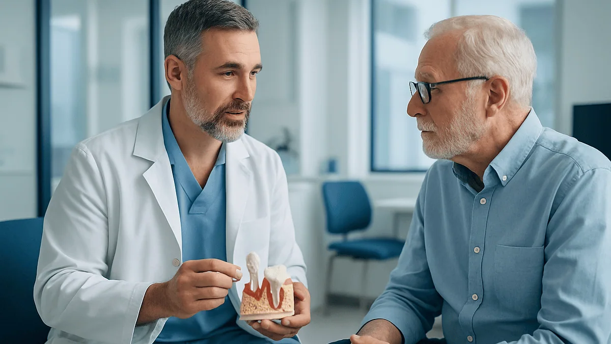
Peri-implantitis is an inflammatory disease of the gum and bone surrounding a dental implant, driven mainly by bacterial plaque that collects at the gum line. It usually starts as peri-implant mucositis, a gum-only inflammation that can be reversed with cleaning, and may progress to loss of the bone that anchors the implant, which does not regrow on its own. A dentist confirms it with probing and X-rays.
The floss came away pink. Not dramatically, not enough to worry about on a Tuesday night, but the same spot around the same implant had bled three days running, and the tooth it replaced had been gone for nine years. The implant itself felt as solid as ever. That, it turns out, is exactly the problem: the part that hurts least is often the part that is quietly changing most.
People who ask what is peri-implantitis are usually somewhere along that arc. A little bleeding, a faint metallic taste, a dental hygienist frowning at a probe reading. The condition rarely announces itself. It creeps, and because a titanium post cannot ache the way a living tooth can, the usual early warning system is missing.
This explainer follows the disease from its first reversible stage to the point where bone is lost, and it is honest about which parts can be undone and which cannot. The decisions about your implant belong to your dental team; the understanding can belong to you.
What is peri-implantitis, in plain language?
Strip away the Latin and peri-implantitis means inflammation around an implant. A dental implant is a screw-shaped post, usually titanium, placed into the jawbone to hold a crown, bridge or denture. Once the bone knits to the surface of that post, a process called osseointegration, the implant can carry chewing forces for decades. Peri-implantitis is the disease that threatens that union.
Two tissues are involved. The first is the soft gum collar, or peri-implant mucosa, that hugs the implant where it emerges into the mouth. The second is the bone beneath it. When only the gum collar is inflamed, dentists use a different term, peri-implant mucositis. When the inflammation has reached the bone and started to dissolve it, the diagnosis becomes peri-implantitis. The distinction matters more than almost anything else in this article, because one stage is reversible and the other leaves permanent marks.
The closest everyday comparison is gum disease around natural teeth. Gingivitis is inflammation of the gum alone; periodontitis is the deeper stage where bone is lost. Peri-implant mucositis and peri-implantitis are the implant versions of that same pair, and the bacteria driving them are broadly similar, according to guidance from the National Institute of Dental and Craniofacial Research on gum disease mechanisms.
Mainstream patient resources such as the Cleveland Clinic list peri-implantitis among the recognized complications of implant treatment, alongside infection and nerve irritation. It is not rare, it is not a sign that something was done wrong, and it is not a reason to avoid implants. It is a reason to understand how it moves.
Peri-implant mucositis vs peri-implantitis: the two stages
Think of the gum around an implant as a turtleneck collar. In a healthy mouth the collar sits snug against the implant, pink, firm and dry of blood when a hygienist runs a probe along it. Peri-implant mucositis is what happens when plaque, the sticky bacterial film that forms on every surface in the mouth, is left undisturbed on that collar for days at a stretch. The body responds the way it responds to any bacterial irritant: small blood vessels widen, immune cells flood in, and the tissue swells and bleeds easily.
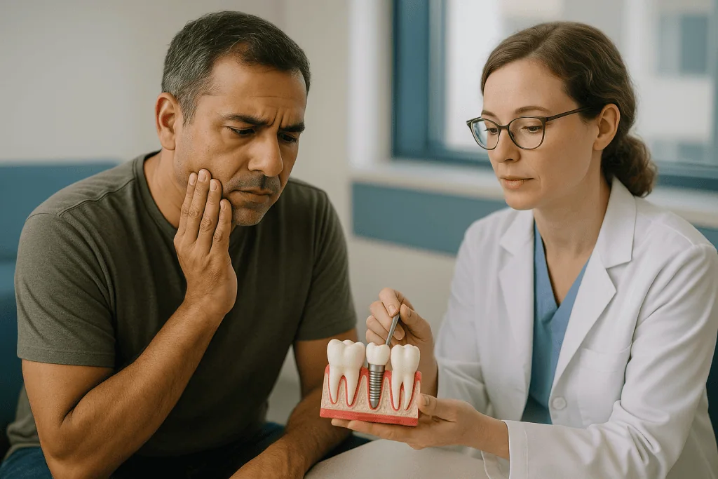
At this stage the bone is untouched. Remove the plaque thoroughly and keep it off, and the collar typically calms down again. That reversibility is the defining feature of mucositis, and it is why the first line of defense against implant loss is a toothbrush, not a surgeon.
Peri-implantitis is the next station on the line. The inflammation deepens, the collar loses its grip on the implant surface, and the gap between them, the peri-implant pocket, becomes a sheltered space where bacteria thrive out of reach of a brush. Inflammatory chemicals released to fight those bacteria also switch on bone-dissolving cells. Bone retreats from the implant surface in a shallow saucer shape that shows up on X-rays as a dark dip around the threads.
The two conditions sit on one continuum, but they are not inevitable steps. Many people with mucositis never progress. What determines who does is the subject of the next two sections: how the tissue around an implant is built, and what keeps feeding the inflammation.
How does gum inflammation around an implant turn into bone loss?
A natural tooth and a dental implant look similar above the gum and behave very differently below it. A tooth is held in its socket by a periodontal ligament, a thin cushion of fibers with its own blood supply, nerve endings and repair cells. The gum attaches to the tooth root through fibers that run perpendicular into the root surface, forming a tight seal.
An implant has none of that. Bone fuses directly to titanium with no ligament between. The gum collar attaches through fibers that run parallel to the implant surface rather than into it, which makes the seal weaker and easier for bacteria to breach. Blood supply to that collar is also poorer, because it comes only from the surrounding bone and gum rather than from a ligament as well. The result is a tissue that defends itself less vigorously and repairs itself more slowly than the gum around a tooth.
Here is the sequence, step by step:
- Plaque matures on the implant collar and the immune system responds with inflammation.
- Swelling deepens the space between gum and implant, sheltering more bacteria.
- Immune signaling molecules, produced to fight the bacteria, also activate osteoclasts, the cells that break down bone.
- Bone recedes from the implant, exposing rough threaded surfaces that plaque clings to more stubbornly than to a smooth tooth root.
- Each loop of the cycle is harder to clean than the last.
Two features make this progression faster than around teeth. There is no ligament to act as a buffer, so inflammation reaches bone sooner. And because implants transmit no fine sensation, the usual signal that would send someone to the dentist, a dull ache, is largely absent. Progression is often silent until a hygienist or an X-ray catches it.
What is the main cause of peri-implantitis?
The short answer is bacteria. The longer answer is bacteria plus everything that helps them stay put or that weakens the body’s response to them. Plaque is the driver in essentially every case; the other factors decide how fast the car travels.

The NHS describes plaque build-up as the cause of gum disease around natural teeth, and the same biofilm is responsible around implants. When it is not removed daily, plaque hardens into calculus, a mineral crust that a brush cannot shift, and calculus gives fresh plaque a rough foothold.
Several contributors sit on top of that foundation:
- A history of periodontitis. People who lost teeth to gum disease carry the same bacterial community and the same immune tendencies into implant treatment.
- Smoking. The Mayo Clinic notes that smoking slows healing after implant surgery and raises complication risk; it also narrows gum blood vessels, muting the inflammation that would otherwise alert you to trouble.
- Poorly controlled diabetes. High blood sugar impairs immune cell function and tissue repair, as the NIDCR notes for gum disease generally.
- Restorations that trap plaque. A crown with an overhanging edge, an awkward emergence angle or residual cement left under the gum at fitting creates a permanent bacterial shelter.
- Too little keratinized gum. Some sites have only thin, loose lining tissue around the implant rather than the firmer band that resists brushing trauma and bacterial entry.
Occlusal overload, meaning excessive biting force on the implant, is often mentioned. The evidence that force alone causes peri-implantitis is weak; force plus plaque appears to be the combination that matters. When you ask your dentist about cause, expect a conversation about biofilm first and everything else second.
Who is more likely to develop peri-implantitis, and who is asked to wait for an implant?
Nobody is immune, but risk is not evenly distributed. The people most likely to develop peri-implantitis are, in practice, the people whose mouths were already struggling before the implant went in. Someone who lost a molar to advanced periodontitis, still smokes, and finds it hard to reach the back of the mouth with a brush starts from a very different place than someone who lost a front tooth to a bicycle accident at nineteen.
That is why the pre-implant assessment matters so much. A thorough team evaluates gum health around the remaining teeth, takes X-rays of bone volume, asks about smoking and medical conditions, and looks at how well plaque is being controlled day to day. The Mayo Clinic describes this planning stage as involving a dental exam, review of medical history and a tailored treatment plan.
Some people are usually asked to wait before implant placement rather than turned away:
- Those with active, untreated periodontitis around remaining teeth. Stabilizing that disease first removes the bacterial reservoir that would otherwise colonize the implant.
- Heavy smokers who are willing to attempt cessation. Healing and long-term tissue health both improve when tobacco exposure drops.
- People whose diabetes is not yet well controlled, until their medical team has adjusted management.
- Anyone with an oral hygiene routine that is not yet reliable, because an implant demands daily maintenance for as long as it is in the mouth.
None of these are moral judgments. They are risk conversations, and they are had precisely because peri-implantitis is easier to prevent than to treat. Some conditions, such as certain bone-affecting medications or radiotherapy history to the jaw, prompt a wider medical discussion before any surgery. The decision to proceed, delay or choose an alternative such as a bridge or partial denture rests with the treating team, informed by that full picture.
Peri-implantitis symptoms: how do I tell if I have it?
The honest answer is that you often cannot, at least not early and not with certainty. Peri-implantitis is largely a clinical diagnosis, meaning it is confirmed by a dental professional using a probe and an X-ray rather than by anything you can see in a bathroom mirror. What you can do is notice the changes that should prompt a visit.
Bleeding is the most common early clue. Gums that bleed when you brush or floss around an implant, or that leave a pink tinge on an interdental brush, are inflamed. Healthy tissue around an implant does not bleed with normal cleaning. Redness and puffiness of the collar, compared with the gum around neighboring teeth, point the same way.
Later signs are more specific. A bad taste or persistent odor localized to one implant suggests a pocket harboring bacteria. Pus, which may appear as a yellowish bead when you press the gum, indicates active infection. Gum that appears to have shrunk back so that metal or the darker threaded part of the implant is visible means bone has already been lost beneath it. Tenderness when chewing on that side is possible but, because implants have no nerve of their own, pain is frequently absent even in advanced disease.
Mobility is a late and serious sign. An implant that feels loose has usually lost a substantial portion of its bone support, though sometimes it is only the crown or a screw that has loosened, which is a different and far simpler problem. Only your dentist can tell the two apart.
These observations are prompts for an appointment, not a way to diagnose yourself. Someone with heavy smoking history may bleed very little despite significant disease, because nicotine constricts gum vessels. The absence of symptoms is not the absence of peri-implantitis, which is the single most important reason regular professional checks around implants exist.
How dentists diagnose peri-implantitis: probing, X-rays and a comparison table
Diagnosis rests on three measurements taken together. The first is bleeding or suppuration on gentle probing, where a fine blunt instrument is slid into the gum collar around the implant. The second is probing depth, how far that instrument travels before meeting resistance, and above all whether that depth has increased since earlier visits. The third is radiographic bone level, read from an X-ray and ideally compared with a baseline image taken when the crown was first fitted.
That word, baseline, is doing a lot of work. Bone around an implant remodels a little in the first year after the crown is placed, so a single X-ray taken years later cannot tell whether a given bone level is stable or slipping. The international World Workshop consensus classification of peri-implant diseases, which most periodontal societies follow, explicitly asks clinicians to compare current findings against earlier records where they exist. If you have had an implant for years and no one has probed around it, asking for that to start is reasonable.
| Finding | Healthy peri-implant tissue | Peri-implant mucositis | Peri-implantitis |
|---|---|---|---|
| Bleeding or pus on probing | Absent | Present | Present |
| Probing depth | Stable since baseline | May be slightly increased by swelling | Increased compared with earlier records |
| Bone on X-ray | Stable after initial remodeling | No loss beyond initial remodeling | Progressive loss beyond remodeling |
| Reversible with cleaning | Not applicable | Yes, in most cases | Inflammation yes; lost bone no |
A few practical points follow from the table. Probing around implants is safe when done gently and does not damage the seal, despite an older belief to the contrary. Two-dimensional X-rays show bone on the sides facing the film but not on the cheek or tongue side, so they can underestimate loss. And because implants cannot respond to the cold or tapping tests used on teeth, the probe and the X-ray remain the core tools. The MedlinePlus entry on periodontitis describes the same probe-and-X-ray approach for natural teeth.
Is peri-implantitis reversible, and what does stable actually mean?
This is the question behind most of the others, so it deserves a precise answer in two halves.
The inflammation is reversible. Bleeding, swelling, pus and pocket tenderness can all resolve when the bacterial load on the implant surface is brought down and kept down. A pocket that stops bleeding on probing and stops deepening is, in clinical language, a stable site, and stable sites can stay that way for years with maintenance.
The bone loss is not reversible in the way most people hope. Bone that has receded from an implant does not grow back on its own once inflammation settles, and the gum usually follows the bone downward, which is why treated sites often look longer than they did. Regenerative surgical procedures, which place bone graft material and sometimes a membrane into the defect after the surface has been cleaned, can refill part of some defects. Whether they do depends heavily on the shape of the defect; contained, bowl-shaped defects with bony walls around them respond better than wide, shallow saucers or exposed surfaces facing the cheek. The evidence for how much bone such procedures regain is mixed and varies between studies, and any surgeon who is honest with you will say so rather than quote a figure.
So the realistic goal of treatment is arrest, not restoration. Stop the disease, keep the implant in function, and accept that the tissue architecture may be permanently changed. Compare it with a knee that has lost cartilage: the pain and swelling can be controlled and the joint can keep working, but the cartilage does not come back.
That framing changes how you think about timing. The difference between reversing mucositis and managing peri-implantitis is the difference between a hygiene visit and a surgical procedure. Catching the process while the bone is still intact is not an optimization; it is the whole game.
Peri-implantitis treatment: how do dentists treat it?
Treatment follows a ladder, and every rung shares one aim: remove bacterial deposits from the implant surface and create conditions where they cannot easily return. Which rung you start on depends on how deep the pockets are and how much bone has gone.
Non-surgical debridement comes first. The hygienist or dentist cleans the implant surface and the pocket using instruments designed not to scratch titanium, such as plastic or titanium-tipped scalers, ultrasonic devices with protective tips, or air-powder polishing that sprays fine particles under the gum. Oral hygiene instruction tailored to the specific implant, including which interdental brush size fits the gaps, is part of the same appointment. The prosthesis may be removed to allow proper access and to check for trapped cement or a poorly fitting margin.
Adjunctive agents are sometimes added. Antiseptic rinses or gels containing chlorhexidine, and locally placed or systemic antibiotics, appear in many protocols. The evidence that they add meaningful benefit beyond mechanical cleaning is inconsistent, and antibiotic use carries resistance concerns, so this is a judgment call for the clinician rather than a standard step.
Surgical treatment is considered when pockets remain deep and bleeding after non-surgical care, or when bone loss is already substantial. The gum is lifted to expose the implant surface for direct cleaning. From there the surgeon may reshape the bone and reposition the gum lower to eliminate the pocket, sometimes smoothing exposed implant threads, an approach called resective surgery. Or, where the defect shape allows, graft material may be placed to encourage bone fill, the regenerative approach described earlier. Some cases combine both.
Implant removal is the last rung, reserved for implants that are mobile or whose bone loss is too advanced for any reconstruction to be worthwhile. It is a treatment, not a failure of one. The site can heal and options are reassessed.
Which rung, and when, sits with your treating team, who can see the defect you cannot.
What do the weeks after peri-implantitis treatment usually look like?
Recovery differs sharply between the non-surgical and surgical rungs, so it helps to picture both.
After non-surgical cleaning, most people notice mild tenderness around the implant for a day or two and sometimes a little sensitivity of neighboring teeth if root surfaces were exposed by the cleaning. Bleeding on brushing typically eases over the following days as the swelling subsides. The gum may look slightly recessed once it tightens, revealing more of the crown or a sliver of implant collar. Your team usually asks you to keep cleaning the area normally from the first day, because leaving it alone lets plaque re-establish. A reassessment appointment some weeks later checks whether pockets have stopped bleeding and whether depths have shallowed; that visit decides whether maintenance is enough or surgery is needed.
After surgical treatment, expect swelling and some bruising for several days, stitches that dissolve or are removed at a follow-up, and instructions to avoid brushing the surgical site directly while the gum heals, using an antiseptic rinse instead if one is prescribed. Chewing is often shifted to the other side for a while. Where graft material has been placed, healing takes longer and X-rays to judge bone fill are typically deferred for months rather than weeks, since new bone mineralizes slowly. The Mayo Clinic describes several months of bone healing after initial implant placement, and regenerative surgery follows a similarly unhurried biological clock.
Across both pathways, the shape of the maintenance schedule changes. Someone who once saw a hygienist twice a year may be moved to more frequent visits for the first year after treatment, with intervals set by how the tissue behaves. The measurements taken at each visit become the new baseline against which future stability is judged.
Pain that worsens rather than eases, swelling that spreads, or fever at any point in this period is not part of normal healing and belongs in the red-flag list later in this article.
Can peri-implantitis be prevented? What daily care and check-ups involve
Prevention is unglamorous and it works better than any treatment, because it acts at the mucositis stage or before it. The NHS advice for gum health is a useful frame: brush twice a day for about two minutes, clean between teeth daily, and attend dental check-ups at the interval your dentist recommends. Around an implant, each of those needs a small adjustment.
Brushing an implant crown is the same as brushing a tooth, but the collar where the crown meets the gum deserves deliberate attention, with the bristles angled toward that junction. Electric and manual brushes both work when used properly; the technique matters more than the technology.
Cleaning between an implant and its neighbors is where most plaque hides and where floss alone often fails, because the gap beneath an implant crown is frequently wider than around a natural tooth. Small interdental brushes sized to the space, or a water flosser aimed at the collar, reach places floss skates over. Your hygienist can show you which size fits which gap; the answer is often different on either side of the same implant.
Implant-supported bridges and full-arch prostheses need their own tools: threader floss or specialized brushes that pass beneath the bridge. If you cannot get an instrument under a prosthesis, the design may need adjusting, and that is a fair thing to raise.
Professional maintenance completes the picture. A hygienist can probe the collar, spot early bleeding you would never notice, remove calculus that has formed below the gum line, and compare readings with the last visit. The MedlinePlus periodontitis page emphasizes regular professional cleanings for anyone with a history of gum disease, and that population overlaps heavily with implant patients.
Smoking cessation is the single lifestyle change with the clearest link to peri-implant tissue health. It is difficult, it is worth asking for help with, and your dental and medical teams can point you to support.
What people often get wrong about what peri-implantitis is and isn't
Myth: Implants cannot get gum disease because they are metal. Titanium does not decay, which is where this idea comes from. But the gum and bone around it are entirely alive, and they respond to bacteria exactly as tissue around teeth does. If anything, as the anatomy section explained, they respond with less resilience.
Myth: If it does not hurt, it is fine. An implant has no pulp and no nerve, so the ache that would warn of trouble around a tooth simply never arrives. Advanced peri-implantitis is frequently painless. Bleeding, not pain, is the early language of this disease.
Myth: Probing around an implant damages it. This was once taught and is still repeated. Gentle probing with an appropriate instrument is the standard way to monitor peri-implant health and does not disrupt a healthy seal.
Myth: Once bone is lost, the implant is finished. Many implants with moderate bone loss are stabilized and remain in function for years after treatment. Removal is one option among several, chosen when bone loss is extensive or the implant is mobile.
Myth: A mouth rinse or an antibiotic will clear it up. Chemical agents cannot remove a mature biofilm from a rough titanium surface. Mechanical cleaning is the core of every protocol; rinses and antibiotics are, at most, adjuncts whose added value remains debated.
Myth: Peri-implantitis means the surgery was botched. Placement errors and cement left under the gum can contribute, but the dominant driver is bacterial plaque interacting with individual risk factors. Well-placed implants in well-maintained mouths develop it too, just less often.
Myth: Regenerative surgery will put the bone back the way it was. Partial fill of favorable defects is possible; full restoration of the original bone contour is not what the evidence describes. Arrest is the realistic goal.
Questions to ask your care team
The appointment where peri-implantitis is first raised is often short and slightly alarming. A written list keeps it useful. These are the questions that tend to matter most, roughly in the order a clinician would find them easiest to answer.
- Is this mucositis or peri-implantitis? In other words, has bone been lost, or only gum inflamed?
- Do you have a baseline X-ray and probing measurements from when the crown was fitted, and how do today’s compare?
- How much bone has been lost around this implant, and on which sides?
- What do you think is driving it in my case: plaque control, a restoration that traps deposits, smoking, a medical condition, or a combination?
- Can the crown or bridge be removed to allow cleaning and to check for residual cement or a poor margin?
- What is the first treatment step you recommend, and how will we judge whether it has worked?
- If non-surgical cleaning is not enough, what surgical options would you consider for this defect shape, and what are their limits?
- Is implant removal on the table now, or only if other steps fail, and what would replacement options look like?
- Which interdental brush sizes or other tools fit the gaps around this specific implant?
- How often should I be seen for maintenance over the next year?
- Should my other implants or teeth be checked more closely given this finding?
- Are there medical issues, such as blood sugar control or medications affecting bone, that you would like me to discuss with my physician?
Bring a phone photo of any bleeding or swelling you noticed at home, since it may not be visible on the day. Ask for a copy of your probing chart and X-rays so that if you ever change providers, your baseline travels with you. And if the answers you receive do not make sense, say so; a good team would rather explain twice than have you leave uncertain about what happens next.
When to call your doctor
Most of peri-implantitis unfolds slowly, and a routine appointment within a few weeks is the right response to bleeding gums or a suspicious taste around an implant. Some signs, though, should move you to contact your dentist the same day, or to seek urgent medical care if a dentist is unavailable.
- Rapidly increasing swelling of the gum, cheek or floor of the mouth around the implant, particularly if it is spreading or making the skin feel tight.
- Fever, chills or feeling generally unwell alongside a swollen or painful implant site.
- Difficulty swallowing, difficulty breathing, or swelling that pushes the tongue upward. These are emergency signs of infection spreading into deeper spaces of the neck and require immediate emergency care.
- Pus draining steadily from around the implant, or a visible abscess.
- An implant, or the crown on it, that has suddenly become loose or moves when touched.
- Severe pain that is not eased by measures your dentist has already suggested, or that wakes you at night.
- After surgical treatment: bleeding that does not stop with gentle pressure, stitches that have come apart with the wound gaping, or swelling and pain that worsen after the third or fourth day rather than easing.
- Numbness or tingling of the lip, chin or tongue that is new or worsening.
Between appointments, a change in the pattern is also worth a call even without drama: bleeding that was occasional becoming daily, a bad taste that has settled in, or gum visibly pulling away from the crown. The Cleveland Clinic lists infection and persistent pain among the complications that warrant contacting your provider after implant treatment, and the same logic applies years later.
Every decision about how to investigate these signs, and what to do about them, sits with your treating team. Your job is to notice and to tell them; theirs is to look, measure and decide.
Frequently asked questions
What is the main cause of peri-implantitis?
Bacterial plaque left on the implant collar is the main cause. The immune response to that biofilm inflames the gum and, if it persists, activates bone-dissolving cells. Factors such as previous gum disease, smoking, poorly controlled diabetes and crowns that trap deposits do not cause the disease alone but determine how fast it progresses once plaque is present.
How do I tell if I have peri-implantitis?
You usually cannot confirm it yourself, because the diagnosis relies on probing and X-rays. Bleeding when you brush or clean around an implant, redness or puffiness of the gum collar, a persistent bad taste, pus, or gum shrinking to expose metal are reasons to book a dental visit. Pain is often absent, so lack of discomfort does not rule the condition out.
Is peri-implantitis reversible?
The inflammation is reversible; the bone loss generally is not. Cleaning can stop bleeding and halt pocket deepening, leaving a stable site that may stay healthy for years. Bone that has receded does not regrow spontaneously, although regenerative surgery can partly refill some favorably shaped defects. The realistic aim of treatment is to arrest the disease, not to restore the original anatomy.
How do dentists treat peri-implantitis?
Treatment begins with non-surgical cleaning of the implant surface using instruments that do not scratch titanium, together with tailored home-care instruction. If pockets keep bleeding or bone loss is substantial, surgery lifts the gum to clean directly and either reshapes the tissue or places graft material. Antiseptics and antibiotics are sometimes added, with debated benefit. Removal is reserved for mobile or severely affected implants.
What is the difference between peri-implant mucositis and peri-implantitis?
Mucositis is inflammation confined to the soft gum collar around an implant, with bleeding on probing but no bone loss beyond normal early remodeling. Peri-implantitis is the deeper stage in which inflammation has reached the bone and X-rays show progressive loss. Mucositis typically resolves with plaque removal; peri-implantitis needs more intensive treatment and leaves permanent changes in the bone.
What are the early peri-implantitis symptoms I should watch for?
Bleeding on brushing or interdental cleaning around one implant is the earliest and most common sign, followed by redness and swelling of the gum collar. A localized bad taste or odor, pus, and gum recession exposing the implant appear later. Because implants have no nerve, aching is unreliable as a warning; smokers may also bleed less despite active disease.
Can peri-implantitis be treated without surgery?
Sometimes, particularly when caught early with shallow pockets and limited bone loss. Non-surgical debridement combined with improved home care can stop bleeding and stabilize the site. Deeper pockets and larger bone defects respond less reliably to cleaning alone, because instruments cannot reach the full implant surface through the gum. Your dentist decides after reassessing the site some weeks after initial treatment.
Does peri-implantitis mean my implant will fail?
Not necessarily. Many implants with moderate bone loss are stabilized by treatment and remain in function for years afterward. Failure, meaning removal or spontaneous loss, becomes likely only when bone loss is extensive or the implant is mobile. The earlier the disease is detected and the better plaque control is afterward, the more favorable the outlook, though no clinician can promise a specific result.
Why do implants get gum disease if they are made of titanium?
The titanium does not become diseased; the living gum and bone around it do. Those tissues respond to bacterial plaque with inflammation just as they do around teeth, and they are structurally more vulnerable because an implant lacks the periodontal ligament and the perpendicular gum fibers that seal a natural tooth. Metal resists decay, but it offers no protection against inflammation.
How can I prevent peri-implantitis from developing?
Remove plaque from the implant collar every day with a brush angled toward the gum line and with interdental brushes or a water flosser sized to the gaps, attend professional maintenance at the interval your dentist sets so early bleeding is caught, and address smoking and blood sugar control with your medical team. Prevention acts at the reversible mucositis stage, which is why it outperforms any treatment.
References
- Cleveland Clinic: Dental Implants
- NHS: Gum disease
- MedlinePlus: Periodontitis
- NIH National Institute of Dental and Craniofacial Research: Periodontal (Gum) Disease
This article is for general information only and is not a substitute for professional medical advice. Please consult a qualified doctor about your individual situation.

More from the Blog
Are Zirconium Teeth Right for You? Bite Force, Smile Goals and When Another Ceramic Fits Better
A good zirconium teeth candidate usually needs a strong restoration for a back tooth, an implant crown, or a bridge, especially with a heavy…

All-on-4 vs All-on-6: When Two More Implants Make Sense
All-on-4 and All-on-6 both anchor a full arch of fixed replacement teeth; the difference is how many implants carry the load. Four implants suit…
What to Ask Before a Dental Crown: Material, Margin Placement and What Happens to the Nerve
Before agreeing to a dental crown, ask what the crown is for, which material is proposed and why, where the edge of the crown…
Dental Implants After Gum Disease: When They Are Possible, and What Comes First
Yes, people with periodontal disease can often get dental implants, but not while the disease is active. Infection and inflammation must be treated first,…

How Long Do All-on-4 Implants Last? Longevity, Care and What Can Go Wrong
With diligent daily cleaning and regular professional maintenance, the four titanium implants supporting an All-on-4 restoration commonly last 20 years or longer, and long-term…
When Removable Dentures Stop Fitting: Relining, Rebasing and When a Remake Is Needed
Removable dentures usually stop fitting because the jawbone and gum underneath slowly shrink, not because the denture itself changes much. Dentists first assess the…