

How Radiotherapy Works and How Long Sessions Take
Key Takeaways
- An external beam radiotherapy session typically lasts about 10 to 30 minutes, but the radiation beam is on for only a few minutes of that time.
- Standard courses run once a day, Monday to Friday, over roughly one to seven weeks, while stereotactic treatments may need only one to five visits.
- Radiation works by breaking DNA; cancer cells repair the damage less effectively than healthy cells, which is why treatment is split into daily fractions with rest at weekends.
- External beam radiotherapy does not make you radioactive, and you can safely hold a baby or share a bed the same day; only certain internal treatments require temporary precautions.
- Side effects usually build gradually and often peak near the end of a course or shortly after, so feeling normal after five sessions is common and not a sign the treatment is failing.
- Roughly half of all people diagnosed with cancer receive radiotherapy at some stage, from early disease treated with curative intent to advanced disease treated for symptom relief.
A single external radiotherapy session usually takes about 10 to 30 minutes, and the beam itself is switched on for only a few minutes of that. A full course is typically given once a day, Monday to Friday, over roughly one to seven weeks, although some modern schedules need only one to five visits. The exact plan depends on the cancer type, its location, and the goal of treatment.
The woman in the corner of the waiting room has a paper cup of coffee, a library book, and a parking ticket she has not yet paid. She is not worried about the meter. By the time her name is called, walked down the corridor, and called again on the way back, she will have been in the treatment room for less time than it takes to brew a pot at home.
That is the part almost nobody expects. Radiotherapy has a reputation built on old films and older fears: long, punishing, mysterious. The reality is closer to a dental hygiene appointment repeated on a schedule, with a machine that hums, rotates, and never touches you.
What follows is the honest timeline, from the first planning scan to the weeks after the last session, and the science that explains why the clock is set the way it is.
How long does radiotherapy take? The two clocks you need to know
Two very different clocks run during radiotherapy, and confusing them is the source of most anxiety about time.
The first clock is the appointment. According to the National Cancer Institute and Mayo Clinic, a standard external beam session usually lasts about 10 to 30 minutes from the moment you lie down to the moment you sit up. Most of that is positioning and imaging. The beam itself is typically on for only a few minutes, delivered in short bursts from several angles.
The second clock is the course. The NHS describes external radiotherapy as a series of daily sessions, usually Monday to Friday with a rest at the weekend, over a course that typically lasts between one and seven weeks. A person having a longer curative course might therefore attend 30 or more appointments; someone having a short palliative course might attend once.
Where you land on that range depends on four things your oncology team weighs together: what kind of cancer it is, where it sits in the body, whether the aim is to eradicate disease or ease symptoms, and how sensitive the surrounding healthy tissue is. Two people with the same diagnosis can have different schedules for perfectly sound reasons.
One practical consequence follows from all this. The disruption of radiotherapy is rarely about the length of any single visit. It is about the rhythm, the daily commute and the way that rhythm nudges everything else in life for a few weeks.
How does radiotherapy actually work on cancer cells?
Radiotherapy is, at heart, a very precise way of breaking DNA.
The machines used today produce high-energy X-rays, or in some centers beams of protons or electrons. When that radiation passes through tissue it knocks electrons loose from atoms, a process called ionization. Some of that energy lands directly on the DNA inside cells, snapping one or both strands of the double helix. More often it strikes water molecules first, creating unstable fragments called free radicals that then attack DNA from within the cell. Either way, the result is damage that a cell must repair before it can divide again.
The National Cancer Institute explains the crucial asymmetry that makes treatment possible. Healthy cells generally have intact repair machinery and can mend much of the damage between sessions. Cancer cells, by contrast, often divide rapidly and carry faulty repair genes, so they accumulate breaks they cannot fix. When a damaged cell attempts to divide, it fails and dies, or it triggers its own self-destruct program.
This is why radiotherapy is not instant. Cells die over days and weeks as they try, and fail, to reproduce. A tumor treated in March may continue to shrink through May. It also explains why the beam is shaped so carefully: the goal is to bathe the tumor in a high dose while the tissue a few millimeters away receives far less.
Nothing about this involves heat, cutting, or a chemical you can feel. The room stays cool and quiet. The work happens at the scale of molecules.
Why is radiotherapy split into daily sessions instead of one big treatment?
If radiation kills cancer cells, why not deliver everything in a single afternoon?
The answer is the repair gap. Dividing the total treatment into smaller portions, known as fractions, gives normal cells time to fix DNA damage between sessions while cancer cells, with their weaker repair, fall progressively further behind. Radiation oncologists sometimes summarize this as giving healthy tissue a nightly head start. The weekend break built into most schedules, described by the NHS, serves the same purpose.
Fractionation also catches cancer cells in different phases of their life cycle. Cells are most vulnerable when they are actively dividing, and a tumor is never fully synchronized. Spreading treatment over days increases the odds that each cell is hit at a sensitive moment. Meanwhile, as the tumor shrinks, oxygen reaches previously starved regions, and oxygen-rich cells are more easily damaged by radiation.
The pendulum has been swinging toward fewer, larger fractions for many cancers, an approach the National Cancer Institute calls hypofractionation. Better imaging and beam shaping mean the surrounding tissue can be protected well enough to tolerate bigger daily portions. For some breast and prostate treatments, courses that once ran six or seven weeks are now often completed in far fewer visits, according to NCI and NHS descriptions of current practice.
Stereotactic techniques take this furthest, delivering a very focused treatment in a single session or a handful of visits. The trade-off is intense precision: those schedules are reserved for tumors that are small, well defined, and not wrapped around a structure that cannot spare any margin.
At what stage of cancer is radiotherapy used?
People often assume radiotherapy belongs to one particular chapter of a cancer story, usually a late one. The evidence says otherwise. The NHS and the National Cancer Institute both note that roughly half of all people with cancer receive radiotherapy at some point, and that it is used from the earliest stage to the most advanced, with different aims at each point.
In early-stage disease, radiotherapy may be the sole treatment, chosen because it can target a tumor without surgery, or because an operation would carry unacceptable risks for that individual. Some early cancers of the prostate, larynx, and lung are approached this way.
After surgery, it is frequently given to the area where a tumor was removed. The intent is to address any microscopic cells that a scalpel cannot see and thereby reduce the chance of the cancer returning in that spot. Breast cancer treated with lumpectomy is the classic example.
Before surgery, a course may be used to shrink a tumor so that an operation becomes smaller or safer, an approach common in rectal cancer. It is also combined with chemotherapy for cancers such as those of the head and neck or cervix, where the two treatments make each other more effective.
In advanced or metastatic disease, radiotherapy shifts roles. Short courses aim to relieve pain from bone metastases, stop bleeding, shrink a tumor pressing on the spinal cord, or ease breathlessness. Here the measure of success is comfort and function rather than cure, and the schedule is typically brief.
None of these uses is a signal about prognosis on its own. The stage determines the goal; the goal determines the schedule.
What are the main types of radiotherapy, and how do their timelines differ?
Radiotherapy is a family of techniques rather than a single procedure, and the type explains much of the variation in how long treatment takes. The National Cancer Institute groups them into radiation delivered from outside the body, radiation placed inside it, and radioactive substances that travel through the bloodstream.
| Approach | How it is delivered | Typical visits | Typical time per visit |
|---|---|---|---|
| Conventional external beam (including intensity-modulated and image-guided techniques) | Machine outside the body shapes beams to the tumor | Daily, Monday to Friday, over about 1 to 7 weeks (NHS) | About 10 to 30 minutes (NCI, Mayo Clinic) |
| Stereotactic radiosurgery or stereotactic body radiotherapy | Very focused, high-precision external beams | A single session or a few visits (NCI) | Often longer per visit because of extra imaging and positioning |
| Palliative external beam | Same machines, simpler planning | Single visit to a short course (NHS) | About 10 to 30 minutes |
| Brachytherapy (internal) | Sealed radioactive source placed in or next to the tumor | One or several procedures; sometimes a short hospital stay (NCI) | Varies; may involve anesthesia |
| Radioactive liquids or capsules (systemic) | Swallowed or injected; travels to target tissue | Usually a single administration (NCI) | Short, but temporary safety precautions afterward |
External beam treatment is by far the most common and is the focus of most of this article. Proton therapy is a form of external beam that uses charged particles rather than X-rays; its schedule resembles conventional treatment, though it is available in fewer centers.
Brachytherapy inverts the usual arrangement. Instead of a beam traveling inward, a small sealed source sits inside the body for minutes, hours, or, with permanent seeds, indefinitely as it fades. That makes the visit itself longer and more procedure-like, but the number of visits smaller.
What to expect at your first radiotherapy appointment
The first appointment is not treatment. It is measurement, and it deserves its own mental category.
This planning visit, sometimes called simulation, is where the team builds the map that every later session will follow. According to the NHS and Cleveland Clinic, it typically involves a CT scan in the exact position you will be treated in, and it usually takes longer than a treatment session. Radiographers may fit you with supports: a molded cushion for a leg, a wing board for arms raised above the head, or, for head and neck treatment, a mesh mask warmed and shaped to your face that hardens within minutes. The mask can feel strange the first time. It is not tight enough to hurt, and you can breathe and swallow normally, but telling staff if you are claustrophobic lets them adapt the process.
Small marks come next. Many departments place two or three permanent ink dots, each roughly the size of a freckle, on the skin so you can be aligned to within millimeters every day. Some use skin pens and clear stickers instead.
You may also receive preparation instructions that matter more than they sound. Pelvic treatments often require a comfortably full bladder or an empty bowel so that organs sit in the same place each day. Breast and chest treatments may involve practicing a held breath that moves the heart away from the beam.
Then comes a pause. Physicists and dosimetrists use the scan to calculate beam angles and shapes, a process that can take days to a couple of weeks before your first real session, as the NHS describes. That gap is not a delay in your care. It is the care.
What happens during a radiotherapy session, minute by minute
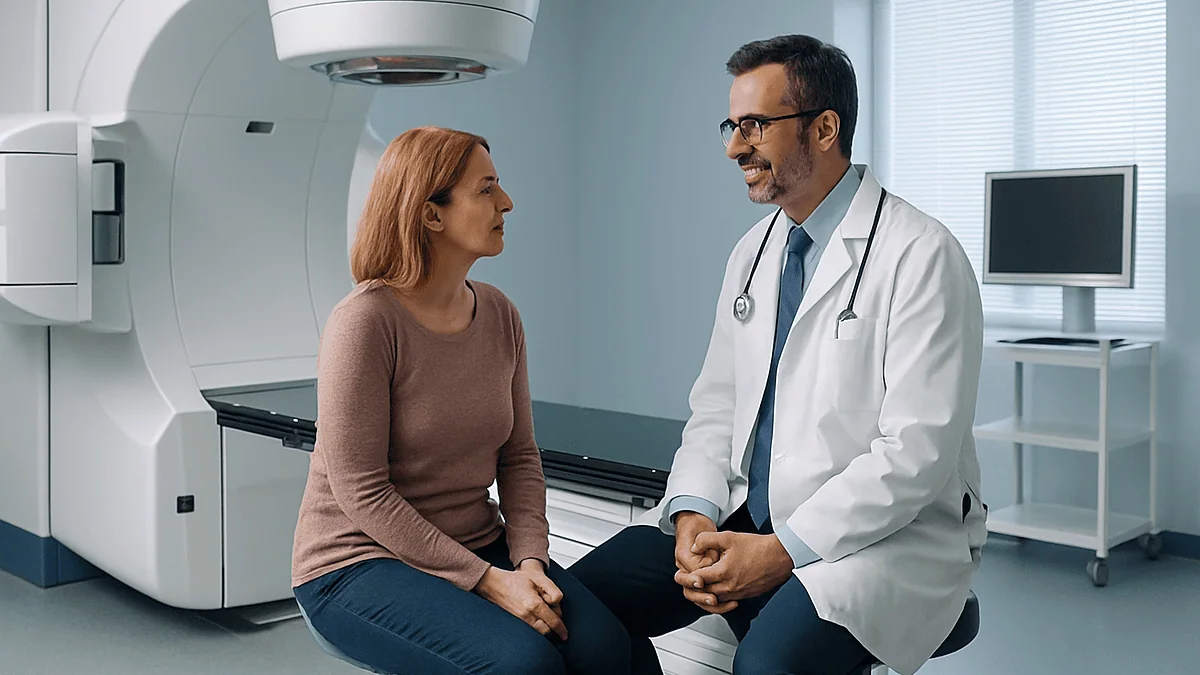
Picture a room the size of a generous living room, dominated by a machine shaped like a giant letter C on its side. That is a linear accelerator, and it will become oddly familiar.
You change into a gown if the treatment area requires it, then lie on a narrow couch in the same position as your planning scan, with the same supports. Two or three radiographers align the marks on your skin with thin laser lines projected from the walls and ceiling. The couch moves a fraction of an inch. It moves again. Precision here is measured in millimeters, so this phase takes most of the appointment.
The team then steps out. This is the moment many people find most exposed, and it helps to know why it happens: staff deliver hundreds of treatments a year and cannot receive scattered radiation from every one. They watch you on cameras and can hear you over an intercom. If you need to stop, you say so, and the beam is off within seconds.
Most modern machines take a quick image before treating, checking your position against the plan. Then the gantry rotates around you, pausing at planned angles or sweeping in a continuous arc. You will hear a buzz or hum while the beam is on. You will feel nothing, as Mayo Clinic and the NCI both emphasize. The whole beam-on period is usually a few minutes.
The couch lowers, someone helps you up, and you leave. Many people walk straight from the treatment room to the parking lot and on to work, school, or lunch.
Why does a few minutes of radiation take a 30-minute appointment?
The mismatch between beam time and appointment time bothers people, and the explanation reveals what actually makes modern radiotherapy safe.
Positioning is the largest slice. A tumor that sits next to the spinal cord, the heart, or the optic nerve leaves little room for error, and the difference between a well-tolerated course and a damaging one can be a few millimeters. Radiographers therefore align, check, adjust, and check again before anything switches on.
Image guidance adds more time. Many departments take a low-dose X-ray or CT image at the start of each session, sometimes daily, sometimes weekly, and compare it with the planning scan. If your bladder is fuller than last week or you have lost weight since planning, the couch shifts to compensate. Internal organs move; the plan does not, so the images bridge the gap. The National Cancer Institute describes this image-guided approach as standard in many centers.
Beam shaping is happening too, invisibly. Inside the machine head, dozens of tungsten leaves slide into position to sculpt each beam to the tumor’s outline from that angle. Between angles they rearrange. It is fast, but it is not instant.
Finally, there is the reality of a busy department. Machines are checked every morning, an earlier patient may need extra time, and occasionally a machine needs an engineer. Waiting-room delays are common and rarely mean anything about your case.
The takeaway for planning your day: budget the appointment length plus travel and a margin, not the beam time. Bring the library book.
Does radiotherapy hurt, and will I be radioactive afterward?
Two questions arrive at almost every planning visit, sometimes whispered. Both have reassuring, well-established answers.
External beam treatment does not hurt during delivery. High-energy X-rays pass through tissue without stimulating the nerves that register touch, heat, or pain, which is why Mayo Clinic and the NCI both compare the experience to having a diagnostic X-ray. Discomfort during a session, when it occurs, comes from lying still in one position, from a mask, or from a sore area that already existed. Tell the radiographers; they can often add padding, adjust the timing, or coordinate with your doctor on comfort measures.
The radioactivity question depends entirely on the type of treatment. With external beam, radiation exists only while the machine is on and you are in front of it. The instant it stops, there is none left in your body. You cannot pass anything to a partner, a grandchild, or a pet. The National Cancer Institute states this plainly, and it applies to every form of external beam treatment, including protons.
Internal treatments are different, and your team will explain the specifics. Temporary brachytherapy sources are removed before you go home, so no precautions follow. Permanent seeds, used mainly for prostate cancer, emit a small amount of radiation that fades over weeks to months; guidance may include limiting prolonged close contact with pregnant women and small children for a defined period. Radioactive liquids or capsules can make body fluids temporarily radioactive, so short-term precautions around bathrooms and close contact are typical for a few days. These are managed safety steps, not signs of danger.
How will I feel after 5 days of radiotherapy?
The first week is often a strange anticlimax. You brace for something dramatic and instead feel, mostly, like yourself. That is expected, and understanding why helps you read the weeks ahead.
Radiation’s biological effects unfold as cells attempt to divide, and the healthy tissues that react most, skin and the lining of the mouth, throat, bowel, or bladder, turn over on a cycle of days to weeks. The NHS and Mayo Clinic note that side effects typically build gradually during a course rather than appearing at once. After five sessions, many people notice nothing physical at all. Some feel a little more tired than usual, which is often as much about the daily travel, the disrupted routine, and the emotional weight of starting treatment as about the radiation itself.
By the second or third week, the picture usually changes. Skin in the treated area may begin to look pinker or darker, feel dry, or itch, much like a mild sunburn that arrives without sun. Someone treated in the pelvis may start to notice a more urgent bladder or looser bowels. Someone treated in the head and neck may find their sense of taste shifting or their mouth drier.
Tiredness tends to accumulate rather than spike. The NHS describes fatigue as one of the most common effects, often peaking toward the end of a course and lingering for weeks afterward.
So the honest answer is that day five rarely tells you much. Keep the first week’s energy in perspective, start the skin care your team recommends early, and expect the course to feel different in week three than in week one.
Can you drive home after radiotherapy?
For most people having external beam treatment, yes. The NHS and Mayo Clinic both describe going home immediately after a session and resuming normal activities, and nothing about the treatment itself impairs coordination, alertness, or vision. The waiting room at any radiotherapy department is full of people who drove themselves in and will drive themselves out.
A few situations call for a different answer, and they are worth knowing about before your first day rather than discovering in the parking lot.
- Treatment to the brain can affect driving eligibility for legal and medical reasons, independent of how you feel. Rules vary by jurisdiction, and your oncology team should advise you specifically.
- Procedures involving sedation or anesthesia, which applies to some brachytherapy, require someone else to take you home, as with any sedated procedure.
- Mounting fatigue later in a course can dull reaction times. Feeling fine in week one does not guarantee feeling fine in week five, so reassess honestly as you go.
- Medications given for pain, nausea, or anxiety alongside treatment may carry their own driving cautions; the prescribing clinician is the right person to ask.
Many people arrange a rota of lifts for the later weeks even if they start out driving alone. Others find the drive a useful pocket of ordinary life between the clinic and home. There is no wrong choice here, only the one that fits your energy on the day.
Parking, incidentally, is one of the most cited practical frustrations of a course. Ask reception on day one whether the department offers reduced-rate or reserved spaces for people attending daily.
What side effects build over a course, and how long do they last?
Radiotherapy side effects follow a geography and a timeline. Unlike chemotherapy, which travels through the whole body, radiation acts where the beam goes. Hair falls out only in the treated area. Nausea is a concern mainly when the stomach or brain is in the field. Knowing your treatment site tells you most of what to watch for.
Early, or acute, effects arise during treatment and in the weeks that follow. The NHS lists tiredness, skin soreness, and problems specific to the treated region, such as a sore mouth or difficulty swallowing with head and neck treatment, or bladder and bowel irritation with pelvic treatment. These typically peak around the end of the course or in the week or two afterward, then gradually settle. The NHS notes that most side effects go away within a few weeks or months after treatment finishes.
Fatigue deserves its own line. It is the most consistent complaint across every treatment site, and it often outlasts everything else. Gentle regular activity, protected sleep, and realistic expectations of yourself help more than pushing through or lying down all day; the American Cancer Society and NHS both point to light exercise as one of the better-supported ways to manage it.
Late effects are a separate category. Months or years after treatment, tissue in the field can become less flexible, skin can stay slightly darker, and specific organs can carry lasting changes. Modern planning is designed to minimize these, and your team will have discussed the ones relevant to your case. Lifelong awareness, not lifelong worry, is the sensible stance.
Report every new symptom, even a trivial one. Departments have review clinics built into the schedule for exactly this reason.
When should you contact your care team or seek urgent help?
Most radiotherapy side effects are managed in the weekly review clinic and never become emergencies. A short list of red flags, however, should prompt a same-day call to your treatment team or, outside clinic hours, the emergency number you were given at the start of the course.
Seek help promptly if you develop a fever or shaking chills, particularly if you are also having chemotherapy, because that combination can lower your infection defenses. Do the same if you cannot swallow enough fluid to stay hydrated for more than a day, if you are vomiting repeatedly, or if pain is escalating beyond what your current plan controls.
Skin that blisters, weeps, cracks open, or shows spreading redness with warmth needs review rather than home management. Any bleeding that is heavy or will not stop, black or bloody stools, or blood in urine should be reported the same day.
Certain symptoms should send you to emergency care without waiting: sudden breathlessness or chest pain, a new seizure, sudden weakness or numbness in the limbs, severe headache with confusion or vomiting, or new difficulty controlling the bladder or bowel alongside back pain. These can signal problems that need hours, not days.
The general principle from the NHS and Mayo Clinic applies throughout: you will never be told off for calling. Radiotherapy teams would far rather field ten unnecessary calls than miss one that mattered. Keep the contact card on the refrigerator and the number saved in your phone from day one.
How long until you know whether radiotherapy has worked?
The last session comes with a peculiar mix of relief and uncertainty. The machine falls silent, the daily rhythm ends, and yet nobody hands you a result.
That is because radiotherapy keeps working after it stops. Cells damaged in the final week continue to die over the following weeks, and a tumor often shrinks for a considerable time after the course. The National Cancer Institute and Mayo Clinic both note that side effects may worsen briefly after the last session for the same reason before they begin to improve. Scans taken too early can be misleading, showing inflammation that looks alarming but is simply tissue responding.
Follow-up is therefore scheduled weeks to months after treatment rather than days. What that follow-up involves depends on the cancer and the aim: imaging for some, blood tests for others, a physical examination and conversation for many. Your oncologist will set out the specific plan and the intervals.
During this stretch, the practical work is recovery. Skin heals. Appetite returns. Fatigue lifts, usually gradually and sometimes unevenly, with good days and flat days. The NHS suggests most people find side effects have largely settled within a few weeks to months, though the exact pace varies with the site treated and how many sessions were given.
What matters most, in the view of this magazine, is not the calendar but the conversation. Ask your team what a good response would look like for your situation, when they expect to be able to judge it, and who to call in the meantime. Radiotherapy is one of the most precisely planned treatments in medicine. Its aftermath deserves the same clarity.
Frequently asked questions
How long does each radiotherapy session take?
A typical external beam session takes about 10 to 30 minutes from lying down to getting up, according to the National Cancer Institute and Mayo Clinic. Most of that time is spent aligning your position and taking a quick verification image. The beam itself is usually on for only a few minutes, delivered in short bursts from different angles. Planning appointments and stereotactic treatments generally take longer than routine sessions.
How many weeks does a course of radiotherapy last?
The NHS describes a typical course as daily sessions from Monday to Friday over one to seven weeks, depending on the cancer type, its location, and whether the goal is cure or symptom relief. Shorter schedules that deliver larger portions in fewer visits are increasingly common for some breast and prostate cancers. Highly focused stereotactic treatments can be completed in a single visit or a small handful of sessions.
How will I feel after 5 days of radiotherapy?
Most people feel much as they did before, perhaps a little more tired from the daily travel and the emotional weight of starting treatment. Radiation’s effects on healthy tissue unfold over days to weeks as cells attempt to divide, so skin changes, soreness, or bowel and bladder symptoms usually appear later in the course, often from the second or third week onward, and tend to peak near the end.
At what stage of cancer is radiotherapy used?
Radiotherapy is used at every stage. In early disease it may be the only treatment or may follow surgery to lower the chance of recurrence in that area. Before surgery it can shrink a tumor. It is combined with chemotherapy for some cancers of the head and neck or cervix. In advanced disease, short courses aim to relieve pain, bleeding, or pressure on nerves. The stage shapes the goal, and the goal shapes the schedule.
Can you drive home after radiotherapy?
Usually, yes. External beam treatment does not affect alertness or coordination, and the NHS and Mayo Clinic describe going home and continuing normal activities straight afterward. Exceptions include treatment to the brain, which may carry legal driving restrictions, procedures done under sedation, and mounting fatigue later in a course. Any medicines prescribed alongside treatment may have their own driving cautions, so ask the clinician who prescribed them.
What happens at the first radiotherapy appointment?
The first visit is a planning session rather than treatment. You have a CT scan in the exact position you will be treated in, often with supports or a molded mask made to fit you. Small permanent ink marks or skin pen marks help staff align you each day. You may receive instructions about bladder or bowel preparation. Treatment starts days to a couple of weeks later, once the team has calculated your individual plan.
Does radiotherapy hurt?
The treatment itself is painless. High-energy X-rays pass through the body without stimulating pain or heat receptors, so the experience resembles a diagnostic X-ray, as Mayo Clinic and the National Cancer Institute describe. Any discomfort during a session usually comes from lying still, wearing a mask, or an area that was already sore. Skin soreness and other side effects can develop over the course and are managed by your care team.
Will I be radioactive after radiotherapy?
Not after external beam treatment. Radiation exists only while the machine is on; the moment it stops, none remains in your body, and you cannot expose family, children, or pets. Internal treatments differ: temporary brachytherapy sources are removed before you leave, permanent seeds emit low levels that fade over months and may warrant brief precautions, and radioactive liquids or capsules can require a few days of specific safety steps.
How long do radiotherapy side effects last?
Early side effects such as tiredness and skin soreness typically peak around the end of a course or in the week or two after, then improve. The NHS notes that most settle within a few weeks or months of finishing treatment. Fatigue often lingers longest. Late effects, which can appear months or years later, are less common with modern planning and are discussed with you before treatment begins.
Why do I have to wait between the planning scan and starting treatment?
The gap exists because physicists and dosimetrists use your planning scan to calculate beam angles, shapes, and intensities that concentrate radiation on the tumor while sparing surrounding tissue. This computational and quality-checking work can take days to a couple of weeks, according to the NHS. It is a deliberate safety step rather than a delay, and the timing is factored into your overall treatment plan from the outset.
References
- NHS: Radiotherapy: What happens
- NHS: Radiotherapy: Side effects
- Cleveland Clinic: Radiation Therapy
- MedlinePlus: Radiation Therapy
This article is for general information only and is not a substitute for professional medical advice. Please consult a qualified doctor about your individual situation.

More from the Blog

Smart Rings Compared: Galaxy Ring, Ultrahuman and Oura: What They Measure Well
The Samsung Galaxy Ring, Oura Ring and Ultrahuman Ring Air all use an optical sensor on the finger to track resting heart rate, nighttime…

Palliative Chemotherapy: What the Term Means
Palliative chemotherapy is chemotherapy given with the aim of controlling a cancer rather than curing it. Its goals are to shrink or slow tumors,…
Whoop vs Oura: What Each Measures Well, Where They Disagree and How Accurate the Sleep Data Are
Whoop and Oura both estimate sleep and recovery from an optical pulse sensor, and both detect whether you are asleep or awake with roughly…

AI Symptom Checkers and ChatGPT Medical Advice: How Accurate Are They, Really?
AI symptom checkers and chatbots such as ChatGPT are reasonably good at flagging emergencies and at listing plausible causes, but they are not reliable…
AI in Radiology: How Algorithms Read Mammograms and CT Scans, and Why a Radiologist Still Signs the Report
AI in radiology means software trained on large image datasets that flags suspicious areas on mammograms and CT scans, sorts urgent cases to the…
Deep Brain Stimulation: What It Is and How Long It Lasts
Deep brain stimulation is a surgical treatment in which thin electrodes are placed in specific areas of the brain and connected to a small…