

How Spinal Fusion Works: Bone Graft, Screws and Rods and What Fusing Actually Means
Key Takeaways
- In spinal fusion the metal screws, rods and cages are temporary scaffolding; the actual fusing is done by bone graft and your own bone-forming cells over several months.
- Fusion treats unwanted motion at an unstable or deformed segment, not back pain in general, and guidelines favor non-surgical care first when no structural cause is visible.
- A laminectomy relieves nerve pressure and preserves motion, while fusion permanently removes motion at the treated level; the two are often combined when decompression would leave the segment unstable.
- MedlinePlus reports a typical operating time of about three to four hours and a hospital stay of a few days, but bone fusion itself continues for months after discharge.
- Smoking, untreated osteoporosis and poorly controlled diabetes measurably impair bone healing and are among the most common reasons a surgeon asks a patient to wait.
- A recliner is generally acceptable for early sleep if it is comfortable and lets you stand safely; most people transition back to a bed within weeks using a log-roll technique.
Spinal fusion is surgery that permanently joins two or more vertebrae so they heal into a single, solid bone. A surgeon places bone graft between the vertebrae, then holds them still with screws, rods or a small cage while the graft slowly knits the bones together over several months. The metal is scaffolding; the graft and your own bone-forming cells do the actual fusing.
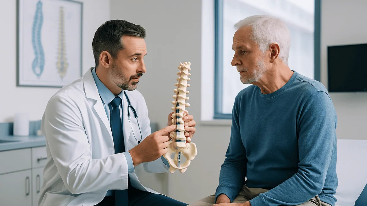
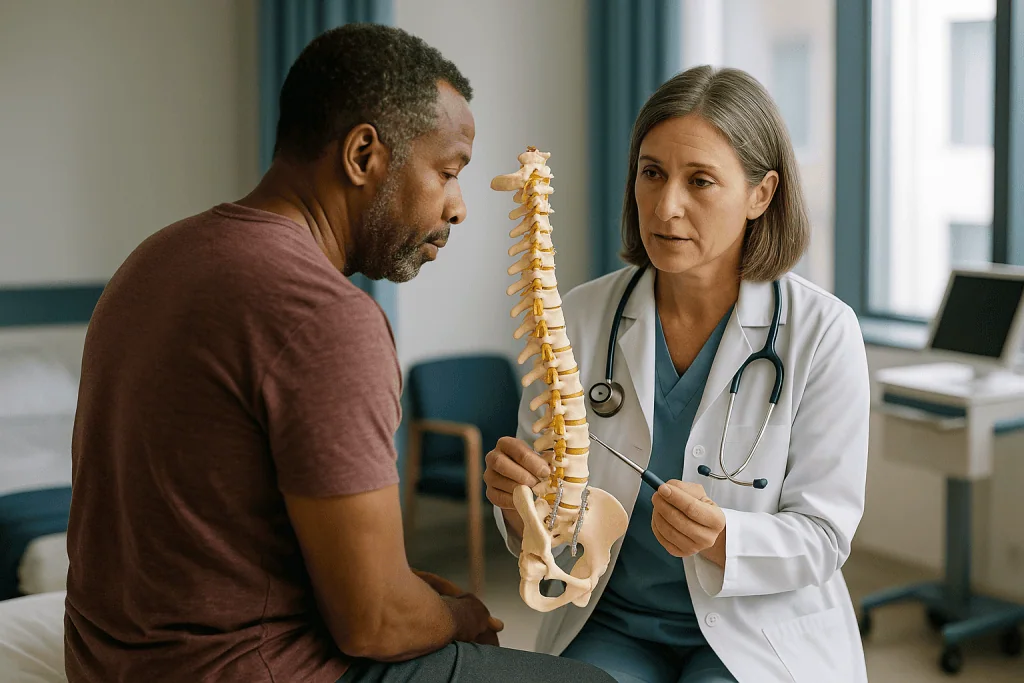
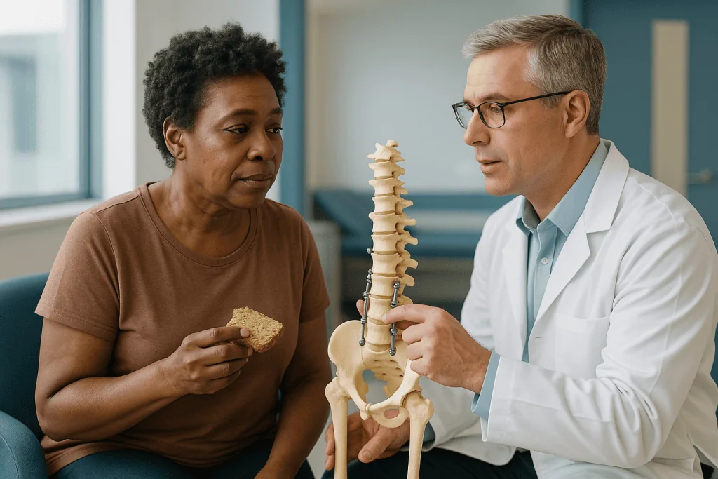
The surgeon slides a plastic model across the desk. Two vertebrae, a pair of screws, a short rod, and a small hollow block sitting where a disc used to be. “We fuse these,” she says, and the patient nods, because that is what you do. On the drive home the real questions arrive. Fuse how? Does the metal do the fusing? Will I ever bend again?
Those questions deserve straight answers, and the honest version is more interesting than the model. Understanding how spinal fusion works means understanding a slow biological event that the hardware merely protects. Screws and rods make headlines; bone graft and bone cells do the job.
This explainer walks through what actually happens in the operating room, what “fusing” means at the level of living tissue, why recovery runs on months rather than weeks, and which worries are justified and which are myths. Every decision about whether fusion is right for you stays with your treating team.
What is spinal fusion surgery for?
Think of the spine as a stack of 24 movable bones, the vertebrae, separated by cushioning discs and linked at the back by small paired joints called facet joints. That arrangement lets you twist, bend and look over your shoulder. Trouble starts when one of those links becomes unstable, slips, collapses or moves in a way that irritates nerves.
Spinal fusion is designed to solve one problem: unwanted motion at a painful or unstable segment. A segment is simply two neighboring vertebrae plus the disc and joints between them. According to Mayo Clinic, surgeons commonly use fusion to treat spinal deformities such as scoliosis, instability from arthritis or fractures, a vertebra that has slipped forward over the one below (spondylolisthesis), and some cases of a damaged or herniated disc.
Notice what is missing from that list: ordinary, non-specific low back pain with no structural cause. Fusion does not treat pain in the abstract. It treats a mechanical fault that a surgeon can point to on imaging and connect to your symptoms. When the fault is clear, removing the motion can relieve pressure on nerves and stop the painful shifting of bone on bone. When the fault is vague, the evidence for fusion becomes much thinner, and most guidelines steer toward non-surgical care first.
Fusion is also frequently paired with decompression, which means removing bone or disc material that is pressing on a nerve. The NHS describes fusion as something added to a decompression when removing tissue would leave the segment too unstable on its own. In that setting the fusion is less the goal than the insurance policy.
How spinal fusion works, step by step
Strip away the variations and nearly every fusion follows the same five moves.
First, access. Under general anesthesia, the surgeon reaches the target segment through the back, the front of the body, or the side. The choice depends on which vertebrae are involved and what else needs doing.
Second, preparation. Any tissue pressing on nerves is removed, and the surfaces that will be fused are roughened or cleared of disc and cartilage. This matters more than it sounds. Bone will not grow across smooth cartilage; it needs raw, bleeding bone to work with, the same way a broken arm needs its two ends in contact to heal.
Third, graft placement. Bone graft, meaning small pieces of bone or bone substitute, is packed between the prepared vertebrae, either in the emptied disc space, along the back of the bones, or both. MedlinePlus notes the graft can come from your own body, a donor, or a synthetic material.
Fourth, fixation. Screws are driven into the thick parts of the vertebrae and connected by rods, or a hollow cage is wedged into the disc space, or both. These hold the segment rigid.
Fifth, closure and waiting. The incision is closed, and the real work begins outside the hospital. Over the following months, the body treats the graft like a fracture site and lays down new bone until the two vertebrae are one.
The point to hold onto: the surgery sets up conditions for fusion. It does not complete it. You leave the operating room with a well-prepared construction site, not a finished building.
What does "fusing" actually mean inside the bone?
Fusing is healing. Specifically, it is the same cascade your body runs when you break a bone, redirected toward joining two bones that were never meant to be joined.
When the surgeon roughens the vertebral surfaces, bleeding begins, and with it a flood of signaling proteins that recruit cells. Some of those cells, osteoblasts, are bone builders. They move into the graft, which acts as a scaffold with just the right porous texture, and begin depositing new bone matrix. Over weeks that matrix hardens as minerals settle into it. Over months, another cell type, osteoclasts, remodels the rough new bone into organized, load-bearing bone with the same internal architecture as the vertebrae on either side.
The end result, on an X-ray or CT scan, is a continuous bridge of bone with no visible gap. Mayo Clinic describes the goal as having the vertebrae heal into “a single, solid bone,” and notes the process takes several months rather than weeks.
Two consequences follow. First, anything that slows bone healing slows fusion: smoking, poorly controlled diabetes, certain long-term medicines, poor nutrition and advanced age all appear in the evidence as factors that weaken the process. Second, the metal hardware has a shelf life of relevance. It carries load while the bridge forms. Once the bridge is solid, the bone carries the load and the screws become passengers.
If the bridge never completes, the condition is called nonunion or pseudarthrosis, literally “false joint.” The hardware then keeps working year after year without relief, and metal that is asked to do that indefinitely can eventually loosen or break. That is why surgeons care so much about the biology, not just the carpentry.
Where does the bone graft come from, and does the type matter?
Graft is the seed from which the fusion grows, and surgeons have three broad sources to choose from. Each has a trade-off, and no single type is right for every patient.
Autograft is your own bone. Traditionally it was harvested from the rim of the pelvis through a separate incision; today it is often collected from bone removed during the decompression itself, since that bone would otherwise be discarded. Autograft contains living cells and natural growth signals, which is why it has long been considered the biological reference standard. The cost is a second surgical site when pelvic bone is used, which can be sore for weeks.
Allograft is donor bone from a tissue bank, processed and sterilized. It provides the scaffold but not living cells. It avoids a second incision and is widely used, often blended with a patient’s own bone chips.
Synthetic or biologic substitutes include ceramic materials that mimic bone mineral and, in some cases, engineered proteins that stimulate bone formation. Cleveland Clinic lists all three categories as options a surgeon may use alone or in combination.
Which matters more, the graft or the patient? The honest reading of the evidence is that patient factors often dominate. A non-smoker with good bone density and a well-stabilized segment tends to fuse well with modest graft; a heavy smoker with osteoporosis may struggle even with the richest graft. That is why your surgeon’s questions about smoking, diabetes and prior bone health are not small talk. They are part of planning the fusion.
What is spine cage surgery, and what do the screws and rods do?
People searching for “spine cage surgery” are usually describing an interbody fusion, where a small hollow implant, the cage, is placed into the space left after a disc is removed. The cage is typically made of a strong plastic or titanium and is packed with bone graft before insertion, so that bone can grow through its hollow center and across the disc space.
A cage does three things at once. It restores the height of the collapsed disc space, which can reopen the tunnels where nerve roots exit. It carries compressive load from the vertebra above to the one below. And it holds the graft exactly where fusion is wanted, under compression, which bone biology favors.
Pedicle screws are threaded into the thick bony pillars at the back of each vertebra, and rods connect the screws on each side, locking the segment. Together they form what surgeons call a construct. Johns Hopkins describes this hardware as holding the vertebrae together while the bone graft heals.
Here is the comparison that helps most patients. Hardware is to fusion what a cast is to a broken wrist. The cast does not heal the bone; it keeps the ends still so the bone can heal itself. The difference is that a spinal cast sits inside, so it is normally left in place for life rather than removed. In most cases the metal causes no trouble once fusion is solid. It is removed only when it loosens, breaks, becomes infected or irritates nearby tissue, decisions that belong entirely to your surgical team.
Cages, screws and rods are often used together, sometimes with a plate at the front of the neck in cervical fusion. The combination the surgeon chooses reflects where instability sits and how much support the segment needs.
Spinal fusion vs laminectomy: what are the differences?
These two operations are often mentioned in the same breath, and they are frequently performed together, yet they aim at different problems. Laminectomy relieves pressure. Fusion removes motion.
A laminectomy removes the lamina, the bony roof at the back of the spinal canal, to make room for compressed nerves. MedlinePlus describes it as surgery to remove the lamina and relieve pressure on the spinal cord or nerves, most often for spinal stenosis. Nothing is joined; the vertebrae keep moving.
| Feature | Laminectomy (decompression) | Spinal fusion |
|---|---|---|
| Main goal | Relieve nerve pressure | Stop motion at an unstable segment |
| What is removed | Lamina, thickened ligament, bone spurs | Disc and cartilage at the surfaces to be fused; often lamina too |
| What is added | Nothing | Bone graft, usually with screws, rods or a cage |
| Motion afterward | Preserved at that level | Permanently lost at the fused level |
| Healing timeline | Soft tissue and bone edge healing, weeks | Bone fusion, several months |
| Typical reasons | Spinal stenosis with leg pain | Instability, slipped vertebra, deformity, fracture |
Why combine them? Removing bone can destabilize a segment, especially when the facet joints are already worn or a vertebra has slipped. The NHS explains that fusion may be added to a decompression to stabilize the spine. The decision rests on imaging, symptoms and how much bone must come out.
A fair summary for a patient: if your main problem is nerve compression and the segment is stable, decompression alone may be enough; if the segment is unstable or likely to become so, fusion enters the conversation. Only your surgeon can judge which description fits your spine.
Who is usually offered spinal fusion, and who is usually asked to wait?
Fusion is a major, irreversible operation, so the bar for offering it is deliberately high. The clearest candidates share a pattern: a structural problem visible on imaging, symptoms that match that problem, and a meaningful trial of non-surgical care that has not worked.
According to Mayo Clinic, the conditions where fusion is most often considered include deformity such as scoliosis, instability from arthritis or injury, a slipped vertebra, and certain disc problems, particularly when nerves are involved. A vertebra that moves visibly on bending X-rays, or a fracture that threatens the spinal cord, makes the case for stabilization much stronger.
Who tends to be asked to wait or to try something else first?
- People whose back pain has no clear structural source. Most guidelines favor exercise-based therapy, education and time before any surgery is discussed.
- People who have not yet completed a reasonable course of physical therapy, activity modification and non-surgical pain management.
- Current smokers, because nicotine measurably impairs bone healing; many surgeons ask patients to stop beforehand, with help from their primary care team.
- People with untreated osteoporosis, since screws need solid bone to hold.
- People with poorly controlled diabetes or active infection elsewhere, which raise wound and healing risks.
None of these is a permanent refusal. They are modifiable conditions, and addressing them often changes the conversation. The Cleveland Clinic frames fusion as an option when more conservative treatments have not helped, which is the right order of operations. A second opinion is a normal part of this process, not an insult to the first surgeon, and your treating team should welcome it.
Front, back, side or minimally invasive: how do the approaches differ?
Surgeons name fusions by the direction they come from and by where they place the graft, which produces a thicket of acronyms. The anatomy underneath is simple.
A posterior approach goes through the back. It gives direct access to the lamina, facet joints and pedicles, which is why it suits combined decompression and fusion. Graft may be laid along the back of the vertebrae (posterolateral fusion) or placed in the disc space after the disc is removed from behind (posterior or transforaminal interbody fusion).
An anterior approach comes through the abdomen for the lower back, or the front of the neck for the cervical spine. It reaches the disc without disturbing the back muscles and allows a large cage to be placed, but in the lumbar spine it requires moving blood vessels aside, often with a vascular surgeon assisting.
A lateral approach enters from the patient’s side, passing through or beside the flank muscles to reach the disc. It avoids both the back muscles and the major vessels at the front, though it has its own nerve pathways to protect.
“Minimally invasive” describes technique rather than approach. Smaller incisions, tubular retractors and X-ray guidance let the surgeon reach the same targets with less muscle cutting. Cleveland Clinic notes it may mean less tissue damage and a shorter stay for suitable patients, while cautioning that it is not possible or appropriate in every case. The fusion itself, the biology that joins the bones, is identical whichever door the surgeon uses.
Which approach is right depends on the level involved, the direction of nerve compression, your body shape and prior surgery. There is no universally superior route, and a surgeon’s recommendation usually reflects where the problem sits and what they can do most safely.
How long does spinal fusion surgery take, and how long is the hospital stay?
Two very different clocks run here: the surgical clock, measured in hours, and the biological clock, measured in months. Patients often conflate them, then feel discouraged when the second one does not keep pace with the first.
On the surgical clock, MedlinePlus gives a typical operating time of three to four hours for spinal fusion. That figure stretches when more levels are fused, when a deformity is being corrected, or when the surgeon must work around scar tissue from a previous operation. A single-level neck fusion may run shorter; a multi-level scoliosis correction runs considerably longer. Your surgeon will give you a range for your specific plan.
Hospital stay follows a similar logic. MedlinePlus describes a stay of around three to four days after fusion, while the NHS cites one to four days for lumbar decompression procedures, which may include fusion. Shorter stays are increasingly common with minimally invasive techniques and good pain control, but discharge is a judgment about whether you can walk safely, manage pain with oral medicines prescribed by your team, and empty your bladder and bowels, not a date on a calendar.
During those days you can expect to be up and walking, usually with physical therapy help, within a day of surgery. Early walking is not bravado; it reduces the risk of blood clots and pneumonia and begins the retraining of your back muscles. Some patients wear a brace, others do not, depending on the surgeon’s assessment of how rigid the construct is.
The biological clock starts when you leave. The hospital stay ends in days. The fusion takes months.
Spinal fusion recovery time: what the first days and weeks usually look like
The first week at home is about three things: moving a little, often; protecting the wound; and managing pain well enough to do the first two. Most people are surprised that walking is encouraged from the start and that lying still is actively discouraged.
Expect soreness at the incision and in the back muscles that were moved aside; many describe it as deep bruising. Nerve-related leg or arm pain may ease quickly, slowly, or fluctuate for weeks as irritated nerves settle, and NHS guidance notes that some symptoms can take time to improve.
Movement rules are usually summarized as avoiding bending, lifting and twisting. The logic is mechanical: bending and twisting load the new construct, and heavy lifting multiplies that load. Walking, by contrast, barely stresses a fused segment while improving circulation and mood. Short walks several times daily are the standard prescription, lengthening as tolerated.
On typical timelines, the NHS suggests many people with lumbar decompression can return to lighter, desk-based work within about four to six weeks, with physically demanding jobs taking longer, and driving resuming somewhere between two and six weeks once you can perform an emergency stop comfortably and are no longer taking medicines that impair driving. Treat these as ranges reported for groups of patients, not a schedule your body has agreed to.
Wound care is simple but matters: keep the dressing clean and dry as instructed, watch for spreading redness or discharge, and ask before soaking in a bath or pool. Constipation is common in the first week, from pain medicines and reduced activity, and your team can advise on preventing it.
Physical therapy usually begins in earnest a few weeks in, once the tissues have calmed, and focuses on walking endurance, posture and the deep trunk muscles that support a healing spine.
Is it okay to sleep in a recliner after back surgery?
Searches for this question spike in the first week after spine surgery, and the answer is usually reassuring: for most people, a recliner is acceptable in the early weeks if it is where you can actually sleep and get up safely. Comfort and safe transfers matter more than the furniture.
Why do so many people gravitate to one? A recliner supports the back in a semi-reclined position that takes load off the incision, and the mechanical lift makes standing up far easier than rolling out of a low bed. For someone whose trunk muscles are sore and whose surgeon has said “no twisting,” that is a real advantage.
A few cautions keep it safe. Avoid positions that leave you slumped with the lower back curled, which can strain the operated area. Change position periodically rather than staying frozen for eight hours. And if a surgeon has fitted you with a brace, follow their instructions about whether it stays on while resting. Your physical therapist can show you the “log roll” technique for getting into and out of bed, which keeps the spine straight and makes a regular mattress workable sooner than you might think.
Sleeping after spinal fusion improves gradually. Many people start in a recliner or propped on pillows and drift back to a bed within a few weeks as soreness settles. Side-lying with a pillow between the knees, or back-lying with a pillow under the knees, are the positions most often recommended because they keep the spine neutral. Stomach sleeping is generally discouraged early on.
If pain prevents sleep for several nights in a row, or if lying flat worsens leg or arm symptoms, that is worth a call to your team rather than a new piece of furniture. Poor sleep slows healing, and your clinicians have options.
Months 2 to 12: how does your team know the fusion is taking?
Around the sixth week, most people feel much better than they did at week one, which creates a trap. The surgical wound has healed, the bruising has faded, and it is tempting to assume the fusion is done. It is not. Bone is still being laid down inside, and this middle phase is when patience pays off.
Mayo Clinic notes that it can take several months for the bones to fuse, and Johns Hopkins similarly describes fusion as a process that continues well after the surgical recovery. Many surgeons regard a year as the point at which they judge whether a fusion is fully mature.
Your team tracks progress in two ways. First, symptoms: steadily improving function, less mechanical pain with movement, and no new instability-type pain. Second, imaging: plain X-rays at intervals, sometimes with bending views to confirm the segment no longer moves, and occasionally a CT scan when the X-rays are ambiguous. Radiologists look for bridging bone across the graft and the absence of a dark line (a lucency) around screws, which could suggest loosening.
Restrictions loosen in stages. Light bending and lifting return first; heavier lifting, impact activity and sports that twist the trunk come last, and only once the surgeon is satisfied the bridge is solid. Physical therapy shifts from protection to strengthening.
Things that help the biology in this phase are unglamorous: not smoking, eating adequate protein and calcium, keeping blood sugar controlled if you have diabetes, and walking daily. Things that hurt it include returning to heavy manual work too soon and ignoring new pain. If you have a long drive or flight planned during this window, discuss timing, movement breaks and clot prevention with your team beforehand.
What are the risks, and what does the evidence actually show?
Every fusion carries the general risks of a major operation and a few that are specific to joining bones. Patients deserve the list in plain language, without either alarm or minimization.
General surgical risks include infection of the wound or deeper tissues, bleeding, blood clots in the legs that can travel to the lungs, and reactions to anesthesia. Mayo Clinic lists infection, poor wound healing, bleeding, blood clots and injury to blood vessels or nerves among the recognized complications.
Spine-specific risks start with nerve injury, which can cause new numbness, weakness or pain, and a tear in the membrane around the spinal cord (a dural tear) that leaks spinal fluid and sometimes needs repair. The NHS describes these alongside the risk that symptoms do not improve as hoped.
Then come the two long-term questions unique to fusion. Nonunion, where the bone fails to bridge, leaves hardware doing a job it was never meant to do permanently, and can mean a second operation. Adjacent segment degeneration describes faster wear at the levels above or below the fusion, because those joints now absorb motion the fused level no longer provides. The evidence shows this happens in a minority of patients and that it is not always symptomatic, but it is real, and it is one reason surgeons fuse the fewest levels that will solve the problem.
Hardware can also loosen, shift or break, most often when fusion is slow or incomplete. And some people develop persistent pain despite a technically perfect fusion, a reminder that pain has sources beyond mechanics.
How these risks weigh against potential benefit is personal. Your age, bone quality, smoking status, diagnosis and goals all shift the balance, and that conversation belongs with your surgeon.
What people often get wrong about how spinal fusion works
Myths about fusion travel faster than the biology, and some of them lead to real decisions made on false premises. A few of the most common deserve a direct correction.
“The screws and rods hold my spine together for life.” They hold it together for months. Once bone bridges the gap, the bone carries the load, and the metal becomes largely redundant. If the bone never bridges, the metal is overworked, which is exactly why surgeons focus on healing, not hardware.
“I will not be able to bend after a fusion.” A single-level lumbar fusion removes motion at one of five lumbar segments; the others, plus your hips, still move. Most people bend, walk and sit normally once healed. Long multi-level fusions for deformity are a different matter and do noticeably limit flexibility, which your surgeon should discuss in advance.
“Fusion fixes back pain.” Fusion treats instability or deformity that happens to cause pain. For non-specific back pain without a structural target, the evidence summarized by sources such as the NHS favors non-surgical management first.
“Minimally invasive means a minor operation.” It means smaller incisions and less muscle disruption. The fusion inside is the same, and so is the months-long healing timeline.
“Once the incision heals, I am recovered.” Skin heals in weeks; bone fuses over months. Feeling good at six weeks is a milestone, not a finish line.
“If I have hardware I cannot have an MRI or go through airport security.” Modern spinal implants are generally compatible with MRI, though the radiology team must be told so they can confirm, and they may or may not trigger security scanners. Ask your surgical team for implant details to keep with you.
Questions to ask your care team before spinal fusion
Good surgeons like informed patients, and the questions below tend to produce the most useful conversations. Bring a notebook or a companion; the answers are hard to hold in memory in a short appointment.
- What exactly is unstable or deformed in my spine, and how does it explain my symptoms? Ask to see it on the imaging.
- What happens if I do not have surgery now? Is there a risk of nerve damage with waiting, or is this a quality-of-life decision I can take time over?
- Which non-surgical options have I not yet tried, and would any of them change the recommendation?
- How many levels do you plan to fuse, and why that number?
- Which approach will you use, and what led you to it for my anatomy?
- What type of bone graft do you intend to use, and will there be a second incision to harvest it?
- Will decompression be done as well, and what does that add to the risks and the recovery?
- What are the specific risks for someone with my health profile, including my bone density, smoking status and other conditions?
- How will we know the fusion is healing, and what imaging will be done, when?
- What activity restrictions should I expect, for how long, and when can I expect to drive and return to my particular job?
- Will I need a brace? Who arranges physical therapy, and when does it start?
- Whom do I call, day or night, if something worries me after discharge?
Two more are worth asking even if they feel awkward. First, would you be comfortable with me seeking a second opinion? A confident surgeon will say yes. Second, if the fusion does not heal or my pain persists, what would the next steps look like? Knowing the contingency plan in advance makes the decision feel less like a leap.
The Cleveland Clinic and Johns Hopkins both emphasize that fusion decisions are individualized. The questions above are how you participate in that individualization rather than receiving it.
When to call your doctor after spinal fusion
Most recovery days are uneventful, and most new sensations after spine surgery are the ordinary noise of healing tissue. Some are not, and the distinction matters because a few complications are time-sensitive. Call your surgical team promptly, or seek emergency care, if you notice any of the following.
- New or worsening weakness in a leg or arm, a foot that drags, or difficulty gripping or lifting. Nerve compression from bleeding or swelling can progress quickly.
- Loss of bladder or bowel control, difficulty passing urine, or numbness in the groin and inner thighs, the so-called saddle area. These can signal pressure on the nerves at the base of the spinal canal and are treated as an emergency.
- Fever, chills, or an incision that becomes increasingly red, hot, swollen or leaks fluid or pus. Clear, watery fluid from the wound can indicate a spinal fluid leak.
- A severe headache that is worse when sitting or standing and better lying flat, another possible sign of spinal fluid leakage.
- Pain, swelling, warmth or redness in a calf, which may indicate a blood clot, or sudden shortness of breath or chest pain, which can mean a clot has traveled to the lungs. The latter requires emergency care.
- Pain that escalates sharply rather than gradually easing, or a sudden “give” or clicking in the back accompanied by new pain, which could suggest hardware or graft problems.
- Persistent vomiting, inability to keep fluids down, or no bowel movement for several days despite the measures your team recommended.
The NHS and MedlinePlus both list infection, nerve injury, spinal fluid leak and blood clots among the complications to watch for after spine surgery, and both advise contacting the care team early rather than waiting to see whether symptoms settle.
A practical rule: if something feels new, feels wrong, or is getting worse instead of better, make the call. Your team would far rather reassure you about a false alarm than treat a complication late. Keep the after-hours number somewhere you can find it without searching.
Frequently asked questions
What is spinal fusion surgery for?
Spinal fusion is used to stop painful or dangerous motion between vertebrae. Common reasons include a vertebra slipping forward over its neighbor, instability from arthritis or fracture, spinal deformity such as scoliosis, and some disc problems affecting nerves. It is typically considered after non-surgical treatments have not helped and when imaging shows a structural fault that explains the symptoms.
What is spine cage surgery?
Spine cage surgery usually means an interbody fusion, where the damaged disc is removed and a small hollow implant called a cage, filled with bone graft, is placed in the space. The cage restores disc height, takes load off nerves and holds the graft in position so bone can grow through it and join the two vertebrae over the following months.
What are the differences between spinal fusion and laminectomy?
A laminectomy removes the bony roof of the spinal canal to relieve pressure on nerves and leaves the vertebrae free to move. Spinal fusion joins two or more vertebrae into one bone to eliminate motion at an unstable level. They are frequently performed together, with fusion added when removing bone would leave the segment too unstable on its own.
Is it okay to sleep in a recliner after back surgery?
For most people, yes, in the early weeks. A recliner supports the back and makes standing up easier, which is why many patients prefer it initially. Avoid slumped positions, change position periodically, and follow any brace instructions. Most people return to a bed within a few weeks using the log-roll technique a physical therapist can teach.
How does spinal fusion recovery time compare with the hospital stay?
They run on completely different clocks. MedlinePlus describes a hospital stay of a few days, and NHS guidance suggests lighter work often resumes in about four to six weeks. The bone fusion itself, however, takes several months, and surgeons commonly reassess at around a year. Feeling well at six weeks does not mean the bones have finished joining.
Does the metal hardware stay in forever?
Usually, yes. Once the bone has fused, the screws and rods carry little load and are generally left alone because removal would mean another operation. Hardware is removed only if it loosens, breaks, becomes infected or irritates nearby tissue. Those decisions are made by your surgical team based on symptoms and imaging.
Will I be able to bend after spinal fusion?
After a single-level fusion, most people regain near-normal bending, because the other spinal segments and the hips still move. Longer fusions across many levels, such as those for deformity, do noticeably reduce flexibility, and your surgeon should explain what to expect for your specific plan before you decide.
Why does smoking matter so much before spinal fusion?
Fusion depends on new bone growing across the graft, and nicotine impairs the blood supply and cell activity that bone healing requires. Smokers have a higher chance of nonunion, where the bones fail to join and the hardware is left overworked. Many surgeons ask patients to stop beforehand, and your primary care team can help with that.
What is adjacent segment degeneration?
It is accelerated wear at the spinal levels directly above or below a fusion, because those joints absorb motion the fused level no longer provides. It occurs in a minority of patients and does not always cause symptoms. Surgeons limit the number of fused levels partly to reduce this risk, and your team will monitor for it over time.
How do doctors know the fusion has healed?
They combine symptoms with imaging. Steady improvement in function and the absence of instability-type pain are reassuring. X-rays taken at intervals, sometimes with bending views, look for a continuous bridge of bone across the graft and no dark lines around screws. When X-rays are unclear, a CT scan can show the bone bridge in more detail.
References
- Spinal fusion – MedlinePlus Medical Encyclopedia
- Lumbar decompression surgery – NHS
- Laminectomy – MedlinePlus Medical Encyclopedia
This article is for general information only and is not a substitute for professional medical advice. Please consult a qualified doctor about your individual situation.

More from the Blog

After Herniated Disc Surgery: Wound Leaks, Fever and New Weakness That Need a Prompt Call
After herniated disc surgery, the signs that need a prompt call are fluid leaking from the wound, a temperature of 100.4°F (38°C) or higher,…
When Is Scoliosis Surgery Recommended? Curve Size, Progression and Symptoms That Matter
Scoliosis surgery is usually recommended when a curve measures about 45 to 50 degrees or more on the Cobb angle, is still progressing, and…
Why Repeat X-Rays Matter During Scoliosis Bracing: Checking Fit, Growth and Curve Control
Repeat X-rays during scoliosis bracing let the care team measure the Cobb angle, check how well the brace is holding the curve, and track…

Who Is a Candidate for Nucleoplasty? Contained Disc Herniations and Who Needs Another Option
A nucleoplasty candidate is typically an adult with leg-dominant pain from a small, contained lumbar disc herniation, confirmed on MRI, whose symptoms have not…

Questions to Ask Before Spinal Stenosis Surgery: Levels, Fusion and the Recovery Plan
Before spinal stenosis surgery, ask your surgeon which spinal levels will be treated and why, whether a fusion is planned or decompression alone is…

Schroth Method Results: What Consistent Practice Can Realistically Change and What It Cannot
Consistent Schroth practice most reliably changes posture, trunk symmetry, breathing mechanics, muscle endurance, and how a person feels about their back. Small randomized trials…