

How X-Rays and CT Scans Guide Trauma Surgery Decisions After a Serious Injury
Key Takeaways
- A portable chest and pelvic X-ray are usually taken on the trauma bed within minutes because a collapsed lung and a broken pelvis are both common and both able to cause rapid deterioration.
- The NHS notes a CT scan typically takes around 10 to 20 minutes, and in trauma the head, neck, chest, abdomen and pelvis are often imaged in a single pass.
- Contrast dye makes blood bright white on CT, so a jet of escaping dye marks active bleeding that may need surgery, while a contained injury without a leak is often observed instead.
- A patient whose blood pressure cannot be held up may go to surgery on bedside ultrasound and X-ray alone, with the detailed CT following once bleeding is controlled.
- Harvard Health puts an abdominal CT at roughly 10 millisieverts, about three years of natural background radiation, compared with about 0.1 for a chest X-ray.
- Guidelines such as NICE advise against routine whole-body CT in children, favoring examination, ultrasound and targeted imaging of the regions that mechanism or symptoms flag.
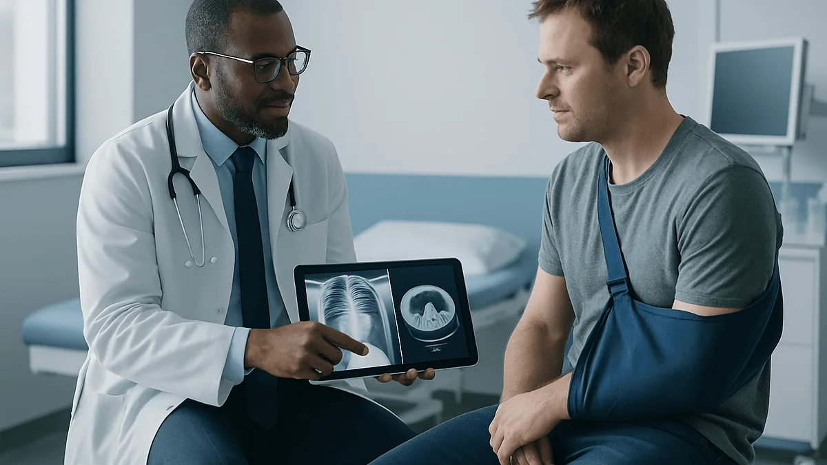
After a serious injury, X-rays and CT scans give trauma surgeons a map of what is broken, bleeding or displaced before anyone decides to operate. A plain X-ray quickly confirms fractures and collapsed lungs; a CT scan builds cross-sectional images that show internal bleeding, organ damage and complex breaks. Surgeons weigh these findings against the patient's blood pressure and breathing, so the same scan may lead to surgery, observation or a wait.
The ambulance doors open and, within seconds, someone in scrubs is already asking about the crash: how fast, which side, was she wearing a belt. A portable X-ray machine rolls toward the bed before the patient’s name is fully entered in the system. Nobody has said the word “surgery” yet. They are not being coy. They genuinely do not know.
That uncertainty is the point of trauma surgery imaging. A body that has taken a hard hit hides most of its damage: a ruptured spleen looks exactly like an intact one from the outside, and a spine fracture can sit quietly behind a bruise. Surgeons need to see inside before they cut, and they need to see fast.
This article walks through what those pictures actually show, why some people are scanned head to toe while others wait, and how a grainy image on a monitor becomes a decision about an operating room.
What does trauma surgery imaging actually mean?
Trauma surgery is the branch of surgery that treats physical injuries from sudden force: crashes, falls, assaults, crush injuries. Imaging is the collective word for tests that make pictures of the inside of the body. Put together, trauma surgery imaging is the set of scans used to find injuries and decide whether an operation is needed, how urgently, and which one.
Two tools do most of the early work. An X-ray passes a small amount of radiation through the body onto a detector; dense tissue such as bone blocks more of the beam and shows up white, while air-filled lungs appear dark. A CT scan, short for computed tomography, spins an X-ray source around the body and lets a computer assemble the readings into thin cross-sectional slices, like looking at a loaf of bread one slice at a time.
The distinction matters because the two tests answer different questions. An X-ray is a quick yes-or-no tool: is the pelvis broken, is a lung collapsed, is there air where it should not be? A CT scan answers the harder follow-up: how badly, where exactly, and is anything bleeding behind it?
Ultrasound often joins them at the bedside, and MRI occasionally follows days later for ligaments or the spinal cord. But in the first hour after a serious injury, X-ray and CT carry the decision. Everything downstream, from a splint in the emergency department to an operation at midnight, traces back to what those images show and, just as importantly, what they rule out.
What happens in the first minutes after a serious injury?
Before any scanner is involved, the trauma team runs a structured check that emergency clinicians worldwide learn in nearly the same order: airway, breathing, circulation, disability (meaning brain and spinal function), and exposure of the whole body to look for hidden wounds. This is often called the primary survey. Its job is to find anything that will kill the patient in the next few minutes.
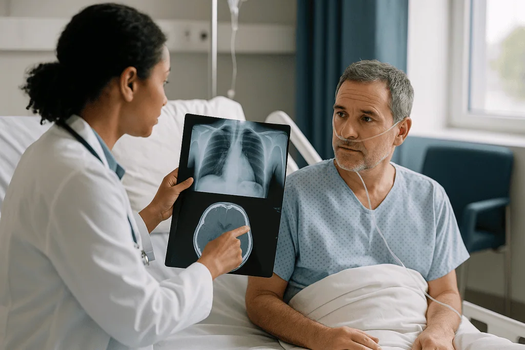
Imaging slots into this sequence rather than interrupting it. A portable chest X-ray and a pelvic X-ray are typically taken right there on the trauma bed, because a collapsed lung and a broken pelvis are both common and both able to cause rapid deterioration. A quick bedside ultrasound, sometimes called a FAST scan, sweeps the abdomen and heart looking for free fluid, which after an injury usually means blood.
These first pictures are deliberately crude. Nobody is trying to catalogue every rib. The team wants to know one thing: is this person stable enough to leave the resuscitation bay for the CT scanner, which is usually down a corridor, or do they need to go straight to an operating room on the strength of the bedside findings alone?
That single question separates two very different pathways. A patient whose blood pressure keeps falling despite fluids, and whose ultrasound shows fluid in the belly, may never see a CT scanner before surgery. A patient with a normal blood pressure and the same mechanism of injury will almost always be scanned first, because the extra detail changes what the surgeon does, or whether the surgeon does anything at all.
How does a CT scan build its picture, and how long does it take?
A CT scanner looks like a large ring with a narrow table sliding through the middle. Inside the ring, an X-ray tube and a row of detectors rotate together many times a second while the table moves the patient slowly through. Each rotation captures the body from hundreds of angles, and software stacks those readings into slices that can be viewed from the front, the side or the top, or rebuilt into a three-dimensional model of a shattered bone.
The scan itself is fast. The NHS notes that a CT scan usually takes around 10 to 20 minutes from lying down to sitting up, and the active scanning portion is far shorter than that. In a trauma setting the team often positions the patient once and images the head, neck, chest, abdomen and pelvis in a single pass, so that a person with several injuries is not moved repeatedly.
Patients are asked to lie still and sometimes to hold their breath for a few seconds while the chest is imaged, because movement blurs the slices. If a person is unconscious or intubated, the anaesthetic team manages breathing during the scan.
The images appear on the radiologist’s screen within moments, and in an emergency a preliminary reading is often given while the patient is still on the table. A radiologist is a doctor who specializes in interpreting scans; the trauma surgeon and the radiologist typically look at the images together, deciding in real time whether something needs an operating room, an interventional radiology suite where bleeding vessels can be sealed from inside, or simply a monitored bed.
X-ray vs CT scan for injury: what each one shows and misses
Asking which test is better misses how surgeons actually use them. They are sequential, and each has blind spots the other covers.
| Question after injury | Plain X-ray | CT scan |
|---|---|---|
| Is a long bone or the pelvis broken? | Usually shows it in seconds at the bedside | Shows the break in detail and any fragments that have rotated |
| Is a lung collapsed or filling with blood? | Detects most large collapses | Detects small collapses and bruised lung tissue an X-ray can miss |
| Is an internal organ bleeding? | Cannot show soft organs directly | With contrast dye, shows liver, spleen and kidney injuries and active bleeding |
| Is the spine fractured? | Misses a meaningful share of neck fractures | Standard test for bony spine injury after significant force |
| Is there bleeding inside the skull? | Not useful | First-line test |
| Radiation exposure (Harvard Health) | Chest X-ray about 0.1 mSv | Abdominal CT about 10 mSv, roughly three years of natural background |
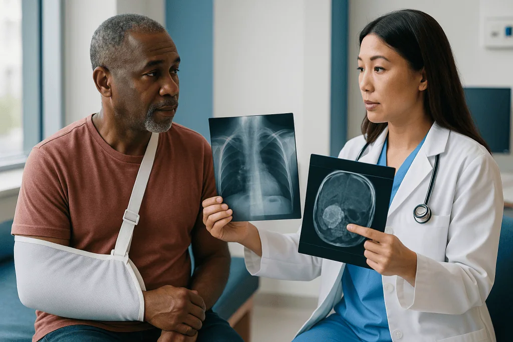
The pattern is clear. X-rays win on speed and portability and lose on soft tissue and fine bony detail. CT wins on detail and loses on dose and the need to move the patient. That is why a stable adult after a high-speed crash is nearly always scanned, while a person who fell from standing onto an outstretched wrist may only ever need two X-ray views.
Surgeons also rely on what a normal CT rules out. A clear scan of the abdomen after blunt force lets the team choose watchful observation over an exploratory operation, a shift that has reshaped trauma care over recent decades. The absence of a finding is, in this field, often the most valuable finding of all.
Whole-body CT trauma scans: who is scanned head to toe, and who is asked to wait?
A whole-body CT, sometimes called a pan-scan, images the head, neck, chest, abdomen and pelvis in one continuous run. Guideline bodies such as NICE in the UK recommend it for adults with suspected multiple injuries after significant force, because piecing the picture together region by region is slower and can miss injuries that sit between the usual zones, such as a torn diaphragm.
The typical candidates are people whose mechanism of injury is alarming even when they look reasonably well: ejection from a vehicle, a fall from a height, a pedestrian struck by a car, or anyone whose examination is unreliable because they are intoxicated, confused or unconscious. In these situations the physical exam simply cannot be trusted to find everything, and the scan substitutes for it.
Two groups are usually asked to wait, for opposite reasons. The first is the person who is too unstable to leave the resuscitation bay. If blood pressure cannot be held up, the surgeon may proceed to the operating room on bedside ultrasound and X-ray alone, and the detailed scan follows once bleeding is controlled.
The second group is the walking wounded: someone with a low-energy fall, a clear head, normal vital signs and a single sore area. For them, targeted X-rays of the painful part are the first step, and CT is added only if the X-ray is unclear or the pain points to a region X-rays image poorly.
Children sit in a third category. NICE guidance advises against routine whole-body CT in children, favoring a clinical assessment and selective imaging of the regions that examination or mechanism flags, because growing tissue is more sensitive to radiation and children’s injury patterns differ from adults’.
CT scan after a car accident: why you may be scanned even if you feel fine
People who walk out of a wrecked car are sometimes puzzled to be lying in a scanner an hour later. The explanation lies in how the body responds to sudden force. Adrenaline blunts pain in the first hours. A spleen can crack and ooze slowly enough that the abdomen feels merely tender. A small pocket of air can leak from a lung and stay small until it does not.
Trauma teams therefore make imaging decisions partly on the mechanism, meaning the physics of the event, rather than solely on how a person feels. Speed of impact, intrusion of metal into the passenger space, whether another occupant died, and whether the patient was thrown or trapped all raise the probability of hidden injury. Decision rules for the neck, such as those that check for midline tenderness, altered alertness and distracting injuries elsewhere, help clinicians decide who needs spine imaging and who can safely have their collar removed without it.
Age and medications matter too. Older adults bruise brains and break ribs with less force, and anyone taking a blood-thinning medicine has a lower threshold for head CT after even a minor knock, because bleeding inside the skull can progress with few early signs. That is a reason to tell the team about every medicine you take, not a reason to stop or change any of them without your prescriber.
Feeling fine is reassuring, and it does count. It is one data point among several, and the scan exists to check the others.
Why is contrast dye injected, and what does it reveal about bleeding?
Many trauma CT scans use an iodine-based contrast agent, a liquid injected into a vein that shows up bright white on the images. Blood carrying the dye lights up, so arteries, veins and well-supplied organs stand out sharply from the tissue around them.
For a trauma surgeon this is the difference between seeing an organ and seeing whether it is bleeding. A bruised liver without contrast looks like a dark smudge. With contrast, a jet of bright dye escaping from a torn vessel marks active bleeding that may need an operation or a catheter procedure to seal it. A spleen laceration that shows no such leak may, in a stable patient, be watched rather than removed.
The team often runs the scanner twice a few seconds apart, once as the dye floods the arteries and again as it reaches the veins, because injuries to each system look different. Kidney and bladder injuries may need a delayed pass as the dye is filtered into urine.
Cleveland Clinic describes the usual sensations: a warm flush spreading through the body and sometimes a metallic taste, both fading within a minute or two. Serious allergic reactions are uncommon but possible, and staff ask about previous reactions and kidney problems because the kidneys clear the dye. In a true emergency the scan usually proceeds regardless, since the information it gives can outweigh those risks; that judgment belongs to the treating team, who weigh it case by case.
How trauma imaging guidelines shape the choice to operate, observe or wait
People often ask what the current guidelines for trauma imaging say, and the honest answer is that they set a framework rather than a script. Bodies such as NICE, the American College of Surgeons and WHO’s emergency care programs share the same core principles: image early in stable patients, do not delay life-saving surgery for a scan in unstable ones, use whole-body CT selectively in adults with multiple suspected injuries, and be more sparing with children.
Within that framework, the scan result feeds a three-way decision. Operate now applies when imaging shows active bleeding the body cannot stop, a perforated bowel, a brain bleed pressing on tissue, or a fracture threatening a nerve or artery. Observe applies when an injury is real but contained: a liver laceration without contrast leak, a small lung collapse, a stable spine fracture. Here the plan is a monitored bed, repeat blood tests and often a repeat scan if anything changes. Wait applies to injuries that need fixing but not tonight, such as a displaced ankle fracture in someone whose brain injury must settle first.
Surgeons frequently describe this as a staged approach. The first operation may be brief and aimed only at stopping bleeding and contamination, with the definitive repair days later once the patient is warm, resuscitated and fully imaged.
None of this is decided by the scan alone. The images set the options; blood pressure, oxygen levels, clotting results and the patient’s other health conditions choose among them. Two people with identical CT findings can legitimately receive different plans.
Reading a fracture on X-ray: what orthopedic trauma surgeons look for
A break that a patient sees as a single crack contains several separate decisions for an orthopedic surgeon, and most of them are read straight off two X-ray views taken at right angles.
The first is displacement: how far the bone ends have moved from where they belong. Bones held in near-perfect alignment often heal in a cast. Bones that have shifted, rotated or shortened may need to be realigned, either by hand under sedation or with plates, screws or rods in the operating room.
The second is whether the fracture line enters a joint. Even a small step in the smooth surface where two bones meet can wear cartilage unevenly for years, so intra-articular fractures are far more likely to be fixed surgically, and CT is often added to map the joint surface in three dimensions.
The third is the skin. An open fracture, where bone has broken through or a wound reaches the bone, carries infection risk and is treated as urgent regardless of how the X-ray looks.
Then come the quieter signs: a fracture pattern that suggests a twisting force and a hidden second injury higher up, bone that looks thin and porous, or a break in a place that rarely fractures without an underlying weakness. Each prompts more questions or more imaging.
MedlinePlus notes that broken bones typically take weeks to months to heal depending on the bone and the person. The X-ray taken in the emergency department is only the first in a series; follow-up films track whether the bone is knitting in position or drifting, and that trend can change the plan even after a cast is on.
What happens when a scan finds something small or unexpected?
Detailed imaging of an entire torso almost inevitably shows things that have nothing to do with the injury: a small kidney cyst, a nodule in a lung, a thyroid lump. Radiologists call these incidental findings. They are common in whole-body trauma CT precisely because the scan is so thorough, and they can be unsettling to hear about while you are still processing a crash.
Most incidental findings are benign and need no treatment, but some need a follow-up scan or a clinic appointment weeks later, when the acute injury is settling. Ask the team to write these down for you and to name who will arrange the follow-up, because the handover from trauma care to routine care is where such items are most easily lost.
Small injury-related findings raise a different question: watch or re-scan? A tiny pocket of air around a lung, a contained bruise on the spleen, or a hairline fracture that does not fully explain the pain may all be managed with observation and a repeat image after a set interval. A repeat CT of the head is commonly considered if a person with a small brain bleed becomes drowsier, develops a new headache or is taking blood thinners.
Occasionally the first scan is simply inconclusive. Swelling can hide a fracture line, and a bowel injury may show only subtle signs on day one. In these cases surgeons rely on serial examinations, repeated at intervals, and a low threshold for imaging again. A normal scan at hour one is reassuring; a stable patient at hour twelve who still has an unexplained finding is not yet finished with imaging.
Radiation, pregnancy and children: how the dose is weighed against the injury
Every X-ray and CT scan uses ionizing radiation, and every trauma team weighs that against the risk of a missed injury. Harvard Health puts the numbers in everyday terms: a chest X-ray delivers about 0.1 millisievert, while a CT of the abdomen delivers roughly 10, close to three years’ worth of the natural background radiation everyone receives from soil, air and cosmic rays. The added lifetime cancer risk from a single trauma CT is small, and it sits against the immediate risk of an undetected bleed, which is not.
For most adults that balance is not close. Where it becomes genuinely delicate is in pregnancy and in childhood.
A pregnant patient after a serious injury is still imaged when the injury demands it, because the greatest threat to the fetus is an unstable mother. Teams shield the abdomen where the target region allows, favor ultrasound for the belly when it can answer the question, and involve obstetric colleagues early. The decision is individual and rests with the treating clinicians in discussion with the patient where she is able to take part.
Children’s tissues are more radiation-sensitive and they have more years ahead in which any effect could appear. Pediatric trauma protocols therefore lean on clinical examination, ultrasound and targeted X-rays, adding CT of a specific region when the exam or mechanism points there, rather than scanning head to toe by default. Modern scanners also adjust dose automatically to body size.
If you or your child are scanned, it is reasonable to ask why that scan was chosen and what it will change. A good team will have an answer that ties directly to a decision.
What do the following days and weeks usually look like?
For a person admitted after a serious injury, imaging does not end when the first CT is read. The first day or two typically bring repeat blood counts and, for contained organ injuries or small brain bleeds, a scheduled repeat scan to confirm nothing has progressed. If surgery happened, an X-ray of the fixed bone or the operated chest is usually taken before discharge as a baseline.
Fractures follow a predictable rhythm of follow-up films. Early X-rays check that alignment has held under a cast or fixation; later ones look for the cloudy bridge of new bone, called callus, that signals healing. MedlinePlus describes healing in terms of weeks to months, and the timeline varies by bone, age, smoking status and blood supply, so any specific expectation should come from your own surgeon rather than a general figure.
Many injuries that were watched rather than operated on still need a clinic visit at a few weeks to close the loop, and incidental findings from the trauma scan are often scheduled for follow-up imaging around the same time.
Physical recovery is only part of it. Poor sleep, flashbacks and a sense of being on edge after a crash are common and are the province of psychological trauma care, a different specialty with its own pathways. If those symptoms persist beyond the first few weeks or interfere with daily life, mention them to your care team; they are treatable and are not a sign of weakness.
Keep a simple folder of appointment letters and scan dates. Trauma care hands you between several teams, and being the one person who knows the whole sequence is genuinely useful.
What people often get wrong about trauma surgery imaging
The first confusion is the word itself. Search results for “trauma” are dominated by psychological trauma, the mind’s response to overwhelming events. Questions such as what the four stages of trauma recovery are, or what symptoms of trauma stored in the body look like, belong to that field. This article is about physical trauma, the kind that breaks bones and tears organs. Both are real; they are simply different conversations with different specialists.
A second myth is that a normal X-ray means nothing is wrong. X-rays are poor at soft tissue and can miss subtle fractures, especially in the neck, wrist and foot. A normal film with ongoing pain is a reason for follow-up, not reassurance to stop looking.
Third, people assume more scanning is always safer. Whole-body CT is powerful when the mechanism warrants it and unhelpful, with real radiation cost, when it does not. Guidelines favor selective imaging in low-energy injuries and in children for good reason.
Fourth, a scan is not a verdict. Some patients hear “there is a laceration on your liver” and expect an operation, then feel confused when the plan is a monitored bed. Contained injuries in stable patients are often observed, and that is evidence-based practice rather than hesitation.
Finally, a normal head CT does not exclude concussion. Concussion is a functional disturbance of the brain that does not show on CT; the scan’s job is to exclude bleeding, and symptoms like headache, fogginess and irritability can be entirely real with a clear scan. Recovery advice for concussion comes from clinical assessment, not from the image.
Questions to ask your care team about your scans
Emergency care moves quickly, and it is easy to leave with a folder of results and no clear picture of what they meant. A handful of questions, asked at any point when the team has a moment, can change that.
- What did the X-ray or CT show, in plain language, and what did it rule out?
- Which findings are from the injury, and which are incidental findings that need separate follow-up?
- If surgery is recommended, what on the scan is driving that, and what would happen if we watched instead?
- If we are observing rather than operating, what change would prompt a repeat scan or a different plan?
- Will I need follow-up imaging, roughly when, and who is responsible for arranging it?
- Was contrast dye used, and is there anything about my kidneys or allergies I should know for future scans?
- Can I have copies of the images and the radiologist’s report for my own records?
Write the answers down or ask a relative to. Sedation, pain and shock make memory unreliable in the first days, and the team will not mind repeating themselves.
It is also fair to ask about the radiation dose and why a particular scan was chosen, especially for a child. The answer should connect to a decision the scan will inform. If the reason offered is only “to be safe,” a follow-up question about what specifically would be missed without it is reasonable and usually welcomed.
None of these questions is a challenge to the team. They are how you become a participant in a plan rather than its subject, and most clinicians would rather explain twice than have a patient leave uncertain about what comes next.
When to call your doctor after a serious injury and imaging
Discharge after trauma imaging, whether from the emergency department or after a hospital stay, comes with a simple rule: injuries can evolve after the scan. The picture was accurate at the moment it was taken, and the body keeps moving.
Seek emergency care immediately, by calling your local emergency number, for any of the following red-flag signs:
- Increasing drowsiness, confusion, repeated vomiting, a worsening headache, seizures or weakness in an arm or leg after a head injury.
- Sudden or worsening shortness of breath, chest pain, or coughing up blood after a chest injury.
- Abdominal pain that intensifies or spreads, a rigid or swelling belly, fainting, or feeling cold, clammy and lightheaded.
- A limb below a cast or injury that becomes numb, blue, pale, cold or unbearably painful, or pain that keeps building despite rest and elevation.
- New difficulty passing urine, blood in the urine, or loss of bladder or bowel control after a back or pelvic injury.
- Fever, spreading redness or pus around a wound or surgical site.
Contact your doctor or the clinic that discharged you, within the day, for pain that is not improving as expected, swelling that keeps increasing, a cast that feels too tight or has cracked, or sleep, mood or memory changes that are interfering with daily life a few weeks after the event.
Keep your discharge letter close; it lists the injuries found, the imaging performed and the follow-up planned, and whoever sees you next will want it. Every decision about repeat scans, surgery or further observation rests with the clinicians assessing you in person, who can combine what the images showed with what your body is doing now.
Frequently asked questions
What are the current guidelines for trauma imaging?
Current trauma imaging guidelines from bodies such as NICE and the American College of Surgeons share core principles: image stable patients early, never delay life-saving surgery for a scan in unstable ones, use whole-body CT selectively in adults with suspected multiple injuries, and image children more sparingly with targeted scans. Decision rules for the neck and head help clinicians choose who needs CT. The treating team applies these to each patient’s situation.
Why did I get a CT scan after a car accident when I felt fine?
Because feeling fine in the first hours is not reliable after significant force. Adrenaline masks pain, a spleen can ooze slowly, and a small lung leak may not cause symptoms at first. Trauma teams base imaging partly on the mechanism, such as speed and intrusion into the vehicle, plus age and blood-thinning medicines. The scan checks the injuries that examination alone can miss.
X-ray vs CT scan for injury: which one do surgeons trust more?
Neither is trusted more; they answer different questions. X-rays are fast, portable and good for long bones, the pelvis and large lung collapses, but miss soft-tissue injuries and subtle fractures. CT shows organ bleeding, brain hemorrhage, spine fractures and fracture fragments in detail, at a higher radiation dose. Surgeons usually start with X-ray and add CT when the stakes or the uncertainty warrant it.
What is a whole-body CT trauma scan and who gets one?
A whole-body CT images the head, neck, chest, abdomen and pelvis in one continuous run, so injuries spanning regions are not missed. It is generally used for adults with suspected multiple injuries after high-energy events, or whose examination is unreliable because of intoxication or reduced consciousness. Stable people with a single low-energy injury usually receive targeted X-rays instead, and children are rarely scanned head to toe.
What to do immediately after a traumatic event such as a crash or fall?
Call your local emergency number, avoid moving anyone with suspected neck or back injury unless they are in danger, and apply firm pressure to visible bleeding. Do not eat or drink in case surgery is needed. Tell responders about the mechanism of injury, medicines taken, especially blood thinners, and any known allergies or kidney problems, since these shape imaging and contrast decisions.
Does a normal head CT mean I do not have a concussion?
No. CT is used to exclude bleeding and skull fractures, not to diagnose concussion, which is a disturbance of brain function that does not show on the scan. Headache, fogginess, sensitivity to light and irritability can be entirely real with a clear CT. Concussion is diagnosed clinically, and recovery guidance comes from your doctor’s assessment rather than the image.
Why is contrast dye used, and is it safe?
Iodine-based contrast makes blood bright on CT, letting surgeons see whether an organ is actively bleeding rather than merely bruised. Cleveland Clinic describes a warm flush and metallic taste that fade within minutes. Serious allergic reactions are uncommon, and staff ask about previous reactions and kidney function because the kidneys clear the dye. In emergencies the benefit usually outweighs the risk, a judgment made by the treating team.
What are the four stages of trauma recovery, and do they apply to injuries?
The stages people search for come from psychological trauma literature and describe emotional recovery, not physical healing. Physical trauma care is often described in phases instead: resuscitation, stabilization, definitive repair and rehabilitation. Both matter after a serious injury; if sleep problems, flashbacks or anxiety persist beyond the first weeks, tell your care team, who can connect you with psychological support.
What are the symptoms of trauma stored in the body, medically speaking?
The phrase usually refers to psychological trauma and how stress can show up as tension, poor sleep or hypervigilance, which is a mental health topic worth raising with a clinician. Physically, injuries can also declare themselves late: increasing abdominal pain, growing drowsiness after a head knock, or new numbness in a limb are signs that an injury has evolved since imaging and need urgent assessment.
Will I need more scans after leaving hospital?
Often, yes. Fractures are followed with X-rays to confirm alignment and then to look for new bone forming, over a timeline MedlinePlus describes as weeks to months. Contained organ injuries and small brain bleeds may have a scheduled repeat CT. Incidental findings from the trauma scan sometimes need separate follow-up imaging. Ask who is arranging each appointment before you leave.
References
This article is for general information only and is not a substitute for professional medical advice. Please consult a qualified doctor about your individual situation.

More from the Blog

How Arthritis Wears a Joint Down: When Damage Reaches the Point Where Replacement Is Discussed
Arthritis wears a joint down by thinning the cartilage that cushions the bone ends, roughening the bone beneath, and inflaming the joint lining, until…

Preparing for Hand Surgery: Nerve Tests, Imaging and the Medicines Your Team Reviews
To prepare for hand surgery, most people complete nerve tests such as a nerve conduction study or EMG, imaging such as X-ray, ultrasound or…

Hip Flexor Pain: Tight, Strained or Something Else, and the Exercises That Help
Hip flexor pain is soreness at the front of the hip or groin where the iliopsoas and rectus femoris muscles lift the thigh. Most…
Does the Robot Operate on Its Own? Robotic Knee Replacement Myths, Corrected
No. In robotic knee replacement, the surgeon performs the operation and the robotic arm acts as a precision tool. The surgeon plans the implant…
How a Pavlik Harness Keeps Your Baby’s Hips in Place So the Socket Can Deepen
A Pavlik harness works by holding a baby's hips bent up and gently spread apart, so the ball at the top of each thigh…
Shoulder Replacement Recovery: Timeline, Sling Life and Real Milestones
Most people wear a sling for about two to six weeks after shoulder replacement, regain everyday reach by three months, and see strength keep…