

Inside the Operating Room During Robotic Thoracic Surgery: Ports, Docking and the Chest Tube

Key Takeaways
- A robotic surgical system has no autonomy: the instruments move only as the surgeon moves the console controls and stop the moment the surgeon looks away from the viewer.
- One-lung ventilation through a double-lumen breathing tube is what deflates the operative lung and creates the working space that makes port-based chest surgery possible.
- Ports are placed along one or two intercostal spaces in a planned arc so the wristed instruments can reach the top of the chest without the robotic arms colliding outside the body.
- Nearly everyone wakes with a chest tube because stapled lung tissue leaks air and cut surfaces drain fluid; MedlinePlus describes a stay of a few days until both stop.
- MedlinePlus quotes roughly five to seven hospital days after an open thoracotomy, with shorter stays typical after keyhole approaches, though individual timelines vary.
- Conversion from robotic to open surgery is a safety decision the surgeon makes for bleeding, dense adhesions or unsafe anatomy, and it is worth asking how often it happens in their practice.
Robotic thoracic surgery procedure steps follow a set sequence: general anesthesia with one-lung ventilation, positioning on the side, three to five small incisions (ports) between the ribs, docking the robotic arms, the surgeon operating from a console to divide vessels, airway and lymph nodes, removal of the specimen in a bag, placement of a chest tube, and closure. The surgeon controls every movement; the machine never acts on its own.
The consent form said “robot-assisted lobectomy,” and the word that stuck was not lobectomy. It was robot. A retired schoolteacher in a paper gown asked her surgeon the question half the waiting room was thinking: who exactly is holding the knife?
The honest answer is more reassuring, and more interesting, than the science-fiction picture. Robotic thoracic surgery procedure steps are a choreographed sequence performed by a team of people, one of whom happens to sit at a console a few feet from the operating table rather than leaning over it. Every cut, every staple, every stitch begins as a movement of a human hand.
This article walks through that sequence in the order it happens: the anesthetic, the ports between the ribs, the docking of the arms, the work inside the chest, and the drain you wake up with. Along the way it answers the questions people actually search, and corrects a few myths that deserve retiring.
Robotic thoracic surgery procedure steps, from first breath to last stitch
Strip away the technology and a robotic lung operation follows the same logic surgeons have used for decades: reach the lung safely, control its blood vessels and airway, remove the diseased part, check for leaks, and leave a drain. The robotic version simply changes how the surgeon reaches in and sees.
Here is the sequence in plain order. You arrive in the operating room awake and are put to sleep with a general anesthetic. A special breathing tube lets the anesthesia team deflate the lung being operated on while the other lung keeps you oxygenated. You are turned onto your side, padded, and secured. The surgeon marks the spaces between your ribs and makes several small incisions, each fitted with a hollow sleeve called a port that instruments pass through. The robotic cart is wheeled in and its arms are attached to those ports, a step called docking. The surgeon then leaves the bedside for the console, while an assistant stays at your side to swap instruments and pass stapler loads.
Inside the chest, the surgeon frees the target lobe or segment, seals and divides its artery, vein and bronchus with staplers, and removes nearby lymph nodes. The specimen is placed in a bag and drawn out through the largest incision. The lung is reinflated and checked under water for air bubbles. A chest tube is placed through one of the existing incisions, the arms are undocked, the wounds are closed, and you wake up in the recovery area.
Each of those steps has its own reasons and its own small risks, which the rest of this article unpacks. Mayo Clinic describes the general robotic setup, and Johns Hopkins outlines what a lobectomy involves; both make the same point that the approach is minimally invasive but the operation itself is major surgery.
Why the surgeon sits across the room: how the robot actually works
The word “robot” suggests autonomy, but a surgical robot is closer to a very sophisticated pair of remote-controlled hands. Cleveland Clinic and Mayo Clinic both describe the same architecture: a patient-side cart with three or four arms, a console where the surgeon sits, and a tower carrying the camera electronics and light source.

At the console the surgeon looks into a viewer that shows a magnified, three-dimensional image from a camera inside the chest. Their fingers rest in loops attached to hand controllers. When the surgeon rotates a wrist, the tiny instrument at the end of the robotic arm rotates in the same way, scaled down and with hand tremor filtered out. A foot pedal switches between arms, another controls the camera, another activates energy devices that seal small vessels.
Three features matter most for chest surgery. First, the instruments have articulating wrists that bend well beyond what a human wrist can do, useful when working around the curved back of the chest cavity. Second, the camera sits a few centimeters from the tissue, so structures such as the pulmonary artery branches appear large and in depth. Third, the surgeon’s view and hands are aligned, so movement feels intuitive rather than mirrored, as it can with conventional keyhole instruments.
What the robot does not do is think, decide or move on its own. If the surgeon lifts their head from the viewer, the instruments freeze. If power fails, the arms can be manually released. Trading places at the console is also easy, which is one reason robotic platforms are widely used for surgical training.
Before the first incision: anesthesia, one-lung ventilation and positioning
The anesthetic for a chest operation is different from the one used for, say, a knee replacement, and the difference is the key that makes everything else possible. Once you are asleep, the anesthesia team places a breathing tube with two channels, called a double-lumen tube, or a bronchial blocker that plugs one main airway. Either device allows them to stop ventilating the lung on the operative side while the other lung keeps your blood oxygenated.
The deflated lung falls away from the chest wall like a collapsed tent, creating room for instruments. Many robotic teams add a gentle flow of carbon dioxide into the chest cavity through one port; the low pressure pushes the lung and diaphragm further out of the way and helps clear surgical smoke from energy devices. Your body absorbs the gas afterwards.
Positioning takes real time and care. You are rolled onto the side opposite the operation, with the table flexed so the space between your ribs opens slightly. The arm on the operative side is supported above your head, padding protects nerves at the elbow and knee, and an axillary roll keeps weight off the shoulder. A urinary catheter and arterial line are common for longer procedures.
Before any incision, the team runs a checklist confirming your identity, the side of the operation, imaging on the screens, antibiotics given, and equipment ready. Marking the correct side is deliberately repeated by more than one person. The WHO surgical safety checklist that underpins this routine exists precisely because wrong-side surgery, while rare, is preventable. Only once every box is answered does the surgeon pick up a marker to plan the ports.
Ports: where the small incisions go and why placement matters
A port is a short hollow tube, usually about the width of a pencil, that sits in an incision between two ribs and acts as a doorway for instruments. Mayo Clinic and Cleveland Clinic both describe robotic operations as being performed through several small incisions rather than one large one; in the chest that typically means three to five, plus one slightly longer “utility” opening used to pass staplers and remove the specimen.
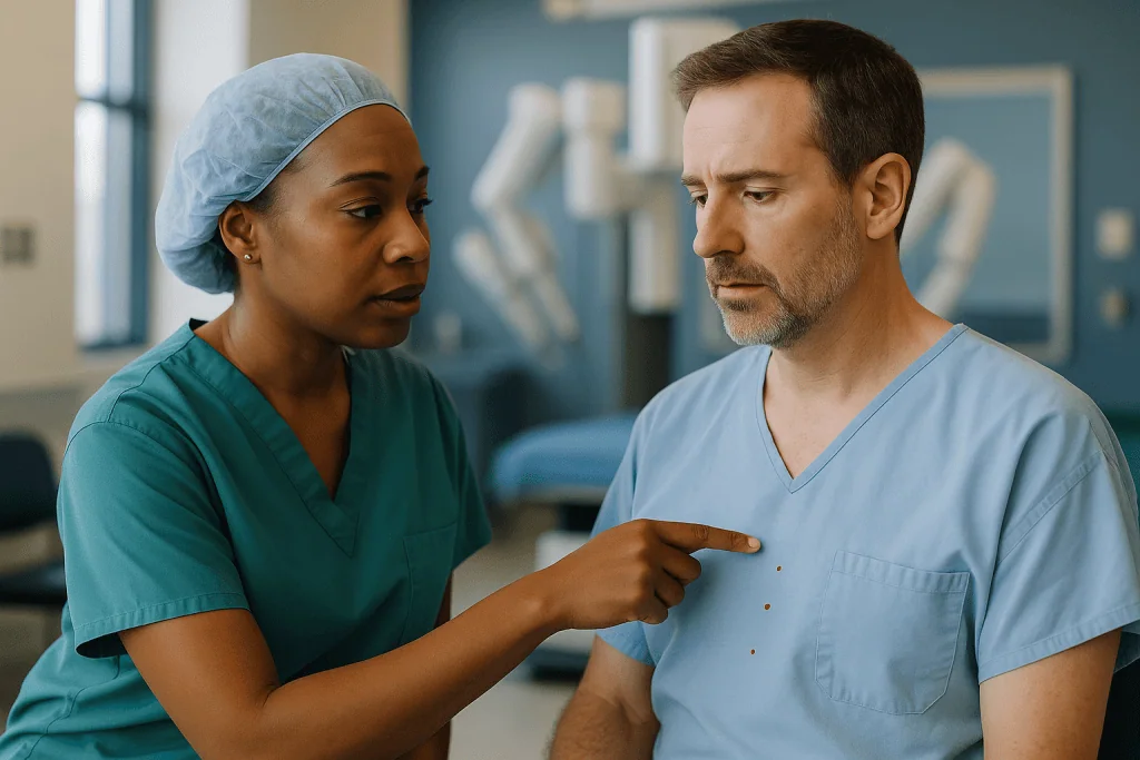
Placement is not random. The surgeon aims for a gentle arc along one or two intercostal spaces, low enough on the chest wall that the instruments can reach up to the top of the chest without colliding, and far enough apart that the robotic arms do not knock into each other outside the body. Anatomy matters here: the diaphragm rises higher on the right, the heart occupies more of the left, and a tall thin person’s ribs sit differently from a shorter, broader frame. Many surgeons mark the ports with a ruler after palpating the ribs.
The first port is usually for the camera. Before committing to the others, the surgeon takes a look inside to confirm the lung has collapsed, that there are no dense adhesions from prior infection or surgery, and that the anatomy matches the scans. The remaining ports are placed under direct vision so no instrument enters blind.
Why so much fuss over a few centimeters? Because the ribs do not move. A conventional open thoracotomy spreads them with a retractor, which is a major source of pain afterwards. Ports avoid spreading, but only if their positions give the instruments the reach they need. Getting the geometry right at this stage prevents awkward angles, extra incisions and, occasionally, the need to convert to an open operation.
Docking: the moment the robotic arms meet the patient
Docking is the step that most visibly separates a robotic operation from other keyhole surgery. The patient-side cart, roughly the size of a large refrigerator on wheels, is driven or guided into position beside the table, and each robotic arm is clipped onto a port. From that moment the ports, the arms and your chest are physically linked.
The team works through a routine. The cart’s position is chosen so the arms can pivot around each port without bumping the table, the anesthesia machine or each other; on newer systems the arms rotate above the patient from a single boom, which gives more flexibility. Each arm is aligned so its “remote center,” the fixed pivot point, sits exactly at the chest wall. That detail is what allows the instrument to move inside without levering against the ribs.
Once docked, the camera is inserted and the surgeon checks the view. Instruments follow, each introduced under vision. A bedside assistant confirms that every arm moves freely through its full range before the surgeon sits down. Mayo Clinic notes that the setup time is one reason robotic procedures can take longer than the conventional equivalent, and docking is a fair share of that.
Two safety points are built in. The table cannot be moved once arms are docked unless the system is designed for it, so positioning is finalized first. And undocking can be done in under a minute if the team needs urgent access to the chest, for example to control bleeding. Teams rehearse this “emergency undock” the way airline crews rehearse evacuations: rarely needed, always ready.
Then the room quiets. The surgeon settles into the console, the assistant stands at the table, and the real work begins.
Inside the chest: dividing vessels, airway and lymph nodes
Every lobe of the lung hangs from a bundle of structures at its root, called the hilum: a branch of the pulmonary artery bringing blood in, a pulmonary vein taking it out, and a bronchus carrying air. Removing a lobe means controlling and dividing each of these, in whichever order the anatomy and the disease make safest.
The surgeon starts by opening the thin membrane over the hilum and separating the lobe from its neighbors along the fissures, the natural clefts between lobes. Small vessels are sealed with an energy instrument; larger ones are dissected free and encircled. For each major artery, vein and bronchus, the assistant passes a stapler through the utility port or a robotic stapler is used. The device places rows of tiny titanium staples and cuts between them in one motion, sealing both ends. The bronchus is often tested afterward by asking the anesthesia team to inflate the remaining lung under a pool of saline and watching for bubbles.
Lymph node removal is part of a proper cancer operation. Nodes sit in predictable stations along the airways and in the space between the lungs, and sampling or dissecting them tells the pathologist whether the disease has spread. That information shapes any treatment after surgery, which is why NHS guidance lists lymph node assessment as a standard part of lung cancer surgery.
The freed lobe is slipped into a plastic retrieval bag inside the chest, the bag’s neck is drawn out through the utility incision, and the specimen follows. Bagging matters: it keeps any tumor cells from touching the wound edges. A final sweep checks for bleeding, the lung is reinflated, and attention turns to the drain.
The chest tube: why you wake up with a drain and what it does
Nearly everyone wakes from lung surgery with a soft plastic tube emerging from one of the incisions and running to a collection unit hanging beside the bed. MedlinePlus describes the chest tube’s job simply: it drains air, fluid or blood from the space between the lung and the chest wall so the lung can stay fully expanded.
Two things collect in that space after an operation. The first is fluid, a mix of blood and tissue fluid from cut surfaces. The second is air leaking from the stapled edge of the lung, because lung tissue is spongy and staple lines do not always seal on day one. Without a drain, either could compress the lung and undo the work of the operation.
The tube is placed before undocking, through the lowest port so gravity helps drainage, with its tip guided toward the top of the chest where air collects. It is stitched to the skin and connected to a system with a water seal, a one-way valve that lets air out but not back in, often with gentle suction. Newer digital units display the air leak as a number on a screen, which takes some of the guesswork out of deciding when the tube can come out.
Nurses will check the drainage volume and watch the water chamber for bubbling when you cough. MedlinePlus notes the tube usually stays for a few days, until drainage falls and any air leak stops; your team’s criteria will be specific to your case. Removal happens at the bedside, takes seconds, and involves a quick pull while you hold your breath or hum. Most people describe it as a strange tug rather than sharp pain. The small hole is closed with a stitch or dressing and heals like the other incisions.
How long does robotic lung surgery take?
Ask three surgeons and you will get three honest ranges, because the answer depends on what is being removed and what the surgeon finds. Johns Hopkins describes lobectomy as an operation lasting several hours; a wedge resection or a biopsy of a small nodule sits at the shorter end, while a segmentectomy or a lobe with dense adhesions runs longer.
A useful way to think about it is in blocks. Anesthesia induction, line placement and positioning take a meaningful stretch before any incision. Port placement and docking add more; Mayo Clinic notes that robotic setup can make these operations longer than the same procedure done conventionally. The work inside the chest, from freeing the fissure to removing lymph nodes, is the main block. Then come leak testing, drain placement, undocking and closure, followed by waking you up and moving you to recovery.
Families waiting outside often hear a total time that includes all of these, not just the cutting. A message that surgery “has started” may mean the room has begun preparing, not that the surgeon has reached the lung. Asking beforehand what the team’s updates will mean can spare an hour of anxiety.
Factors that lengthen operations include previous chest surgery or infection, which leave scar tissue that must be released carefully; a tumor close to the main vessels; anatomy that differs from the scan; and larger body size, which changes angles. None of these are signs that something is wrong. Surgeons routinely say a slow, careful hour beats a fast complication.
What you will not feel is any of it. From the anesthetic taking hold to waking in recovery, the interval passes in what most patients describe as a blink.
VATS vs robotic surgery vs open thoracotomy: what actually differs
Three routes lead into the chest, and the choice among them is usually about the surgeon’s training, the operation required and your particular anatomy rather than one being universally best. VATS, or video-assisted thoracoscopic surgery, uses a camera and long straight instruments through small incisions, with the surgeon standing at the table watching a screen. Robotic surgery uses articulating instruments through similar ports, controlled from a console. Open thoracotomy uses one long incision and spreads the ribs to allow direct hand access.
| Feature | Open thoracotomy | VATS | Robotic-assisted |
|---|---|---|---|
| Incision | One long incision, ribs spread | Several small ports, no spreading | Several small ports, no spreading |
| Surgeon’s position | At the table, direct view | At the table, 2D screen | At console, magnified 3D view |
| Instrument movement | Human hand and wrist | Straight, limited angles | Wristed, tremor-filtered |
| Tactile feedback | Full | Partial through instruments | Little to none |
| Typical hospital stay (MedlinePlus) | Roughly 5 to 7 days | Usually shorter | Usually shorter |
| Setup | Standard | Standard | Extra docking time |
Mayo Clinic and Cleveland Clinic summarize the evidence on minimally invasive approaches in general: smaller wounds, less blood loss, less pain and quicker recovery than open surgery, with complication rates comparable to conventional methods. High-quality head-to-head trials comparing VATS with robotic lobectomy are fewer, and most show broadly similar recovery, with differences that vary by study. When your surgeon recommends one route, the most useful question is not “which is best” but “which do you do most, and why this one for me.”
Who robotic thoracic surgery is usually for, and who is asked to wait
Most people who are offered a robotic chest operation have a lung nodule or early-stage lung cancer that a surgeon has judged removable, a lump in the mediastinum such as a thymic tumor, a persistent collapsed lung needing pleural treatment, or occasionally a diaphragm or esophageal problem. NHS guidance describes surgery as the usual treatment for non-small-cell lung cancer when the tumor is confined to one part of the lung and the person is well enough to withstand it.
That second condition matters as much as the first. Before any decision, teams assess breathing capacity with spirometry, which measures how much air you can move, and often a gas transfer test, which measures how well oxygen crosses into the blood. If a lobe is coming out, the lungs left behind must be able to do the job. Heart function, kidney function and general fitness are reviewed, and a multidisciplinary meeting of surgeons, oncologists, radiologists and pathologists weighs the imaging together.
Some people are asked to wait rather than told no. A chest infection at the time of surgery raises anesthetic risk and is usually treated first. Uncontrolled diabetes, a recent heart event or active smoking may prompt a delay while these are addressed; quitting even a few weeks before surgery measurably improves lung function and wound healing. Blood-thinning medicines are often paused or adjusted under the prescribing clinician’s direction; never change them yourself.
Others are steered toward a different route. Very large tumors, extensive scar tissue from previous surgery, or a tumor invading the chest wall may make an open operation safer. People whose lungs cannot spare a lobe may be offered a smaller segmentectomy or a non-surgical treatment such as stereotactic radiotherapy, which NHS guidance lists as an option when surgery is unsuitable. The treating team makes these calls case by case, and it is entirely reasonable to ask them to explain the reasoning.
What is the downside of robotic surgery? Risks, limits and conversion
Every operation carries risk, and honest patient information lists it. Mayo Clinic is direct: robotic surgery has risks similar to those of conventional open surgery, including infection and bleeding, and it is not an option for everyone. The robot does not remove the ordinary hazards of chest surgery; it changes the route in.
Chest-specific complications include prolonged air leak from the staple line, which keeps the drain in longer; pneumonia in the remaining lung, made more likely by shallow breathing after surgery; abnormal heart rhythm, particularly atrial fibrillation, which is common after lung resection and usually settles; bleeding; and, rarely, injury to nearby structures such as the nerve that runs to the diaphragm or the vocal cords. MedlinePlus lists similar risks for lung surgery of any kind. Blood clots in the legs or lungs are a risk after any major operation, which is why compression stockings, injections to thin the blood and early walking are routine.
The robot adds its own considerations. Surgeons lose most tactile feedback and rely on visual cues, so a delicate structure can be pulled harder than intended. Setup adds time under anesthesia. Instruments are expensive to replace and the system requires a trained team; this is a health-system question rather than one for you at the bedside, but it explains why not every hospital offers it.
Conversion deserves plain explanation. If bleeding cannot be controlled through the ports, adhesions are too dense, or the anatomy is unsafe to approach robotically, the surgeon will undock and open the chest. This is not a failure; it is the plan working. Ask before surgery how often it happens in your surgeon’s practice and what it would mean for your recovery.
Alternatives exist for those who cannot or prefer not to have surgery, and your team should describe them in the same neutral terms.
How painful is thoracic surgery, and how the pain is managed
Chest surgery has a reputation for pain, and it is earned mostly by the open operation. Spreading the ribs stretches the joints where they meet the spine and can bruise the intercostal nerves that run beneath each rib. Minimally invasive approaches avoid the spreading, and Cleveland Clinic and Mayo Clinic both list less pain among the advantages over open surgery. Less does not mean none.
What people typically describe after robotic lung surgery is soreness at the port sites, a deep ache where the chest tube sits, and a sharp catch when coughing or taking a full breath. Shoulder pain on the operated side is common and often surprising; it is referred pain from irritation of the diaphragm, not a shoulder injury. Some experience numbness or a burning strip along a rib for weeks as the intercostal nerve recovers.
Management is layered rather than relying on one drug. Before the incisions close, many surgeons inject local anesthetic around the intercostal nerves or place a small catheter beside the spine that bathes the nerves continuously for a day or two, a technique called a paravertebral or erector spinae block. Afterwards, scheduled non-opioid pain medicines form the base, with opioid medicines reserved for breakthrough pain. The aim is not zero pain but pain low enough to breathe deeply, cough and walk, because those three actions prevent pneumonia and clots. Your team will set the plan, and any adjustment belongs to them.
Simple measures help. Hugging a pillow while coughing splints the chest. Sitting upright rather than lying flat eases breathing. Walking loosens the ache. Pain that escalates rather than settles, or that arrives with fever or breathlessness, is a signal to call rather than endure.
Robotic lobectomy recovery time: the first days and weeks
Recovery unfolds in fairly predictable chapters, though the pace varies with age, fitness and how much lung was removed. The figures below are typical ranges from patient guidance, not promises.
The first day is about breathing and moving. Nurses will help you sit up within hours and walk, often the same day. A respiratory therapist may bring an incentive spirometer, a plastic device that rewards slow deep breaths with a rising marker, to keep the remaining lung open. The chest tube stays connected to its unit, which you wheel with you on walks. A chest X-ray each morning tracks lung expansion.
Days two to four usually revolve around the drain. When fluid output falls and the air leak has stopped, the tube comes out; MedlinePlus describes a few days as usual. Many people go home within a day of removal. MedlinePlus quotes roughly five to seven days in hospital for an open thoracotomy, with shorter stays after keyhole approaches, and Johns Hopkins gives similar guidance for lobectomy. Some are discharged with a small portable drain if a slow leak persists.
The first weeks at home bring fatigue that surprises many, along with breathlessness on stairs as the remaining lung adjusts to doing more of the work. Johns Hopkins advises no heavy lifting and gradual return to activity, with driving resuming once you can twist and brake without pain and are off opioid medicines. Most people return to desk work within a few weeks and to full physical activity over one to three months, according to MedlinePlus, which notes recovery can take several weeks to months.
Pathology results typically arrive at the first clinic visit, one to two weeks after discharge, and shape any next steps. Airline travel is usually discussed at that visit; the residual air in the chest and clot risk mean your team’s clearance matters more than the calendar.
What people often get wrong about robotic thoracic surgery procedure steps
Myths gather around new technology, and this one has collected a fair set. Correcting them is not pedantry; it changes what you consent to and what you expect afterwards.
The robot operates on you. It does not. Every movement of every instrument is a scaled, filtered version of the surgeon’s own hand movement. There is no autopilot, no programmed sequence, and the instruments stop the instant the surgeon looks away from the viewer. Cleveland Clinic and Mayo Clinic both describe the platform as a tool under complete surgeon control.
Smaller cuts mean minor surgery. The incisions are small, but the operation inside is the same removal of lung tissue, vessels and lymph nodes that an open operation performs. Recovery is generally faster than after thoracotomy, yet weeks of fatigue are normal, and the risks listed by MedlinePlus apply.
Robotic is always better than VATS. The evidence, as Mayo Clinic frames it, is that minimally invasive approaches beat open surgery on pain and recovery; comparisons between robotic and VATS show broadly similar outcomes that vary by study and by surgeon. The more important variable is usually the team’s experience with the approach they are proposing.
You will not need a chest tube. Almost everyone does, because stapled lung tissue leaks air and cut surfaces weep fluid. The drain is a normal part of the plan, not a sign of trouble.
Conversion to open surgery means the robot failed. It means the surgeon made a judgment that direct access was safer at that moment. Surgeons who never convert are either not operating on difficult cases or not telling the whole story.
You cannot feel anything afterward because the nerves were spared. Ports still pass between ribs and can irritate intercostal nerves, so a strip of numbness or burning is common and usually eases over weeks.
Questions to ask your care team before robotic chest surgery
A good consultation leaves room for questions, and the most useful ones are specific. Take this list, cross out what has already been answered, and add your own.
- Exactly what will be removed, and why that amount rather than more or less?
- Which approach are you recommending for me, and why that one over VATS or an open operation?
- How often do you perform this procedure robotically, and how often do you convert to open surgery?
- How will my breathing tests translate into what I can expect to do afterward?
- What kind of anesthetic block or pain plan will be used, and who manages my pain medicines after discharge?
- How long do you expect the chest tube to stay, and might I go home with a portable drain?
- What is the plan if the pathology shows something different from the scan?
- Which of my current medicines should be paused before surgery, and who gives me those instructions?
- When can I drive, lift, return to work and fly, and who signs off on each?
- What symptoms after discharge should prompt a phone call, and what number do I ring at night?
- Will a multidisciplinary team review my case, and when will I hear the outcome?
- Are there non-surgical options for my situation, and what are their trade-offs?
Bring someone with you if you can; two sets of ears catch more. Ask permission to record the conversation or request a written summary. NHS guidance encourages people facing lung cancer treatment to ask about all options, and no reasonable surgeon minds being asked how experienced they are with a technique. If an answer feels rushed, it is fair to ask for a second appointment before signing anything. Consent is a process, not a signature.
When to call your doctor after robotic thoracic surgery
Most recovery days are unremarkable: sore, tired, a little breathless on stairs, steadily better. A few symptoms fall outside that pattern and deserve a same-day call to your surgical team, or emergency services if severe. MedlinePlus and Johns Hopkins list similar warning signs after lung surgery.
Call emergency services immediately for sudden severe breathlessness, chest pain that is new or different from your incision pain, coughing up more than a streak of blood, fainting, blue lips, or confusion. These can signal a collapsed lung, a clot traveling to the lung, bleeding or a heart problem, and none should wait for morning.
Contact your surgical team the same day for a fever above the threshold your team gave you, chills, or a wound that becomes increasingly red, hot, swollen or leaks pus. Do the same for calf pain or swelling in one leg, which can indicate a blood clot; a racing or irregular heartbeat that you can feel, which may be the atrial fibrillation common after lung surgery; a productive cough with green or brown sputum; or pain that is climbing rather than easing despite your medicines.
If you went home with a portable drain, call if the tubing disconnects, the dressing soaks through, the drainage suddenly turns bright red or stops entirely, or the skin around the tube bubbles or crackles when pressed.
Less urgent but worth a call within a day or two: persistent shoulder pain that is not settling, difficulty passing urine, no bowel movement for several days on opioid medicines, or a mood that has sunk low enough to worry you or those around you. Recovery is not only physical.
Keep the ward’s phone number, the surgeon’s clinic number and the out-of-hours line somewhere visible. Nobody on your team will think less of you for calling about something that turns out to be nothing; they think far more about the calls that came too late.
Frequently asked questions
How long does robotic lung surgery take?
Several hours in total, according to Johns Hopkins guidance on lobectomy, though the exact time depends on what is removed and what the surgeon finds. That total includes anesthesia, positioning, port placement and docking before any lung tissue is touched, as well as leak testing, drain placement and closure at the end. Mayo Clinic notes robotic setup can add time. Ask your team what their progress updates will actually mean.
What is the difference between VATS and robotic surgery?
Both are keyhole approaches through small ports without spreading the ribs; the difference is how the surgeon controls the instruments. In VATS the surgeon stands at the table using long straight instruments and watching a two-dimensional screen. In robotic surgery the surgeon sits at a console with a magnified three-dimensional view, controlling wristed, tremor-filtered instruments. Evidence shows both recover faster than open surgery; comparisons between the two show broadly similar results that vary by study.
How painful is thoracic surgery done robotically?
Less painful than open thoracotomy because the ribs are not spread, according to Mayo Clinic and Cleveland Clinic, but not painless. Expect soreness at port sites, a deep ache where the chest tube sits, a catch when coughing, and often referred shoulder pain from diaphragm irritation. Nerve blocks placed during surgery plus scheduled non-opioid medicines form the usual plan, set by your team, with the goal of breathing deeply and walking comfortably.
What is the downside of robotic surgery for the chest?
Mayo Clinic lists risks similar to conventional surgery, including infection and bleeding, and notes it is not suitable for everyone. Specific limits include the surgeon’s loss of tactile feedback, extra time under anesthesia for docking, the need for a trained team, and the possibility of converting to an open operation if bleeding or adhesions make the robotic approach unsafe. Chest-specific risks such as air leak, pneumonia and irregular heart rhythm apply to all approaches.
Why do I need a chest tube after lung surgery?
Because the space between the lung and chest wall fills with air and fluid after an operation, and either can compress the lung. MedlinePlus explains that the tube drains this so the lung stays expanded. Air leaks from the stapled edge of lung tissue and fluid weeps from cut surfaces; both usually settle within a few days. The tube is removed at the bedside once drainage falls and bubbling stops, using criteria your surgical team sets.
What is robotic lobectomy recovery time at home?
Typically several weeks to a few months for full activity, according to MedlinePlus guidance on lung surgery, with fatigue and breathlessness on exertion common early on. Johns Hopkins advises avoiding heavy lifting and resuming driving only when you can move freely without pain and are off opioid medicines. Desk work often resumes within a few weeks. Timelines differ by person, and your surgeon’s clearance for each activity is the one that counts.
Does the robot ever operate on its own?
No. The system translates the surgeon’s hand movements at the console into scaled, tremor-filtered movements of the instruments, and there is no programmed or autonomous action. Cleveland Clinic and Mayo Clinic describe the platform as a tool under complete surgeon control. Safety features freeze the instruments if the surgeon’s head leaves the viewer, and the arms can be released manually if power is lost. A trained assistant also stands at the bedside throughout.
What happens if the surgeon has to convert to open surgery?
The robotic arms are undocked and the surgeon makes a longer incision to reach the chest directly. Reasons include bleeding that cannot be controlled through the ports, dense scar tissue, or anatomy that proves unsafe to approach robotically. Conversion is a planned safety option rather than a failure, and teams rehearse rapid undocking for this purpose. Recovery after conversion is closer to that of an open thoracotomy, which MedlinePlus associates with a longer hospital stay.
How many incisions are made in robotic thoracic surgery?
Usually three to five small port incisions between the ribs, plus one slightly longer utility opening for passing staplers and removing the specimen in a bag. Mayo Clinic and Cleveland Clinic describe robotic procedures generally as being done through several small incisions rather than one large cut. The chest tube is typically brought out through the lowest existing port so no additional wound is needed. Exact numbers vary with the operation and the surgeon’s technique.
Who is not a good candidate for robotic lung surgery?
People whose breathing tests suggest the remaining lung could not cope with removal of a lobe, those with very large tumors or tumors invading the chest wall, and those with extensive scar tissue from prior chest surgery or infection may be steered toward an open operation or non-surgical treatment. Active infection, a recent heart event or uncontrolled medical conditions often prompt a delay rather than a refusal. NHS guidance describes radiotherapy as an alternative when surgery is unsuitable.
References
- MedlinePlus: Lung surgery
- MedlinePlus: Chest tube insertion
- Cleveland Clinic: Robotic surgery
- NHS: Lung cancer: Treatment
This article is for general information only and is not a substitute for professional medical advice. Please consult a qualified doctor about your individual situation.

More from the Blog

What Pulmonary Rehabilitation Offers People With Bronchiectasis: Realistic Expectations
Pulmonary rehabilitation for bronchiectasis is a supervised program of exercise training, airway clearance coaching, and education that usually runs for several weeks. Evidence from…

Daily Life With Pulmonary Disease: Air Quality, Vaccinations and Staying Active Under Treatment
Daily life with chronic lung disease goes more smoothly when three habits become routine: checking local air quality and adjusting outdoor plans on poor…

Pneumothorax in Young, Tall Adults and Athletes: Why It Happens and How Care Differs
Spontaneous pneumothorax, a collapsed lung with no obvious injury, occurs most often in tall, slim people in their late teens to thirties, usually because…

Preparing for a Pulmonology Consultation: Scans, Breathing Tests and Records to Bring
To prepare for a pulmonology appointment, gather your actual chest X-ray and CT images (not only the written reports), any previous spirometry or lung…

What Does Daily Cystic Fibrosis Treatment Involve? Airway Clearance, Inhalers and Nutrition
Daily cystic fibrosis treatment usually combines airway clearance to loosen and cough out thick mucus, inhaled medicines that open the airways, thin secretions or…

Is It Bacterial or Viral Pneumonia? How the Cause Changes Your Treatment Plan
Bacterial and viral pneumonia are treated differently because the cause decides which medicines can help. Bacterial pneumonia is usually treated with antibiotics chosen by…