Robotic Thoracic Surgery
Robotic thoracic surgery is a minimally invasive approach for selected chest and lung procedures, using robotic instruments for precision, smaller incisions, and faster recovery.

Quick answer
Robotic thoracic surgery is minimally invasive chest surgery performed through several small incisions between the ribs. The surgeon controls precision robotic instruments and a magnified three-dimensional camera from a console, removing lung tumours, lung lobes or segments, thymic and mediastinal masses, or diseased pleural tissue. For suitable patients it typically involves less tissue trauma and a shorter hospital stay than open chest surgery.
Robotic Lung Surgery: A Less Invasive Approach to Chest Operations
Robotic lung surgery is a minimally invasive way of operating inside the chest. Instead of one long incision with the ribs spread apart, the surgeon works through several small openings between the ribs, controlling slender robotic instruments and a magnified three-dimensional camera from a console in the operating theatre. It is used for selected conditions of the lungs, mediastinum, thymus, pleura and diaphragm, most often lung nodules, early-stage lung cancer, thymic tumours and certain benign chest diseases. For appropriately selected patients, the approach usually means less surgical trauma, a shorter hospital stay, less postoperative discomfort and an earlier return to daily life than traditional open thoracic surgery.
Being told you may need chest or lung surgery raises understandable concerns. Most people worry about pain, about how they will breathe afterwards, about time away from family and work, and about whether the operation will affect their long-term quality of life. If you are travelling from abroad for treatment, you will have practical questions on top of the medical ones: how the diagnosis will be confirmed, who coordinates the care, whether the hospital manages complex thoracic cases routinely, and what happens once you are back home. This page answers those questions as directly as the evidence allows.
One point deserves to be stated plainly at the start. Robotic thoracic surgery is still major surgery. Small incisions do not make the operation inside the chest any smaller. It requires an accurate diagnosis, experienced thoracic surgeons, modern anaesthesia and intensive care support, precise imaging, and a team prepared to change plan, including converting to an open operation, if that becomes the safer path during surgery. The aim is never to use advanced technology for its own sake. The aim is to choose the operation that best fits your disease, your anatomy, your lung function and your overall health.
What is robotic assisted thoracic surgery?
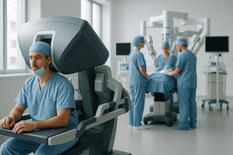
Robotic assisted thoracic surgery is chest surgery in which the surgeon operates through small incisions using robotic instruments, rather than by hand through a large opening. The robotic platform does not operate independently and makes no decisions of its own. It translates the surgeon’s hand movements into precise, scaled movements of slender, wristed instruments inside the chest, while filtering out natural hand tremor. The surgeon remains in full control from the first incision to the last stitch.
The approach belongs to the same family of techniques as video-assisted thoracoscopic surgery, known as VATS. The practical differences lie in the tools. Robotic systems provide three-dimensional depth perception on a high-definition monitor, a stable magnified view controlled directly by the operating surgeon, and instruments that bend and rotate beyond the range of the human wrist. This can make dissection steadier in narrow or delicate areas of the chest, such as the mediastinum and the lymph node stations around the airways. An assistant surgeon and the operating theatre team remain at your side throughout, handling instrument exchange, suction, stapling, specimen retrieval and safety monitoring.
What type of thoracic surgery is done by robotics?
Robotic platforms are used for both diagnostic and therapeutic chest operations. The procedures most commonly performed robotically are:
- Wedge resectionremoval of a small, wedge-shaped piece of lung tissue, often to diagnose or remove a nodule.
- Segmentectomyremoval of one anatomical segment of a lung lobe, preserving the rest of the lobe.
- Lobectomyremoval of an entire lobe of the lung, most often for lung cancer.
- Lymph node sampling or dissectionevaluation of the lymph node stations in the chest, essential for accurate cancer staging.
- Thymectomyremoval of the thymus gland for thymoma, thymic hyperplasia or thymic disease associated with myasthenia gravis.
- Mediastinal mass excisionremoval of cysts and tumours lying between the lungs, including bronchogenic cysts and neurogenic tumours.
- Pleural proceduressurgery for recurrent pneumothorax, pleural biopsy and removal of abnormal pleural tissue.
- Diaphragm and selected oesophageal proceduresperformed in appropriate centres and carefully chosen cases.
Whether your particular operation can be done robotically depends less on the name of the diagnosis than on its specifics: where the lesion sits, how large it is, whether it involves major blood vessels or the chest wall, whether you have had chest surgery before, and how well your heart and lungs would tolerate the procedure.
How Robotic Thoracic Surgery Differs from Open and VATS Approaches
Robotic thoracic surgery is one of three main ways of entering the chest, alongside open thoracotomy and video-assisted thoracoscopic surgery. An open thoracotomy uses a long incision between the ribs, usually with rib spreading, and gives the surgeon direct hand access; it remains the right choice for some large or invasive tumours, for certain complex reconstructions and for many emergencies. VATS uses small incisions and rigid instruments guided by a camera held at the bedside. The robotic approach also works through small incisions, but the surgeon operates from a console, directing wristed instruments and controlling the camera personally.
Inside the chest, the work is the same whichever access is used: separating tissue planes, sealing and dividing blood vessels, dividing airways, removing tumours or diseased lung, and sampling or clearing lymph nodes. What changes is the access route and the view. On the robotic console, the surgeon sees the operative field magnified in three dimensions, which helps when dissecting around the pulmonary vessels, the airways and the delicate structures of the mediastinum.
Not every patient is a candidate. Previous chest surgery with dense adhesions, tumours invading the chest wall or major vessels, advanced disease, severe cardiopulmonary risk or an emergency presentation may make an open operation safer or more appropriate. A capable thoracic team weighs two questions separately: can this operation be done robotically, and should it be. The second question matters more, because the medical objective of the operation must never be compromised for the sake of smaller incisions.
Who May Be Considered for Robotic Lung Surgery
You may be considered for robotic lung surgery after imaging, symptoms or biopsy results point to a chest condition that needs tissue diagnosis or surgical treatment. Many patients feel entirely well when the process begins: a lung nodule or mediastinal mass is discovered on a scan performed for an unrelated reason. Others come to a thoracic clinic because of symptoms that have not settled with time or standard treatment.
Symptoms that commonly lead to thoracic evaluation include a cough that does not resolve, coughing up blood, shortness of breath, recurrent chest infections, unexplained chest discomfort, hoarseness, difficulty swallowing, persistent fatigue and unintentional weight loss. Some thymic and mediastinal conditions announce themselves indirectly, through muscle weakness or an associated autoimmune condition. Pleural disease may cause fluid to collect around the lung, producing breathlessness that worsens gradually.
Diagnosis follows a stepwise path. It begins with a careful history and physical examination, followed by imaging. A chest X-ray may detect an abnormality, but detailed evaluation usually requires computed tomography of the chest. Depending on what the CT shows, your doctors may recommend PET imaging, magnetic resonance imaging, bronchoscopy, endobronchial ultrasound, pulmonary function testing, cardiac assessment, blood tests or an image-guided biopsy. When cancer is suspected, staging is decisive: whether the disease is localised, regionally advanced or metastatic determines whether surgery is the right first step at all.
Patients who may be candidates for robotic thoracic surgery include those with early-stage lung cancer suitable for surgical removal, suspicious lung nodules that require diagnosis or treatment, benign lung lesions that are growing or causing symptoms, selected mediastinal tumours, thymoma or thymic hyperplasia, pleural disorders, recurrent pneumothorax, and certain chest wall and diaphragm conditions. The decision is always individual. It weighs disease biology, imaging findings, surgical risk, lung reserve, age, general fitness and your own priorities.
Many international patients seek a second opinion before committing to an operation, and that is a reasonable instinct. A fresh review of your imaging, pathology reports, pulmonary function tests and previous treatment can confirm whether robotic surgery is feasible, whether further diagnostic steps are needed first, or whether another route, open surgery, radiotherapy, systemic therapy or active surveillance, would serve you better.
Conditions Treated with Robotic Thoracic Surgery
Robotic access is chosen when the location of the disease and the patient’s health allow the operation to be completed safely and completely through small incisions. The range of conditions is broad, but the logic behind each decision is the same: the disease sets the operation, and the operation sets the access.
Robotic surgery for lung cancer
Robotic surgery for lung cancer is usually considered when the disease appears localised and your heart and lung function can support removal of lung tissue. The surgical plan typically involves removing a lobe or an anatomical segment of the lung together with an evaluation of the lymph nodes in the chest. The extent of resection depends on tumour size and position, tissue diagnosis, staging results and how much breathing capacity you can afford to lose.
Precise dissection matters here for two reasons. First, the tumour must be removed completely, with the vessels and airway supplying it divided safely. Second, the lymph node stations must be assessed properly, because the pathology of those nodes shapes every treatment decision that follows surgery. Robotic instruments are well suited to this kind of careful work around vessels, airways and nodal stations. The same approach can also be used for metastatic lesions confined to the lung and for suspicious nodules, where diagnosis and treatment can sometimes be completed in a single operation.
What is a robotic lobectomy?
A robotic lobectomy is the removal of one of the five lobes of the lung, together with its supplying blood vessels, its airway branch and the nearby lymph nodes, performed through small incisions with robotic instruments. It is one of the standard operations for early-stage lung cancer and is also used for some benign conditions that have destroyed a lobe, such as chronic infection or bronchiectasis in selected cases.
Not every lung resection is a lobectomy. In carefully selected small cancers, or in patients with limited breathing reserve, a segmentectomy may remove the disease while preserving more functioning lung. A wedge resection removes a smaller, non-anatomical piece of tissue and is often used for diagnosis or for small benign and metastatic lesions. Your surgeon balances two principles when recommending the extent of resection: removing enough tissue to treat the disease properly, and preserving as much breathing capacity as the disease allows.
Mediastinal tumours and robotic thymectomy
The mediastinum: the space between the lungs that contains the heart, great vessels, thymus, oesophagus and major nerves, is a natural fit for robotic instruments, because it is narrow, deep and unforgiving of clumsy movement. Robotic techniques are used for selected thymomas, thymic cysts, bronchogenic cysts, neurogenic tumours and other masses in this region. For patients with myasthenia gravis associated with thymic disease or thymic enlargement, removal of the thymus may form part of the treatment plan when the neurology and thoracic surgery teams recommend it together. The wristed instruments allow the surgeon to separate the gland or tumour from the heart, the great vessels and the phrenic nerves with a controlled, magnified view.
Pleural, diaphragm and other chest conditions
Minimally invasive thoracic surgery is also used for diseases of the pleura, the membrane lining the lungs and chest wall. This includes surgery for recurrent pneumothorax, pleural biopsies, pleural cysts and, in selected cases, removal of abnormal pleural tissue. Patients with fluid repeatedly collecting around the lung may need a procedure both to improve breathing and to obtain tissue for diagnosis. Other indications include chest infections requiring drainage or decortication at the appropriate stage, certain diaphragm procedures, selected oesophageal operations and removal of benign chest tumours. In each case, the deciding factor is whether the surgeon can achieve the full medical objective of the operation through small incisions without compromise.
Where robotic cardiothoracic surgery fits
Robotic cardiothoracic surgery is the umbrella term for robot-assisted operations across the whole chest, and it covers two distinct fields. One is general thoracic surgery: the lung, mediastinal, thymic and pleural procedures described on this page. The other is cardiac surgery, where robotic techniques are used in specialised centres for selected heart operations. The distinction matters when you read about the subject or compare hospitals, because the teams, training and case selection differ. You can read more about the broader field of cardiothoracic surgery and about robotic cardiac surgery on their dedicated pages.
How Robotic Thoracic Surgery Is Performed
Preparation and evaluation before surgery
Preparation begins with confirming the diagnosis and understanding your overall condition. The thoracic team reviews your imaging, pathology if available, medical history, medications, allergies, smoking history, previous operations and current symptoms. When cancer is suspected or confirmed, cases are commonly discussed in a multidisciplinary tumour board that may bring together thoracic surgeons, pulmonologists, medical oncologists, radiation oncologists, radiologists, nuclear medicine physicians, pathologists, anaesthesiologists and intensive care specialists. The purpose is simple: to make sure surgery is being recommended in the right clinical context, and that alternatives have been considered honestly.
Pulmonary function testing estimates how well your lungs will tolerate the operation, which matters most when part of a lung must be removed. Cardiac evaluation may be recommended if you have heart disease, diabetes, reduced exercise tolerance, advanced age or abnormal findings. Blood tests, electrocardiography and an anaesthesia assessment identify risks that should be addressed before the procedure rather than discovered during it.
You will usually be advised to stop smoking before surgery, because smoking raises the risk of pneumonia, poor wound healing, prolonged air leaks and breathing complications afterwards. The team also reviews every medicine you take, including blood thinners; decisions about pausing or adjusting any of them belong to your treating doctors, and you will receive specific instructions for your situation rather than general rules. Before admission you are told about fasting, arrival time, the expected hospital stay and breathing exercises worth practising in advance. International patients also receive guidance on travel timing, medical documentation, interpretation support and how follow-up will be organised after discharge.
What happens during lung cancer robotic surgery?
Lung cancer robotic surgery follows a well-rehearsed sequence, and most other robotic chest operations follow the same broad pattern:
- 1. General anaesthesia and lung isolation. A specialised breathing tube lets the anaesthesia team ventilate one lung while the surgeon operates on the other side of the chest, creating the working space needed for safe visualisation.
- 2. Positioning. You are positioned carefully to protect nerves, joints and pressure points for the duration of the operation.
- 3. Port placement. The surgeon makes several small incisions between the ribs and introduces the camera and robotic instruments. Carbon dioxide may be used in selected cases to enlarge the working space.
- 4. The operation itself. Working from the console, the surgeon dissects the target tissue, a wedge, a segment, a lobe, the thymus or a mediastinal mass, sealing and dividing vessels and airways with energy devices and surgical staplers, and sampling or removing lymph nodes where the diagnosis requires it.
- 5. Specimen retrieval. The removed tissue is placed in a protective retrieval bag and taken out through one of the small incisions, which may be slightly enlarged if needed. This protects the incision from contact with tumour tissue.
- 6. Chest drain and closure. A chest tube is usually placed to drain air and fluid and help the lung re-expand, and the incisions are closed.
- 7. Recovery transfer. You are moved to the recovery area or the intensive care unit, depending on the procedure and your medical condition.
For thymic and mediastinal operations, the central work is the careful separation of the lesion from the heart, great vessels, nerves and lungs. For pleural procedures, abnormal tissue can be biopsied or removed, and additional techniques may be used to reduce the chance of a pneumothorax or fluid collection returning.
Technology used during the operation
Several categories of technology work together in a robotic thoracic operation. High-definition three-dimensional imaging gives the surgeon depth perception and magnification of fine structures. Wristed robotic instruments provide controlled movement in narrow spaces, most valuable around the pulmonary vessels, the airways, the lymph node stations and the mediastinum. Energy devices seal tissue, surgical stapling systems divide vessels and airway branches, and lung isolation techniques keep the operative field still and clear.
Around the operation itself sit further tools. Small or deep lung nodules can be difficult to find once the lung is deflated, so preoperative image-guided marking or bronchoscopic localisation may be used to pinpoint them. In selected cases, pathologists examine tissue during the operation to help guide the extent of resection before the chest is closed. Modern anaesthesia monitoring runs continuously throughout.
The honest caveat is that technology only delivers what the team around it can deliver. A robotic system extends a surgeon’s capability; it does not replace accurate diagnosis, disciplined technique, experienced anaesthesia care or the judgement to convert to an open operation when that is the safest course. Acibadem’s robotic surgery programme is built on that principle: the platform serves the operation, not the other way round.
How long does the operation take, and how long is the hospital stay?
Operating time varies with the procedure, the location of the disease, previous surgery, your anatomy and whether lymph node assessment or additional steps are needed. A diagnostic biopsy or wedge resection is generally shorter; a lobectomy, segmentectomy, thymectomy or complex mediastinal operation takes longer. Treat any estimate you are given as a guide, not a commitment: the team takes the time the operation needs.
The hospital stay afterwards depends on pain control, breathing, chest tube drainage, lung expansion, mobility and your overall health. Many patients having minimally invasive thoracic surgery leave hospital earlier than they would after an open operation, but discharge happens only when the team confirms it is medically safe. Some patients need longer observation, particularly those with underlying lung disease, a persistent air leak or other conditions that deserve extra caution.
Recovery After Robotic Thoracic Surgery
Recovery after robotic lung surgery begins within hours of the operation. Nurses and respiratory therapists help you cough, breathe deeply, use a breathing exercise device, sit up and walk as early as is medically appropriate. Early movement is not a formality: it reduces the risk of blood clots, pneumonia and muscle deconditioning, and it is one of the strongest things you can contribute to your own recovery. Pain is managed with a tailored plan that may combine oral medication, regional anaesthesia techniques and non-opioid options where suitable.
The chest tube is removed once air leak and fluid drainage have settled and imaging confirms the lung has expanded properly. Before you leave hospital, the team walks through wound care, showering, activity limits, breathing exercises and which changes in how you feel should be reported to your treating doctor rather than watched at home.
Most patients gradually resume light daily activities within days to a few weeks, depending on the procedure and their baseline health. Strenuous exercise, heavy lifting, long flights and the return to work should each be discussed with your surgeon, because the right timing differs from patient to patient. For cancer patients, the final pathology report guides whether additional treatment, chemotherapy, immunotherapy, targeted therapy or radiotherapy, coordinated through oncologyis recommended after surgery. Follow-up may include clinic visits, imaging surveillance, pulmonary rehabilitation and, for international patients, coordination with your doctors at home.
| Time period | What you can generally expect |
|---|---|
| Day 1 | Close monitoring after anaesthesia. Breathing exercises, pain control, sitting up and early walking begin as soon as medically appropriate. A chest tube is usually in place. |
| First week | Walking a little further each day, continuing breathing exercises, caring for the incisions and moving to oral pain medication. Many patients are discharged during this period once recovery is stable and the chest tube is out. |
| First month | Energy gradually improves and light daily activities become easier, while heavy lifting and strenuous exercise usually remain restricted. Pathology results and any additional treatment plan are reviewed with you. |
| Longer term | Stamina and breathing capacity continue to rebuild. Cancer patients enter a surveillance programme or begin further therapy if recommended. Follow-up imaging and specialist visits are scheduled according to the diagnosis. |
Benefits of Robotic Thoracic Surgery
For selected patients, robotic lung surgery offers practical advantages over traditional open chest surgery. Two caveats keep these honest. The benefits are comparative, not absolute, every chest operation involves recovery, discomfort and risk. And they depend on selection: a well-chosen open operation serves you better than a poorly chosen robotic one.
| Benefit | What it means for you |
|---|---|
| Smaller incisions | The operation is performed through small access points, so there is typically less tissue disruption and a more discreet scar pattern than after a large thoracotomy incision. |
| Less postoperative discomfort for many patients | Avoiding rib spreading and reducing muscle trauma can make breathing exercises, coughing and walking easier in the days after surgery. |
| Enhanced visualisation and precision | Magnified three-dimensional imaging and wristed instruments help the surgeon work carefully around blood vessels, airways, lymph nodes and delicate mediastinal structures. |
| Shorter hospital stay in appropriate cases | Many patients spend less time in hospital than is typical after open surgery, although the actual discharge date always depends on safety, chest tube status and overall recovery. |
| Earlier mobility and breathing recovery | Reduced surgical trauma can make it easier to walk, cough, breathe deeply and take part in respiratory recovery exercises soon after the operation. |
| A strong role in multidisciplinary cancer care | When used for lung cancer or other thoracic tumours, robotic surgery integrates with staging, pathology review, systemic therapy, radiotherapy and structured follow-up. |
Why Acting Early Matters
Early evaluation matters because many thoracic conditions are more treatable before they progress. A small lung cancer may be suitable for complete surgical removal, while a more advanced tumour usually requires a longer and more complex treatment strategy. A growing mediastinal mass can eventually press on nearby structures, and recurrent pleural problems can lead to repeated infections, scarring or reduced lung expansion.
Delay also makes diagnosis less straightforward. A chronic cough, breathlessness or chest discomfort is easily attributed to infection, smoking history, asthma, reflux or age, and sometimes rightly so. But persistent or unexplained symptoms deserve proper imaging and specialist review. When a suspicious nodule is found, the timing of follow-up scans or biopsy should be guided by the nodule’s imaging characteristics, your risk factors and international recommendations, not by anxiety and not by avoidance.
For patients who already have a diagnosis, delay can narrow the options. Tumours grow, lymph nodes can become involved, lung function can decline, and the window for a less invasive operation can close. In benign disease, repeated inflammation or infection creates scar tissue that makes eventual surgery technically harder and recovery more demanding.
Acting early does not mean rushing into an operation. It means obtaining a timely, accurate diagnosis and a clear plan from an experienced team. Sometimes careful surveillance is exactly the right answer. Sometimes surgery should proceed without unnecessary delay. The point is to make that decision with complete information and proper specialist input, rather than by default.
What Shapes the Outcome of Robotic Thoracic Surgery
Outcomes depend on a combination of disease-related, patient-related and treatment-related factors, and it helps to understand them before surgery rather than after. The first is accurate diagnosis. A suspicious lung nodule has to be judged in the context of its imaging features, growth pattern, your smoking history, any prior cancer and your overall risk. When cancer is suspected or confirmed, staging determines whether surgery alone is appropriate or whether chemotherapy, immunotherapy, targeted therapy or radiotherapy should be part of the plan.
The second is the extent of surgery. Removing too little tissue can fail to treat the disease adequately; removing more lung than necessary costs breathing capacity you may want later. Thoracic surgeons balance oncological principles against preservation of lung function: in selected early lung cancers a segmentectomy may be considered, while in other cases a lobectomy remains the more appropriate operation. Thorough lymph node evaluation matters in either case, because it underpins accurate staging and every treatment decision that follows.
The third is your own health. Lung function, heart disease, diabetes, kidney function, nutrition, smoking status, weight, physical conditioning and frailty all influence surgical risk. Patients with chronic obstructive pulmonary disease or reduced respiratory reserve may still be candidates, but they need careful assessment and preparation. Stopping smoking, pulmonary rehabilitation and better nutrition before surgery are all within your reach and all support a smoother recovery.
The fourth is the team. Robotic surgery involves a genuine learning curve and depends on coordinated teamwork, not just the surgeon at the console, but anaesthesia, nursing, respiratory therapy, intensive care, pathology, imaging and postoperative monitoring. A hospital that manages complex thoracic patients routinely is better placed to anticipate and handle issues such as air leaks, heart rhythm disturbances, pneumonia, bleeding or difficult pain control.
The fifth is follow-up. Surgery is not complete when the incision closes. You need a clear interpretation of the pathology, wound review, imaging surveillance, guidance on medication and activity, and a plan for travel and the return to normal life. For international patients, coordination with doctors at home keeps the care continuous after you fly back.
When is robotic lung cancer surgery the right choice?
Robotic lung cancer surgery is the right choice when three things line up: the disease is at a stage where surgery is the recommended treatment, the tumour’s size and position allow complete removal through small incisions, and your heart and lungs can carry the operation and the recovery. When any of the three does not hold, a different plan serves you better: an open operation, systemic therapy or radiotherapy first, or in some low-risk nodules, structured surveillance. A team that recommends against robotic surgery in your case is applying exactly the judgement you want from it.
Robotic Thoracic Surgery at Acibadem
International patients considering robotic thoracic surgery usually need more than an operation. They need a reliable diagnostic pathway, specialists who can interpret complex findings, and a care structure that understands the practical realities of travelling for treatment. At Acibadem, patients with suspected or confirmed thoracic disease are evaluated through a structured process: imaging, pathology, pulmonary function results, previous records and treatment history are reviewed before any recommendation is made.
When cancer or complex disease is involved, cases are commonly discussed in multidisciplinary tumour boards or specialist meetings. This keeps the surgical recommendation in its proper clinical context and ensures that alternatives, surveillance, systemic therapy, radiotherapy, interventional pulmonology or open surgery, are weighed openly. The thoracic surgeons of the Thoracic Surgery Department use minimally invasive approaches, including robotic surgery, for selected chest and lung procedures, and the choice of approach is individual in every case: it depends on the diagnosis, the location of the lesion, your anatomy, your lung function, previous treatment and your overall health.
The diagnostic and surgical infrastructure supports that decision-making. High-resolution imaging defines the size and position of chest lesions and their relationship to nearby structures. PET imaging, bronchoscopy, endobronchial ultrasound, image-guided biopsy, pulmonary function testing and advanced pathology techniques are used when clinically indicated. In the operating theatre, three-dimensional visualisation, robotic instrumentation and refined anaesthesia monitoring support precise work through small incisions, with the team prepared to modify the plan whenever safety requires it.
For patients travelling from abroad, communication and coordination sit at the centre of the experience. Acibadem International provides support in more than 20 languages, helping patients prepare medical records, understand treatment plans, coordinate hospital appointments and plan the practical side of travel. For thoracic surgery this matters more than for many treatments, because the pathway typically includes preoperative testing, an inpatient stay, pathology review and clear guidance on when flying home is medically sensible.
Personalised planning is especially important in chest disease because two patients with similar scans can need very different care. One small lung nodule warrants surveillance; another needs biopsy, wedge resection, segmentectomy or lobectomy depending on its features. A mediastinal mass may need removal, tissue diagnosis first, or treatment by another specialty entirely. A lung cancer patient may need surgery alone or a combination of therapies. Because thoracic care draws on pulmonology, oncology, radiology, pathology, nuclear medicine, anaesthesia, intensive care, pain management and physiotherapy, having these disciplines working within one system produces clearer recommendations and more coordinated care before, during and after the operation.
Making a Considered Decision
Robotic thoracic surgery is a valuable option for selected patients who need chest or lung surgery and want to understand whether a minimally invasive approach fits their situation. It can offer smaller incisions, precise dissection and a recovery pathway that helps many patients return to normal life sooner than after open surgery. But the best operation is always the one that fits the diagnosis, the stage of disease, the anatomy, the lung function and the overall health of the person having it, and sometimes that operation is not robotic.
Whatever the diagnosis: a lung nodule, lung cancer, a mediastinal mass, thymic disease, recurrent pneumothorax or a pleural disorder: a sound decision rests on an accurate, unhurried evaluation of imaging and test results by an experienced thoracic team. Where recommendations differ, or where it is unclear whether surgery is necessary at all, a second specialist opinion is a legitimate and common part of making the decision well. Good thoracic care starts with a correct diagnosis and a plan you fully understand, everything else follows from that.
Preparation
- Evaluation usually includes chest imaging, pulmonary function tests, blood tests, anesthesia assessment, and review of current medications. Patients may be asked to stop smoking, adjust blood thinners, and fast before surgery. The surgical team explains the planned approach, risks, and expected recovery steps.
Aftercare
- After surgery, patients are monitored for breathing, pain control, and chest tube drainage if used. Early walking, breathing exercises, and wound care help reduce complications and support recovery. Follow-up visits review pathology results when relevant and guide return to normal activities.
Frequently Asked Questions
What affects the cost of robotic thoracic surgery?
The final cost depends on the exact procedure, complexity of the case, robotic system use, surgeon and hospital fees, anaesthesia, imaging, pathology, hospital stay, intensive care needs and follow-up plan. Travel, translation and accommodation support may also affect the overall budget.
How can I get a personalised quote?
A personalised quote usually requires medical records, recent imaging, biopsy or pathology results if available, lung function tests and a summary of previous treatments. Acibadem can review your documents through a free consultation and provide guidance on the likely treatment plan and package scope.
What is usually included in an international patient package?
Package content varies, but it may include specialist consultation, standard preoperative tests, surgery, anaesthesia, hospital stay, routine medications, nursing care and translation support. Items such as extra diagnostics, extended stay, intensive care, advanced pathology or additional treatments may be quoted separately.
Is robotic thoracic surgery always more suitable than other approaches?
No. Robotic surgery is suitable only for selected patients and conditions. A thoracic surgeon decides the safest approach after assessing the diagnosis, tumour or lesion location, lung function, previous surgery, general health and expected treatment benefit.
Can international patients plan surgery without long waiting times?
Scheduling depends on medical urgency, the need for additional tests, surgeon availability and operating room planning. International patient teams can help coordinate record review, appointments, translation and travel logistics once the case is assessed.
Is this information medical or financial advice?
No. This is general educational information and does not replace consultation with a qualified specialist or a formal written quote. For an individual assessment, a free consultation is recommended.
Medically reviewed by the Acıbadem International Medical Board September 1, 2026
See our medical review board →
Update history
- PublishedJune 8, 2026
- Medical review approvedSeptember 1, 2026
- Last content updateSeptember 1, 2026
Trusted care for international patients
From the Acibadem Blog

What Does Faster Recovery Mean After Robotic Thoracic Surgery? Drains, Pain Control and Walking
After robotic thoracic surgery, faster recovery usually means smaller incisions and no spread ribs, so many people have less early pain, a chest drain…