

Living With Cardiac Sarcoidosis: Repeat Imaging, Rhythm Monitoring and Daily Routines

Key Takeaways
- Clinically obvious heart involvement affects roughly 5% of people with sarcoidosis, but autopsy studies find it in about a quarter, which is why scans often detect disease before symptoms.
- FDG-PET shows active inflammation while cardiac MRI shows scar, so a quiet PET does not mean earlier scar has disappeared or that rhythm risk has gone.
- The Heart Rhythm Society consensus recommends an ICD after cardiac arrest, sustained ventricular arrhythmia, or an ejection fraction of 35% or lower despite treatment.
- Sarcoidosis can raise blood calcium, so vitamin D and calcium supplements should not be started without checking with your team, even when taking corticosteroids.
- Unexplained high-grade heart block in someone under about 60 is a recognized trigger for screening for cardiac sarcoidosis.
- Sarcoidosis is not directly inherited, but relatives carry a modestly increased risk through shared genetic susceptibility plus an environmental trigger.
Living with cardiac sarcoidosis usually means a long partnership with a heart team: repeat scans such as cardiac MRI and FDG-PET to check whether inflammation is quieting, regular rhythm monitoring because the disease can disturb the heart's electrical wiring, medicines that calm the immune response, and steady daily routines around activity, sleep and appointments. Many people live full lives; the plan is individual and set by the treating team.
The clinic letter arrives with a line that reads almost like a school timetable: PET scan in the spring, device check in the summer, echo before the winter. For someone newly told they have cardiac sarcoidosis, that calendar can feel like the disease itself, a series of waiting rooms stretching out for years.
Then the ordinary questions surface. Can I still coach my daughter’s soccer team? Why do I need another scan when the last one was fine? Is this tiredness the illness, the medicine or just life? Living with cardiac sarcoidosis is less about a single dramatic event than about learning a rhythm of surveillance and adjusting to it without letting it run everything.
This explainer walks through what those scans and monitors are actually looking for, why the schedule tends to be lighter than people fear once inflammation settles, and which daily habits have real evidence behind them. It is written to inform, not to replace the plan your own cardiology team builds with you.
What is actually happening inside the heart in cardiac sarcoidosis?
Sarcoidosis is a condition in which the immune system forms tiny clusters of inflammatory cells, called granulomas, in body tissues for reasons that remain only partly understood. In most people those clusters settle in the lungs or lymph nodes. When they form in heart muscle, the condition is called cardiac sarcoidosis.
Heart muscle is a poor place for clutter. The heart depends on a precise electrical circuit that starts in the upper chambers, passes through a narrow junction and fans out through the lower chambers. Granulomas, and the scar tissue that can replace them, may sit directly on that circuit. The result is one of three broad problems. The signal can be blocked at the junction, a problem called heart block, producing a slow or irregular pulse. Scarred muscle can generate its own fast, chaotic rhythms in the lower chambers, called ventricular arrhythmias. And when enough muscle is inflamed or scarred, the heart pumps less forcefully, which is heart failure.
Those three problems explain nearly every part of the monitoring plan. Imaging looks for active inflammation and for scar. Rhythm monitoring looks for block and for fast rhythms. Blood tests and symptom reviews track pumping strength and the side effects of treatment.
The Heart Rhythm Society’s expert consensus statement estimates that clinically obvious heart involvement affects roughly 5% of people with sarcoidosis, while studies of hearts examined after death have found involvement in around a quarter, which is why so many cases are picked up on scans before symptoms appear (see the PubMed reference). Understanding this gap helps explain why teams sometimes treat findings that never caused a symptom.
What does living with cardiac sarcoidosis look like day to day?
For most people, living with cardiac sarcoidosis quickly becomes less about the heart and more about the calendar and the medicine cabinet. Appointments cluster in the first year, when the team is working out how active the inflammation is and how the heart is responding. After that, if things are stable, visits usually thin out.
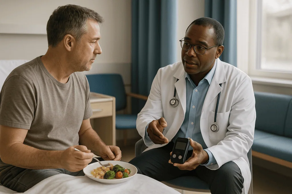
A typical week may contain nothing medical at all. A typical month might include a blood test to check for medicine side effects, or a remote transmission from an implanted device that happens overnight without any effort. A typical year usually includes at least one imaging study and an in-person device or rhythm review. The frequency is set by the treating team and shifts as the picture changes.
The daily reality that people describe most often is fatigue, which the NHS and Mayo Clinic both list as a common feature of sarcoidosis in general. Fatigue can come from the inflammation itself, from a slow or irregular heart rhythm, from reduced pumping strength, from the medicines, or from disrupted sleep. Untangling those threads is genuinely difficult and worth raising rather than accepting as inevitable.
Emotionally, many people describe a phase of hypervigilance, checking their pulse repeatedly or interpreting every flutter as danger. That tends to ease as the surveillance plan proves itself and as normal palpitations, which everyone has, are recognized for what they are. Where anxiety persists, cardiology teams often have access to psychological support, and it is reasonable to ask.
The overarching message from mainstream sources is consistent: sarcoidosis is a manageable long-term condition for most people, and the aim of monitoring is to catch problems early rather than to wait for symptoms.
Who is monitored closely, and who is usually asked to wait?
Not everyone with sarcoidosis needs a cardiac workup, and not everyone with cardiac involvement needs the most intensive surveillance. The Heart Rhythm Society consensus describes several groups where closer attention is usually recommended.
People with sarcoidosis elsewhere in the body who develop symptoms suggesting heart involvement, such as unexplained fainting, palpitations or breathlessness out of proportion to their lung disease, are typically referred for cardiac imaging. People under about 60 who develop unexplained high-grade heart block are another group; the consensus recommends screening them for cardiac sarcoidosis because it is a recognized cause of conduction problems in younger adults. Anyone with a ventricular arrhythmia of uncertain cause, or with confirmed cardiac sarcoidosis on biopsy or imaging, sits firmly in the close-monitoring group.
Who is usually asked to wait? People with sarcoidosis confined to the lungs or skin, a normal electrocardiogram (a quick tracing of the heart’s electrical activity) and no cardiac symptoms are often followed with periodic clinical checks rather than repeated advanced imaging. The rationale is practical: scans carry radiation or contrast exposure, cost time and anxiety, and are most useful when there is a reasonable chance of finding something that changes management.
Waiting is not the same as being ignored. Teams generally ask this group to report new symptoms promptly and may repeat a baseline electrocardiogram at intervals. If a scan done for another reason shows an unexpected finding, the person usually moves into the closer-monitoring pathway.
The decision about which group you belong in, and when you might move between them, rests with your treating team and depends on the full picture rather than any single test.
Which scans are used, and how often is cardiac sarcoidosis imaging repeated?
Three imaging tools do most of the work, and each answers a different question.

An echocardiogram is an ultrasound of the heart. It measures how strongly the heart pumps, usually expressed as the ejection fraction, the percentage of blood the main pumping chamber pushes out with each beat. It is quick, radiation-free and easy to repeat.
Cardiac MRI uses magnetic fields to picture the heart in fine detail. With a contrast agent, it shows scar as bright patches, a finding called late gadolinium enhancement. Scar tells the team where past damage sits and helps estimate arrhythmia risk.
FDG-PET uses a radioactive sugar tracer that inflamed cells absorb. It shows active inflammation rather than old scar, which makes it the tool most often used to judge whether immune-calming treatment is working. Before the scan, people are usually asked to follow a low-carbohydrate preparation so that normal heart muscle does not absorb the tracer and blur the picture.
| Test | What it shows | Typical role in follow-up |
|---|---|---|
| Echocardiogram | Pumping strength, chamber size, valve function | Routine repeat check, often yearly or with symptom change |
| Cardiac MRI | Scar and tissue swelling | Baseline, then repeated when the team needs to reassess scar burden |
| FDG-PET | Active inflammation | Diagnosis, then repeated after a period of treatment to gauge response |
How often these repeat is individualized. The Heart Rhythm Society consensus describes repeating PET after a course of immunosuppression to assess response, and many teams do this after several months of treatment, then again if the plan changes. Echocardiograms tend to be the most frequent because they are simple. MRI is repeated less often because scar changes slowly. The exact interval is set by your team based on how active the disease has been and how well you are.
Rhythm monitoring: from wearable monitors to implanted devices
Because the electrical system is where cardiac sarcoidosis does much of its damage, rhythm monitoring runs alongside imaging throughout follow-up.
The simplest tool is the electrocardiogram, a tracing taken in a few minutes in clinic. It catches persistent problems such as heart block but can miss rhythms that come and go. For those, teams use ambulatory monitors: a small recorder worn for 24 hours to two weeks, or a patch monitor that sticks to the chest. If symptoms are rare, a tiny implantable loop recorder can be placed under the skin to record for years.
Two implanted devices treat rather than record. A pacemaker delivers electrical impulses when the heart’s own signal is blocked or too slow. An implantable cardioverter-defibrillator, or ICD, does the same and also detects dangerous fast rhythms and delivers a corrective shock. The American Heart Association explains that an ICD is offered when the risk of a life-threatening rhythm is judged high enough to justify the device and its drawbacks.
In cardiac sarcoidosis, the Heart Rhythm Society consensus recommends an ICD for people who have survived a cardiac arrest, had sustained ventricular arrhythmia, or whose ejection fraction is 35% or lower despite treatment. It also describes situations where an ICD may be reasonable, such as when a pacemaker is needed anyway or when imaging shows extensive scar. Because heart block in sarcoidosis can improve with treatment in some people, the decision often involves weighing device benefits against the chance of recovery.
Once a device is in, follow-up usually combines in-clinic checks with remote monitoring, where the device sends data automatically. Batteries typically last years and are replaced in a shorter procedure.
How do medicines for cardiac sarcoidosis work?
Treatment aims at two separate targets: the inflammation and the electrical or pumping consequences. Nothing here is a recommendation; every decision belongs to the prescribing team.
Corticosteroids are the longest-established treatment for active cardiac sarcoidosis. They dampen the immune response broadly and can reduce granuloma activity, which is why FDG-PET often looks quieter after a course. The Heart Rhythm Society consensus notes that treatment is typically continued for months and often longer, with gradual reduction guided by imaging and symptoms. Long courses bring well-known side effects, including weight gain, raised blood sugar, mood change and bone thinning, which is why teams monitor blood tests and often arrange bone protection.
Steroid-sparing agents, such as methotrexate, are immune-modulating medicines sometimes added so the corticosteroid can be reduced. They act more slowly and need their own blood monitoring. Biologic medicines that block a signaling protein called tumor necrosis factor are used in some resistant cases; the evidence base is smaller and mostly observational, and they carry infection risks that require screening beforehand.
The second group of medicines addresses the heart itself. Standard heart failure medicines, which relax blood vessels or slow the heart, are used when pumping strength is reduced. Antiarrhythmic medicines may be used to suppress fast rhythms, often alongside an ICD rather than instead of one. In selected people with recurrent ventricular arrhythmia, catheter ablation, a procedure that targets the tissue generating the rhythm, may be considered.
What the evidence does not yet settle is the optimal duration of immunosuppression or which combination works best. Trials are ongoing, and honest teams will say the plan is partly individual judgment.
What do the weeks and months after diagnosis usually look like?
The first few weeks are usually the busiest. Baseline imaging is completed if it has not been already, blood tests establish a starting point for kidney function, blood sugar and liver tests, and any urgent rhythm problem is dealt with, sometimes with a device before immune treatment begins. If corticosteroids are started, people often notice their effects within days, both welcome and unwelcome: more energy in some, restless sleep and appetite change in others.
Around the second to fourth month, the pattern shifts to checking. Blood tests continue, and if the team is planning a repeat FDG-PET to gauge response, it often falls in this window or a little later, consistent with the Heart Rhythm Society consensus describing reassessment after a course of treatment. A quieter scan usually supports gradual medicine reduction; a scan that still glows may prompt a change of approach.
By six to twelve months, many people describe settling into a routine. Appointments become less frequent, device follow-up runs largely by remote transmission, and the questions turn from “what is happening” to “how do I keep this stable.” An echocardiogram at around a year is common practice to track pumping strength.
Beyond the first year, follow-up depends on how things have gone. Sarcoidosis can flare after a quiet period, so most teams continue periodic reviews indefinitely, with imaging repeated when symptoms change or medicines are altered.
None of these timeframes is a promise. They describe a typical arc drawn from guideline documents and mainstream patient information; your team may move faster or slower for good reasons.
Daily routines: exercise, sleep, salt and alcohol
People often expect a long list of forbidden activities. The evidence supports a shorter, more specific one.
Exercise is generally encouraged for people with sarcoidosis, and the American Heart Association recommends regular moderate activity for most people with heart conditions. In cardiac sarcoidosis the main caveat is arrhythmia risk. Teams typically ask people with recent ventricular arrhythmia, an unsettled rhythm or a new device to avoid intense or competitive exertion until stability is confirmed, then build back gradually. Brisk walking, cycling on flat ground and swimming are the kinds of activities usually discussed first. A cardiac rehabilitation program, where available, offers supervised exertion with monitoring.
Sleep matters more than it might seem. Poor sleep worsens fatigue, and corticosteroids taken later in the day are a common cause of insomnia; timing is worth discussing with the prescriber rather than adjusting alone. Sleep apnea, where breathing pauses during sleep, is common in people with heart disease and can aggravate arrhythmias, so loud snoring or unrefreshing sleep deserves a mention at clinic.
Salt becomes relevant when pumping strength is reduced. Heart failure guidance from the American Heart Association favors limiting sodium and watching for fluid buildup, such as rapid weight gain over a few days or swollen ankles.
Alcohol can trigger arrhythmias and interacts with several medicines used in sarcoidosis, particularly those that affect the liver. Moderation, or abstaining if advised, is a reasonable baseline.
Vaccination is a quieter but important routine: people on immune-suppressing medicines are more vulnerable to infections, and the CDC advises staying current with recommended vaccines, noting that some live vaccines are avoided during immunosuppression. Your team can confirm which apply to you.
How to calm sarcoidosis flares: what the evidence supports
“How to calm sarcoidosis” is one of the most common searches around this condition, and the honest answer has two halves: what medicine does, and what the person can do.
The medical half is clear. Active inflammation in the heart is treated with immune-suppressing medicines, and the evidence that these reduce inflammation on imaging is reasonably consistent, even though large randomized trials are lacking. There is no reliable evidence that any supplement, diet or herbal product reduces cardiac granulomas. The NIH Office of Dietary Supplements has no listed supplement with proven benefit for sarcoidosis, and some products interact with immune-modulating medicines or affect the heart rhythm.
One caution deserves emphasis. Sarcoidosis can disturb the body’s handling of vitamin D and calcium, sometimes raising blood calcium. Mayo Clinic and the NHS both note this, and it is why people with sarcoidosis are usually advised not to take vitamin D or calcium supplements without checking with their team, even though these supplements are widely marketed for bone health.
The personal half is about reducing the load that makes flares harder to live with. Regular sleep, steady activity, treating infections promptly and not smoking all have general cardiovascular evidence behind them, even if none has been tested specifically as a way to prevent sarcoidosis flares. Stress management is often mentioned; the evidence that stress causes flares is weak, but the evidence that stress worsens how symptoms feel is solid, so techniques that help you cope are worth pursuing for that reason.
If you notice a return of breathlessness, palpitations or dizziness after a stable period, the right response is to contact your team rather than to try to calm it at home.
Is cardiac sarcoidosis hereditary?
Many people ask whether they have passed something to their children. The short answer, from mainstream sources, is that sarcoidosis is not a straightforwardly inherited disease, but genetics play a part.
The NHS describes sarcoidosis as not passed directly from parent to child, while acknowledging that it appears to run in some families slightly more than chance would predict. Mayo Clinic frames the likely cause as a combination of genetic susceptibility and an environmental trigger, such as an infection or an inhaled substance, that sets off the immune reaction in someone predisposed to it. Several genes involved in immune regulation have been associated with sarcoidosis in research studies, but none acts like a single inherited switch.
What does that mean in practice? Relatives of someone with sarcoidosis carry a modestly increased risk, not a high one. There is no routine genetic test for sarcoidosis, and mainstream guidance does not recommend screening healthy family members with heart scans. What relatives can reasonably do is be aware of the condition, so that unexplained symptoms such as persistent cough, fainting or an unusually slow pulse prompt an earlier conversation with a doctor.
Cardiac involvement specifically does not seem to follow a separate hereditary pattern beyond the overall susceptibility to sarcoidosis. Research groups continue to look for genetic markers that predict which organs the disease will affect, but nothing has yet reached routine clinical use.
If you have a strong family history, or several relatives with autoimmune or inflammatory conditions, mention it to your team. It may not change your treatment, but it helps build a fuller picture and may be relevant to research they are involved in.
Cardiac sarcoidosis life expectancy: what the evidence actually shows
This is the question people most want answered and the one that responsible sources answer most carefully.
For sarcoidosis overall, the picture is reassuring. The NHS notes that many people with sarcoidosis improve without treatment, and Mayo Clinic describes a condition that most people live with for a normal lifespan. Cardiac involvement is different because the heart’s electrical and pumping problems can be serious, and mainstream sources including Cleveland Clinic identify heart complications as one of the more important causes of sarcoidosis-related death.
What the evidence does not support is a single life expectancy figure. Published outcome studies vary widely in how patients were selected, how the diagnosis was made and what treatments were used, so their numbers cannot be transferred to an individual. Guideline documents such as the Heart Rhythm Society consensus deliberately avoid quoting survival percentages for this reason.
What the same evidence does show is which factors matter most. A reduced ejection fraction, sustained ventricular arrhythmias and extensive scar on MRI are consistently associated with worse outcomes. Preserved pumping strength, absence of dangerous rhythms and inflammation that quiets with treatment are associated with better ones. Much of the modern surveillance plan exists precisely to identify and manage the first group of features early, and ICDs are offered because they address the most sudden of those risks.
An honest way to think about it: cardiac sarcoidosis is a condition where the individual details, rather than the label, drive prognosis. Your team, who can see your scans and rhythm data, is the only reliable source for how those details apply to you, and even they will speak in likelihoods rather than certainties.
What people often get wrong about living with cardiac sarcoidosis
Some misunderstandings come up so often that correcting them is worth a section of their own.
“A normal PET scan means it’s gone.” A quiet scan means inflammation is not active at that moment. Sarcoidosis can flare later, and scar from earlier inflammation remains and can still cause rhythm problems. This is why device follow-up and periodic reviews continue even when scans look good.
“If I feel fine, I can skip the monitoring.” The Heart Rhythm Society consensus exists partly because dangerous rhythms in cardiac sarcoidosis can occur without warning symptoms. Remote device checks and scheduled scans catch what you cannot feel.
“Steroids are the whole treatment.” Immune-calming medicines address inflammation; pacemakers, ICDs, heart failure medicines and sometimes ablation address the consequences. Both arms matter, and one does not replace the other.
“I should take vitamin D for my bones because of the steroids.” Sometimes appropriate, sometimes harmful. Sarcoidosis can raise blood calcium, so supplement decisions need blood tests and a prescriber’s input, as Mayo Clinic and the NHS both caution.
“Exercise is dangerous now.” Unrestricted intense exertion may be discouraged during unstable phases, but regular moderate activity is generally recommended and supports heart health. The details depend on your rhythm status and device.
“There is exciting news of a breakthrough.” Research is genuinely active, including trials comparing immune-modulating regimens and studies of imaging-guided treatment. None has yet produced a treatment that eliminates the disease, and any source promising that should be treated with caution.
Getting these right removes a surprising amount of daily anxiety, because it replaces guesswork with a clear understanding of what each part of the plan is for.
Questions to ask your care team
A good consultation is a two-way exchange, and people who arrive with written questions usually leave with clearer answers. The following are drawn from what patients most often say they wish they had asked earlier. Pick the ones that fit your situation; you do not need to work through them all in one visit.
- Is my heart inflammation currently active, and how do you know?
- What is my ejection fraction now, and how has it changed since my last scan?
- Which scan will you repeat next, roughly when, and what result would change the plan?
- Do I have heart block or ventricular arrhythmia on any of my monitoring, and how serious is it?
- Am I someone for whom a pacemaker or ICD is being considered, and what are the reasons for and against?
- How long do you expect me to stay on immune-suppressing medicine, and what will guide the reduction?
- Which blood tests are you monitoring, how often, and what should I do if I miss one?
- Are there activities or intensities of exercise you would like me to avoid for now, and when will that be reviewed?
- Which vaccines should I have, and are there any I should avoid while on this treatment?
- Should I check with you before starting any supplement, including vitamin D or calcium?
- What symptoms should prompt a same-day call, and what number do I use out of hours?
- Is there a cardiac rehabilitation program or psychological support I can access?
Writing down the answers, or bringing someone to take notes, is a small habit that pays off across a long follow-up. It also helps you notice when the plan changes, which is the moment to ask why.
When to call your doctor
Most of the time, living with cardiac sarcoidosis is uneventful. Knowing the exceptions is what makes that possible.
Call emergency services immediately if you faint or nearly faint, especially during exertion; if you have chest pain or pressure that does not settle within a few minutes; if you have sudden severe breathlessness; or if your ICD delivers a shock and you feel unwell afterward, or delivers more than one shock in a short period. These can signal a dangerous rhythm or acute heart failure and need urgent assessment.
Contact your team the same day for a new or noticeably slower pulse, persistent palpitations that feel different from your usual pattern, new dizziness or lightheadedness, breathlessness on activities that were previously comfortable, swelling of the ankles or abdomen, or a weight gain of a few pounds over a few days, which can indicate fluid retention. A single ICD shock after which you feel well should also be reported promptly, as the American Heart Association advises, so the device can be checked.
Also call if you develop a fever or signs of infection while taking immune-suppressing medicines, since your ability to fight infection may be reduced; if you notice new mood changes, excessive thirst or blurred vision on corticosteroids; or if you cannot take your medicines for any reason, because stopping some of them abruptly can be unsafe.
A useful rule: if you are unsure whether something is serious, that uncertainty is itself a reason to call. Teams would far rather hear about a false alarm than miss an early warning. The decision about what to do next always rests with them, and having their contact details saved in your phone is one of the simplest routines you can build.
Frequently asked questions
What is the average life expectancy for someone with cardiac sarcoidosis?
There is no reliable single figure, and guideline documents deliberately avoid quoting one. Most people with sarcoidosis live a normal lifespan, but cardiac involvement carries added risk from arrhythmias and reduced pumping strength. Prognosis depends on individual details such as ejection fraction, scar extent and rhythm history, which your team can interpret for you far better than any published average.
How to calm sarcoidosis when it flares?
Active inflammation is calmed medically, usually with immune-suppressing medicines chosen and adjusted by your prescriber. No supplement, diet or herbal product has evidence of reducing cardiac granulomas. Good sleep, steady activity, prompt treatment of infections and not smoking support overall heart health. New palpitations, dizziness or breathlessness after a stable period should prompt a call to your team rather than home remedies.
Is there any exciting news about new treatments for sarcoidosis?
Research is active, including trials comparing immune-modulating regimens and studies using imaging to guide treatment intensity. Biologic medicines that block tumor necrosis factor are being studied for resistant cases. None has yet produced a treatment that eliminates the disease, and mainstream sources describe sarcoidosis as a condition that is managed and controlled rather than removed. Treat any claim of a breakthrough with caution.
Is cardiac sarcoidosis hereditary?
Not directly. The NHS describes sarcoidosis as not passed from parent to child, though it appears slightly more often in some families. Mayo Clinic attributes it to genetic susceptibility combined with an environmental trigger. Relatives carry a modest increase in risk, no routine genetic test exists, and healthy family members are not usually screened with heart scans; awareness of symptoms is the practical step.
Why do I need a repeat cardiac sarcoidosis PET scan when I feel fine?
Because inflammation in heart muscle does not reliably cause symptoms, and the scan is the main way to see whether treatment has quieted it. The Heart Rhythm Society consensus describes repeating PET after a course of immunosuppression to assess response and guide medicine reduction. A quiet scan supports tapering; a scan that still shows activity may prompt a change in approach.
How long will I need to take steroids for cardiac sarcoidosis?
Typically months and often longer, with gradual reduction guided by imaging and symptoms, according to the Heart Rhythm Society consensus. The exact course is individual and set by your prescriber. Steroid-sparing medicines are sometimes added to allow lower corticosteroid exposure. Never change or stop these medicines on your own; abrupt stopping can be unsafe, and your team will plan any taper.
Can I exercise with cardiac sarcoidosis?
Usually yes, within limits your team sets. Regular moderate activity is generally recommended for heart health. During unstable phases, such as after a recent ventricular arrhythmia or a new device, intense or competitive exertion is often discouraged until stability is confirmed. Brisk walking, cycling and swimming are commonly discussed first, and cardiac rehabilitation offers supervised, monitored exertion where available.
Will I definitely need a pacemaker or ICD?
Not necessarily. Devices are offered based on specific findings: a pacemaker for heart block that is slow or symptomatic, and an ICD after cardiac arrest, sustained ventricular arrhythmia or an ejection fraction of 35% or lower despite treatment, per the Heart Rhythm Society consensus. Because heart block can improve with treatment in some people, the decision weighs benefits against the chance of recovery.
Can I take vitamin D and calcium to protect my bones on steroids?
Only after checking with your team. Sarcoidosis can alter how the body processes vitamin D and may raise blood calcium, so supplements that are routine for other people can be harmful here. Mayo Clinic and the NHS both advise caution. Your prescriber can check blood levels and decide whether bone protection is needed and which form is safe for you.
How often will I have follow-up appointments?
Visits usually cluster in the first year while the team judges disease activity and treatment response, then thin out if things are stable. Blood tests may be monthly early on, imaging is repeated after a period of treatment and roughly yearly thereafter, and device checks often run remotely. The schedule is individual and adjusts whenever symptoms, scans or medicines change.
References
This article is for general information only and is not a substitute for professional medical advice. Please consult a qualified doctor about your individual situation.

More from the Blog

Flying Home After TAVI: What International Patients Ask the Heart Team Before Booking
Flying home after TAVI is usually possible once the heart team confirms the access site has sealed, the heart rhythm is stable, and the…

Who Should Review a Cardiac Amyloidosis Treatment Plan? Why Hematology Joins Cardiology
A cardiac amyloidosis treatment plan is usually reviewed jointly by a cardiologist with amyloid experience and, when light-chain (AL) disease is possible, a hematologist,…

Cardiac Rehabilitation After Coronary Artery Disease Treatment: What the Program Includes
Cardiac rehabilitation after a stent is a medically supervised program that usually combines an individual assessment, monitored exercise training, education about medicines and heart-healthy…

Questions to Ask a Cardiothoracic Surgeon Before the Operation: Approach, Stay and Rehab
Before heart or chest surgery, ask your cardiothoracic surgeon four things: why this operation and why now; which approach is planned (open, minimally invasive…

Preparing for Aortic Aneurysm Repair: CT Angiography, Stopping Smoking and Blood Pressure
Preparing for aortic aneurysm surgery usually means three things in the weeks beforehand: a CT angiogram to map the aneurysm and plan the repair,…

Cardiac Rehab for Older Adults: How Sessions Adapt to Joints, Balance and Stamina
Cardiac rehab for older adults is the same supervised program of monitored exercise, education and risk-factor coaching offered after a heart attack, stent, bypass…