

Neurophysiology Tests: EMG, EEG and Nerve Conduction in Plain Language
Key Takeaways
- Neurophysiology tests measure function, not structure: a nerve can look normal on MRI yet conduct signals too slowly, which is why doctors often order both kinds of testing.
- Healthy arm nerves conduct impulses at roughly 50 to 60 meters per second, and cold hands can slow that enough to distort results, so arrive warm for a nerve conduction study.
- EMG and nerve conduction studies are usually done together in one 60- to 90-minute visit because each detects problems the other misses.
- EEG electrodes only record, no current enters your head, and the test cannot read thoughts, only the collective rhythm of brain activity.
- A single routine EEG detects epilepsy-related patterns in only about 25 to 50 percent of people who have the condition, which is why sleep-deprived and multi-day recordings exist.
- A normal EMG does not rule out small-fiber neuropathy, and EMG changes after a fresh nerve injury can take two to three weeks to appear, so timing affects results.
Quick Answer
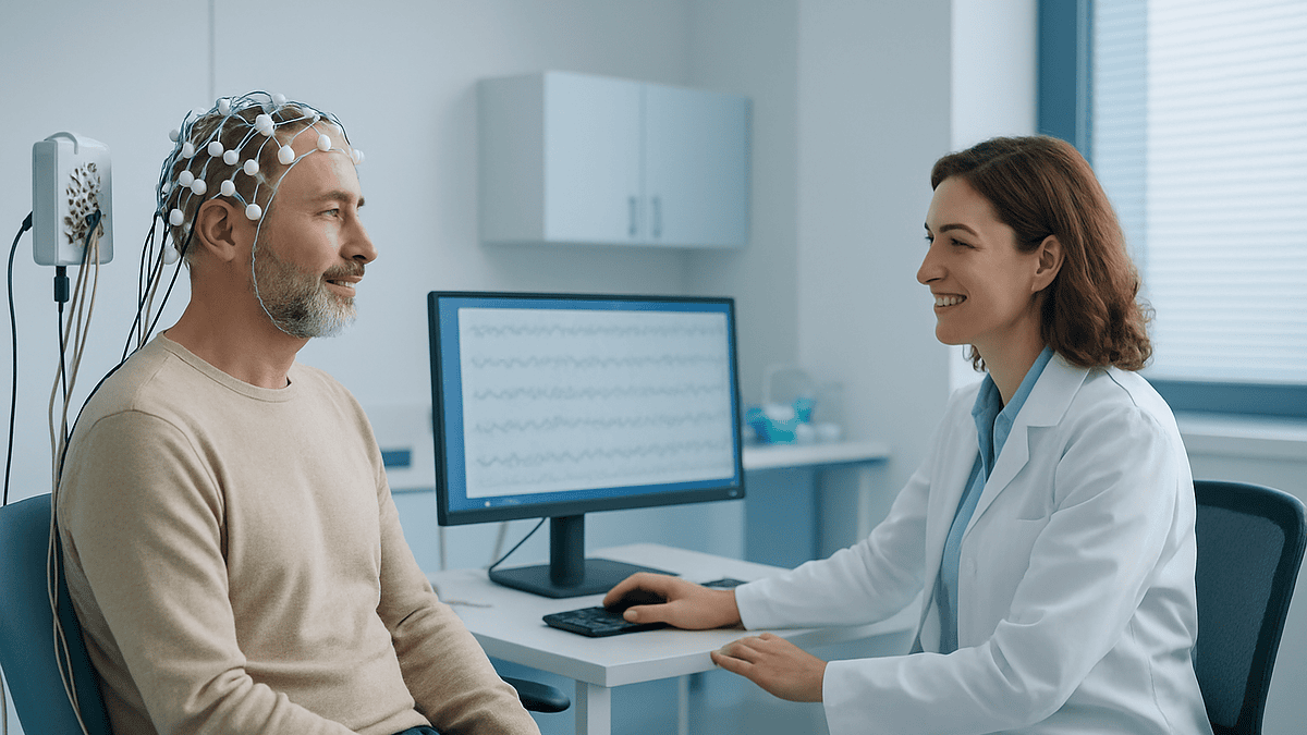
Neurophysiology tests measure the electrical activity of nerves, muscles, and the brain. An EMG records signals inside muscle, a nerve conduction study times impulses traveling along nerves, and an EEG traces brain-wave patterns through scalp electrodes. Doctors use these tests to investigate symptoms such as numbness, weakness, tingling, or seizures. They are generally safe, take 30 to 90 minutes, and cause mild, brief discomfort at most.
The referral letter arrives with a word most people have never said out loud: neurophysiology. Maybe it started with a hand that goes numb at 3 a.m., or a foot that catches on the stairs, or an unexplained blackout at the kitchen table. Now there’s an appointment for something called an EMG, or an EEG, and a quiet worry about needles, wires, and what the machine might find.
Here’s the reassuring part: these tests are among the gentlest investigations in medicine. Nothing is injected, nothing is imaged with radiation, and nothing is altered in your body. The equipment simply listens, to the faint electrical chatter your nerves, muscles, and brain produce every second of your life.
What follows is a plain-language tour of the three most common tests, what each one actually feels like, and, just as important, what the results can and cannot tell you.
What does neurophysiology actually measure?
Your nervous system runs on electricity, real, measurable electricity. When you decide to wiggle a toe, a signal leaves your brain, travels down the spinal cord, and races along a motor nerve at roughly 50 to 60 meters per second, about the speed of a car on a highway. When you touch a hot pan, sensory nerves fire the alarm back up the same kind of wiring. And the brain itself hums constantly with rhythmic waves measured in millionths of a volt.
Clinical neurophysiology is the branch of medicine that records these signals and asks whether they look healthy. If you’ve ever had an EKG for your heart, the concept is familiar: electrodes pick up natural electrical activity and turn it into a tracing a specialist can read. The nervous-system versions do the same thing for different tissues.
- EEG (electroencephalogram): records brain-wave rhythms through the scalp.
- Nerve conduction study: times how fast and how strongly signals travel along individual nerves.
- EMG (electromyography): listens to the electrical activity inside muscles at rest and during movement.
The power of this approach is precision. An MRI shows what the nervous system looks like; neurophysiology shows how well it works. A nerve can appear structurally normal on a scan and still conduct signals too slowly, and the reverse is true, too. That’s why doctors often order both kinds of testing rather than choosing one over the other.
Why would you be referred to a neurophysiologist?
Referrals usually follow a pattern: you describe a symptom, your doctor examines you, and the exam raises a question that only electrical testing can answer. The most common triggers fall into two groups.
Nerve and muscle symptomsthe territory of EMG and nerve conduction studies:
- Numbness or tingling in the hands, especially at night (a classic pattern for carpal tunnel syndrome, which affects an estimated 3 to 6 percent of adults)
- Burning or pins-and-needles in the feet, common in people with diabetes
- Weakness: a foot that drags, a grip that fails, trouble rising from a chair
- Persistent muscle twitching, cramping, or shrinking muscle bulk
- Pain radiating from the neck or lower back into an arm or leg, where the question is whether a spinal nerve root is being pinched
Brain-related eventsthe territory of EEG:
- A suspected seizure or an unexplained blackout
- Episodes of confusion, staring spells, or unusual movements
- Monitoring known epilepsy when symptoms change
Notice what these have in common: the symptom could have several different causes, and the treatment differs depending on which one is real. Tingling fingers might come from a compressed nerve at the wrist, a pinched root in the neck, or a generalized neuropathy. The physical exam narrows the list; neurophysiology testing often settles the question. That’s the honest reason for the referral, not because anyone assumes the worst, but because guessing is a poor basis for treatment.
What is the difference between a neurologist and a neurophysiologist?
Think of it as the difference between your ongoing physician and the specialist who runs one specific kind of investigation. A neurologist is a medical doctor who diagnoses and manages conditions of the brain, spinal cord, nerves, and muscles, everything from migraine to epilepsy to neuropathy. This is the person who takes your history, examines you, orders tests, explains the diagnosis, and oversees treatment over time.
A clinical neurophysiologist focuses on performing and interpreting the electrical tests themselves. In the United States, this is typically a neurologist who completed an additional fellowship year in clinical neurophysiology; in some other health systems, including parts of the UK and Europe, clinical neurophysiology is its own specialty, and testing teams may include specially trained scientists and technologists alongside physicians.
In practice, the roles overlap and cooperate. A typical journey looks like this:
- Your primary care doctor or neurologist identifies a question the exam can’t answer.
- The neurophysiology team performs the EMG, nerve conduction study, or EEG and writes a detailed report.
- Your referring doctor combines that report with your symptoms, exam, and any imaging to reach a diagnosis and plan.
One practical consequence worth knowing: the person testing you may discuss what the tracings show, but the full interpretation, what it means for you, usually comes from the doctor who referred you. If you leave the appointment without a verdict, that’s normal, not evasive.
What do neurophysiologists actually treat?
Here’s an answer you won’t find in most search results: strictly speaking, neurophysiologists diagnose more than they treat. Their central tool is measurement, pinpointing where in the nervous system a problem lives, how severe it is, and what type of damage is present. Treatment decisions then flow back to the referring neurologist, surgeon, or primary care doctor.
That said, the diagnostic work directly shapes care for a wide range of conditions:
- Entrapment neuropathies such as carpal tunnel syndrome and ulnar nerve compression at the elbow, where testing grades severity and helps decide between splinting and surgery
- Peripheral neuropathy from diabetes, vitamin deficiencies, or other causes, where the pattern of nerve involvement points toward the underlying culprit
- Radiculopathypinched nerve roots in the neck or lower back
- Muscle diseases (myopathies) and disorders of the nerve-muscle junction that cause fatigable weakness
- Motor neuron conditions, where careful EMG evidence is part of the diagnostic criteria
- Epilepsy and other seizure disorders, where EEG helps classify the type of seizure: a distinction that changes management
There’s also a role most patients never see: intraoperative monitoring. During certain spine and brain surgeries, neurophysiologists track nerve signals in real time so the surgical team gets an early warning if a pathway is at risk. It’s the same science, applied while you’re asleep on the table.
What is an EMG test and what does it feel like?
Electromyography answers a specific question: is the muscle itself healthy, and is the nerve supplying it delivering proper instructions? To find out, the examiner inserts a very thin needle electrode, closer in gauge to an acupuncture needle than to the one used for blood draws, into selected muscles. Nothing is injected and nothing is removed; the needle is purely a microphone.
The recording happens in two phases. First, the muscle is examined at rest. A healthy resting muscle is electrically quiet; spontaneous crackles or discharges can signal an irritated nerve or a muscle disorder. Then you’re asked to contract the muscle gently, press down, lift, squeeze, while the machine displays and plays the activity. Yes, plays: the signals are converted to sound, so you’ll hear static-like popping that rises as you push harder. Many patients find the soundtrack oddly fascinating.
As for sensation, honesty helps: each insertion feels like a brief pinch, followed by a dull, pressure-like ache while the needle sits in the muscle. Most people rate it uncomfortable rather than painful, and each muscle takes only a minute or two. Depending on the question being asked, the examiner may sample anywhere from a few muscles to a dozen, so the whole study runs roughly 30 to 60 minutes. Afterward you might notice small bruises or a day of mild soreness, similar to the feeling after an unaccustomed workout. Normal activities can resume immediately.
What is a nerve conduction study?
If the EMG listens to muscles, the nerve conduction study times the wiring. Flat electrodes are taped to the skin over a nerve, say, the median nerve running through your wrist, and a small electrical pulse is delivered at one point while the response is recorded farther along. The machine measures two things with millisecond precision:
- Speed (conduction velocity): healthy arm nerves typically conduct at about 50 to 60 meters per second, leg nerves slightly slower. Marked slowing suggests damage to myelin, the insulating sheath around the nerve, like a frayed cable leaking signal.
- Strength (amplitude): a weak response suggests that some of the nerve fibers themselves have been lost, a different type of injury with different implications.
This speed-versus-strength distinction is one of the most useful facts in the entire field, because it separates conditions that primarily strip insulation from conditions that destroy the fibers, and those two categories are investigated and managed differently.
The pulses feel like brief static shocks or the snap of a rubber band: startling for a half-second, then gone. There is no lingering pain, and the currents are far too small to cause harm. Skin temperature genuinely matterscold hands conduct measurably slower and can make a healthy nerve look sluggish, so the technologist may warm your hands or feet first. A typical study covers several nerves and takes 20 to 45 minutes, often in the same appointment as the EMG.
EMG vs. nerve conduction study: what’s the real difference?
The two tests are booked together so often that many people assume they’re one procedure. They’re complementary halves of a single investigation: the nerve conduction study evaluates the cables, and the EMG evaluates the destination, the muscle, plus the health of the connection between them. Here’s the side-by-side view:
| Nerve conduction study | EMG | |
|---|---|---|
| What it measures | Speed and strength of signals along a nerve | Electrical activity inside muscle, at rest and during effort |
| Electrodes | Flat pads taped to the skin | Thin needle inserted into muscle |
| Sensation | Brief static-shock pulses | Pinch on insertion, dull ache during recording |
| Typical duration | 20–45 minutes | 30–60 minutes |
| Best at detecting | Compressed or damaged nerves; neuropathy patterns | Muscle disease; nerve-root problems; how muscles are reinnervating |
Why do both? Because each test sees what the other misses. A pinched nerve root in the spine, for example, often produces a normal nerve conduction study, the compression sits closer to the spinal cord than the study can easily reach, while the EMG picks up telltale changes in the muscles that root supplies. Conversely, early carpal tunnel syndrome may show up on conduction timing before any muscle changes appear. Together, the pair locates the problem, grades its severity, and often estimates how long it has been developing.
What is an EEG test and how does it work?
The electroencephalogram is the oldest trick in clinical neurophysiology, in continuous use since the 1930s, and its premise hasn’t changed: billions of brain cells firing together produce rhythmic electrical waves faint enough to be measured in microvolts, millionths of a volt, yet strong enough to reach the scalp.
A technologist attaches about 20 small metal discs to your head using a conductive paste (some clinics use a snug electrode cap instead). This is the part worth demystifying: the electrodes only record. No current enters your head, nothing is felt, and the test cannot read thoughts, only the collective rhythm of brain activity, like hearing the roar of a stadium without catching any single conversation.
A routine recording lasts 20 to 40 minutes. You’ll rest with your eyes closed, then open them on cue. Two standard maneuvers may follow, because they can coax out abnormalities that hide during quiet rest:
- Hyperventilation: a few minutes of deep, fast breathing, which can make you briefly lightheaded
- Photic stimulation: a lamp flashing at varying frequencies while you keep your eyes closed
What the specialist reads afterward is pattern and rhythm, for instance, the healthy alpha rhythm of about 8 to 12 cycles per second that appears when a relaxed adult closes their eyes. Disruptions in these patterns, or sharp discharges where smooth waves should be, are the clues the report describes. The entire experience is painless; the most common complaint is paste in the hair afterward.
Which type of EEG might you have?
Not all EEGs are the routine 30-minute version. Because brain-wave abnormalities can be intermittent, present one hour, absent the next, clinicians choose the recording format that gives the best odds of catching what they’re looking for.
- Routine EEG: the standard outpatient test described above. Quick and informative, but a snapshot.
- Sleep-deprived EEG: you’re asked to sleep much less than usual the night before, because drowsiness and light sleep make certain epilepsy-related patterns more likely to surface. If you doze during the recording, that’s a feature, not a failure.
- Ambulatory EEG: a portable recorder worn at home for one to three days, electrodes secured under a head wrap. It captures brain activity through ordinary life, meals, sleep, work, and you keep a diary of any symptoms so events can be matched to the tracing.
- Video EEG monitoring: the most thorough option, done during a hospital stay lasting several days. Continuous brain-wave recording is paired with continuous video, so if an episode occurs, doctors can see exactly what happened and what the brain was doing at that moment. This is often how teams distinguish epileptic seizures from other causes of collapse or unusual episodes: a distinction that fundamentally changes treatment.
The pattern to notice: each step up trades convenience for sensitivity. A routine EEG is a photograph; video monitoring is a documentary. Your doctor’s choice reflects how elusive the answer has been so far, not how serious your condition is presumed to be.
What conditions can neurophysiology tests help investigate?
It’s worth being precise here, because the internet often oversells what any single test proves. Neurophysiology findings are evidence, usually strong evidence, that a doctor weighs alongside your story, exam, blood work, and imaging. With that caveat, the tests contribute to investigating:
- Carpal tunnel syndrome and other nerve entrapmentsnerve conduction studies are the standard confirmatory test and help grade severity
- Peripheral neuropathytesting maps which nerves are affected and whether the damage targets insulation or fibers, narrowing the list of causes
- Pinched nerve roots (radiculopathy) from the neck or lower back
- Muscle diseases, from inflammatory myopathies to inherited conditions
- Disorders of the nerve-muscle junction that cause weakness worsening with use
- Motor neuron diseaseEMG findings form part of the formal diagnostic criteria
- Epilepsy and seizure classificationEEG helps determine seizure type and, in surgical candidates, where seizures begin
- Unexplained blackouts and altered consciousness, including distinguishing seizures from fainting or other episodes
- Encephalopathygeneralized brain dysfunction from metabolic or other causes, where EEG tracks severity
A related family of tests, evoked potentials, measures how quickly signals travel through sensory pathways, flashing a checkerboard pattern to time the visual pathway, or clicking tones for hearing pathways. These are less commonly ordered now that MRI is widespread, but they still answer questions imaging can’t, particularly about how well a pathway functions rather than how it looks.
How do you prepare for an EMG, nerve conduction study, or EEG?
Preparation is refreshingly simple, but a few details genuinely affect the quality of the recording, and skipping them can mean a repeat visit.
For EMG and nerve conduction studies:
- Skip lotions, oils, and creams on your arms and legs that day; residue interferes with electrode contact.
- Arrive warm. Cold limbs slow nerve conduction enough to distort results, so bring layers in winter: the team may warm your hands or feet before starting.
- Tell the team in advance if you take medicines that affect bleeding or if you have a pacemaker, implanted stimulator, or lymphedema. None of these automatically rules out testing, but the approach may be adjusted.
- Wear loose clothing that gives access to the limbs being studied.
For an EEG:
- Wash your hair the night before and skip gels, sprays, oils, and conditioner; clean, dry hair helps electrodes grip.
- Eat normally beforehand, low blood sugar can alter brain-wave patterns.
- Bring a list of everything you take; some medicines influence the tracing, and the reader needs that context. Do not stop anything on your own.
- If a sleep-deprived study is ordered, follow the specific instructions about how little to sleep, and arrange a ride: you shouldn’t drive while severely sleep-deprived.
For all three tests, one preparation matters more than any other: come with your story straight. When did symptoms start, what makes them better or worse, which fingers, which episodes? The tracing is only half the report; your history is the other half.
Do neurophysiology tests hurt? An honest answer
Patients deserve a straight answer, so here it is, test by test, without minimizing or catastrophizing.
The EEG doesn’t hurt at all. Electrodes record; they deliver nothing. The genuine downsides are sitting still, the flashing-light portion (briefly odd, occasionally unpleasant), and paste in your hair. If deep breathing is part of the protocol, expect a minute or two of lightheadedness.
The nerve conduction study surprises more than it hurts. Each pulse feels like a static shock or a rubber-band snap, sharp, then instantly over. Some people barely react; others flinch every time. The sensation does not build or linger, and there is no soreness afterward.
The EMG is the one people ask about, and it’s fair to call it uncomfortable. The needle insertion pinches, and holding a contraction while the needle records produces a deep ache. On the usual zero-to-ten scales, most patients land in the mild-to-moderate range, and each muscle takes only a minute or two. Small bruises and a day of muscle soreness are common afterward; serious complications are rare.
Two practical notes. First, anxiety amplifies everything, patients consistently report the tests were easier than they’d imagined, which is exactly why reading an honest description beforehand helps. Second, you’re in control throughout: examiners can pause between muscles or nerves, and telling them you’re struggling is normal and welcome. No one benefits from a recording made while you’re rigid with tension, least of all the tracing itself, since tense muscles add electrical noise.
What do the results mean, and what can’t they tell you?
This is where a plain-language guide earns its keep, because the honest limits of these tests rarely make it into search results.
What an abnormal result means: the report localizes the problem (which nerve, which root, which pattern of brain activity), characterizes it (insulation damage versus fiber loss, for instance), and grades severity. That combination is often what converts a vague symptom into a specific diagnosis, and a specific plan.
What a normal result means, and doesn’t:
- A normal EMG and nerve conduction study does not prove your symptoms are imagined. Standard testing measures large nerve fibers; small-fiber neuropathy, which causes burning pain and altered temperature sensation, can produce entirely normal results and requires different investigations.
- A normal routine EEG does not rule out epilepsy. Between seizures, a single routine recording detects epilepsy-related patterns in only roughly a quarter to half of people who genuinely have the condition, which is precisely why sleep-deprived, ambulatory, and video studies exist.
- Timing matters. After a fresh nerve injury, some EMG changes take two to three weeks to appear, so a very early test can look deceptively clean.
The deeper point: these are physiological snapshots, interpreted in context. A finding that would alarm one patient may be an incidental footnote in another. Resist the urge to decode the report line by line at home; the numbers only acquire meaning next to your symptoms and exam. Ask your referring doctor to walk you through three things, what was found, what it explains, and what happens next.
When should you see a doctor about nerve or brain symptoms?
Most of the symptoms that lead to neurophysiology testing develop gradually and can safely wait for a scheduled appointment. A few cannot. Knowing the difference matters more than any test.
Call emergency services immediately for:
- Sudden weakness or numbness on one side of the body, face drooping, or trouble speaking, possible stroke, where every minute counts
- A first-ever seizure, a seizure lasting more than five minutes, or one seizure following another without recovery
- Sudden severe back pain with new leg weakness, numbness in the groin or inner thighs, or loss of bladder or bowel control
See a doctor promptly (within days) for:
- Weakness that is clearly progressing: a grip failing week by week, a foot dragging more than it did last month
- Numbness spreading up a limb rather than staying put
- Unexplained blackouts, staring spells, or episodes of confusion, even if brief
Book a routine appointment for:
- Tingling or numbness persisting beyond a few weeks, especially hand numbness that wakes you at night
- Persistent burning in the feet, particularly if you have diabetes
- Frequently dropping objects, or muscle twitching that doesn’t settle
One habit worth adopting before any appointment: keep a short symptom diary. Which fingers, which times of day, what triggers, what relieves. Nerve symptoms follow anatomical patterns, and a precise description often does as much diagnostic work as the tests themselves. It also helps your doctor decide whether neurophysiology testing is needed at all, sometimes the story alone is answer enough.
Frequently asked questions
Why would you be referred to a neurophysiologist?
You’d typically be referred when a symptom, numbness, tingling, weakness, muscle twitching, or unexplained blackouts, has several possible causes that only electrical testing can separate. Common triggers include suspected carpal tunnel syndrome, peripheral neuropathy, pinched spinal nerve roots, muscle disease, or possible seizures. The referral doesn’t mean your doctor suspects something serious; it means the physical exam raised a question that an EMG, nerve conduction study, or EEG can answer with precision.
What is the difference between a neurologist and a neurophysiologist?
A neurologist diagnoses and manages nervous-system conditions over time, taking your history, examining you, and overseeing treatment. A clinical neurophysiologist specializes in performing and interpreting the electrical tests: EMG, nerve conduction studies, and EEG. In the US, this is usually a neurologist with an extra fellowship year in clinical neurophysiology; in some other countries it’s a separate specialty. In practice, the neurophysiologist reports findings and your referring doctor explains what they mean for you.
What do neurophysiologists treat?
Strictly speaking, neurophysiologists diagnose more than they treat: their tool is measurement. Their testing shapes care for carpal tunnel syndrome, peripheral neuropathy, pinched nerve roots, muscle diseases, nerve-muscle junction disorders, motor neuron conditions, and epilepsy. Treatment itself usually stays with the referring neurologist, surgeon, or primary care doctor. Neurophysiologists also monitor nerve signals in real time during certain spine and brain surgeries, giving the surgical team early warning if a pathway is at risk.
What does neurophysiology do?
Neurophysiology records the natural electrical activity of nerves, muscles, and the brain to check whether they’re working properly. An EEG traces brain-wave rhythms, a nerve conduction study times signals along nerves, and an EMG listens to muscles at rest and during effort. While imaging like MRI shows what the nervous system looks like, neurophysiology shows how it functions, locating problems, characterizing the type of damage, and grading severity.
Does an EMG test hurt?
It’s uncomfortable rather than truly painful for most people. Each needle insertion feels like a brief pinch, followed by a dull ache while the thin electrode records the muscle, usually a minute or two per muscle. Nothing is injected. Afterward, small bruises and a day of mild soreness are common, similar to post-workout muscle ache. Most patients report the test was easier than they’d feared, and you can ask the examiner to pause at any point.
Can I drive home after an EMG or EEG?
Usually yes, neither test involves sedation, and there are no lasting physical effects, so most people drive home and resume normal activities immediately. The main exception is a sleep-deprived EEG: severe sleep deprivation itself makes driving unsafe, so arrange a ride for that appointment. If you’re being evaluated for seizures or blackouts, ask your doctor about driving separately, since local rules about driving after such episodes apply regardless of the test.
How long does a nerve conduction study take?
A nerve conduction study typically takes 20 to 45 minutes, depending on how many nerves need testing. It’s usually paired with an EMG in the same appointment, bringing the total visit to roughly 60 to 90 minutes. Each measurement is quick, a brief static-shock pulse followed by recording, but studying several nerves at multiple points along each one adds up. Arriving with warm hands and feet helps, since cold limbs slow conduction and can require rewarming time.
Can an EEG detect epilepsy if I don’t have a seizure during the test?
Sometimes, but not reliably in a single session. Between seizures, many people with epilepsy show brief telltale discharges on EEG, but a single routine recording catches these in only roughly 25 to 50 percent of cases. A normal routine EEG therefore does not rule out epilepsy. That’s why doctors escalate to sleep-deprived EEGs, ambulatory recordings worn at home for one to three days, or multi-day video monitoring in hospital when the answer stays elusive.
Can an EMG and nerve conduction study be normal even if I have real symptoms?
Yes, and it doesn’t mean your symptoms are imagined. Standard testing measures large nerve fibers, so small-fiber neuropathy, which causes burning pain and altered temperature sensation, can produce entirely normal results. Timing matters too: after a fresh nerve injury, some EMG changes take two to three weeks to develop. A normal study is genuinely useful information, since it rules out several conditions and redirects the investigation toward other causes.
Do I need to stop my medications before an EMG or EEG?
Don’t stop anything on your own, most medicines don’t need to be paused for these tests. What matters is telling the team everything you take in advance, since some medicines influence brain-wave tracings or affect bleeding at EMG needle sites, and the specialists factor this into how they perform and interpret the study. Also mention pacemakers or implanted stimulators before a nerve conduction study. Your care team will give specific instructions if anything needs adjusting.
References
This article is for general information only and is not a substitute for professional medical advice. Please consult a qualified doctor about your individual situation.

More from the Blog

Recovery After a Stereotactic Biopsy: Headache, Pin-Site Soreness, Driving and Results
Stereotactic biopsy recovery is usually short. After a breast biopsy, most people go home the same day and have bruising or soreness for a…
After a PET-CT Scan: Clearing the Tracer, Contact Precautions and When Results Arrive
After a PET-CT scan, most people go home the same day, drink extra fluids and empty their bladder often to help clear the radioactive…
Preparing for an MRI: Eating, Metal, What to Wear
For most MRI scans you can eat and drink normally beforehand, because the magnet images tissue, not stomach contents. The exceptions are scans of…

What Does a Comprehensive Check-Up Include? Blood Work, Imaging and the Follow-Up Plan
A comprehensive check-up usually includes a medical and family history, a physical examination with blood pressure and body measurements, and blood and urine tests…

What a CT Scan Shows, and Why Doctors Order One
A CT scan shows detailed cross-sectional images of bones, organs, blood vessels and soft tissue by combining many X-ray views into slices. Doctors most…

Can an Ultrasound Detect Cancer? What It Can Show, Its Limits and the Next Steps
An ultrasound can detect abnormal lumps, masses and changes in organs that may be cancer, and it is especially good at telling a fluid-filled…