

Ophthalmic Lasers Explained: Retina, Glaucoma and Corneal Lasers and What Each One Does
Key Takeaways
- Retinal, glaucoma and corneal lasers share only the light source: the first two protect existing sight while corneal lasers aim to reduce dependence on glasses.
- Scatter laser for diabetic retinopathy works by lowering the retina's oxygen demand, which shrinks abnormal vessels but can reduce peripheral and night vision.
- Selective laser trabeculoplasty lowers eye pressure gradually over several weeks, its effect can fade over time, and many people still need some drops afterward.
- Posterior capsule opacification is not a returning cataract; a YAG capsulotomy clears it in a few painless minutes and rarely needs repeating.
- LASIK and PRK reach similar final vision for the same prescription, but LASIK recovers in days while PRK takes weeks and preserves more corneal tissue.
- New floaters, flashes, a spreading shadow, severe pain or a sudden vision drop after any eye laser warrant a same-day call to the treating team.
Eye lasers fall into three broad groups. Retinal lasers seal leaking or abnormal blood vessels and barricade retinal tears. Glaucoma lasers improve fluid drainage or reduce fluid production to lower eye pressure. Corneal lasers such as PRK, LASIK and SMILE reshape the cornea to reduce dependence on glasses. A separate YAG laser clears clouding that can form after cataract surgery. Each treats a different problem, and suitability is decided by an ophthalmologist.
Maya’s mother had “laser” for her diabetic eyes years ago. Her brother had “laser” so he could ditch his glasses. Now Maya, sitting in a dim examination room with dilated pupils and a pressure reading she does not like, has just been told she might benefit from “a laser” too. Same four letters. Three entirely different procedures, done on three different parts of the eye, for three different reasons.
That confusion is understandable. The types of eye laser treatment share a technology, focused light, and almost nothing else. One works on a light-sensing tissue at the back of the eye that is thinner than a sheet of paper. Another opens a microscopic drain. A third sculpts the clear window at the front so that light lands where it should.
This guide walks through what each laser does, who it usually suits, what the following weeks look like, and which questions are worth bringing to your next appointment.
Why the types of eye laser treatment are not one thing
A laser is simply light of a single wavelength, aimed with extraordinary precision. In ophthalmology that precision is put to work in three ways: to heat tissue, to punch a tiny hole through it, or to remove it a fraction of a micrometer at a time. Which effect the surgeon wants depends entirely on the problem.
At the back of the eye sits the retina, the layer of light-sensing cells that converts an image into nerve signals. Retinal lasers are mostly about heat: they create small, controlled scars that seal leaking vessels or weld a torn retina to the wall behind it. Nobody sees better the moment they leave; the goal is to stop things getting worse.
Glaucoma is a group of conditions where pressure inside the eye damages the optic nerve, the cable carrying vision to the brain. Glaucoma lasers aim at the eye’s plumbing. Some nudge the drainage meshwork to work harder, some open a hole in the iris to let fluid move freely, and some quiet the tissue that makes fluid in the first place.
The cornea is the clear dome at the front. Corneal lasers are the ones most people picture when they hear “laser eye surgery”: PRK, LASIK and SMILE reshape that dome so light focuses on the retina without glasses. These are the only lasers in this article chosen for convenience rather than to protect sight.
Then there is the YAG capsulotomy, a quick laser that clears a film that can form months or years after cataract surgery. It belongs in its own category because it is neither refractive surgery nor disease control, just tidying up.
Keeping these groups separate matters. A friend’s story about LASIK tells you almost nothing about what to expect from a retinal laser, and vice versa.
How does an eye laser actually work?
Ordinary light scatters. Laser light travels in a tight, coherent beam, so a surgeon can deliver energy to a spot smaller than a pinhead while leaving the tissue next to it untouched. The eye is unusually suited to this because most of it is transparent: the beam passes through the cornea, the fluid-filled front chamber, the lens and the jelly-like vitreous before landing exactly where it is meant to.

Different wavelengths interact with tissue differently, and that is why one machine cannot do every job. The retinal lasers and the glaucoma trabeculoplasty lasers rely on pigment. Dark cells absorb the energy, warm up and either form a scar or, in the case of selective laser trabeculoplasty, are gently stimulated without visible burning. The YAG laser used for capsulotomy and iridotomy works by photodisruption: an ultrashort pulse creates a microscopic shock wave that opens a hole in tissue rather than heating it. The excimer laser used in PRK and LASIK breaks molecular bonds in the cornea with ultraviolet light, vaporizing tissue in layers thinner than a human hair, which is what allows a prescription to be carved into the surface with such accuracy. The femtosecond laser, used to create the LASIK flap and to perform SMILE, fires pulses lasting a quadrillionth of a second, separating tissue planes with minimal heat.
In practical terms, most of these procedures happen in a clinic chair or on a reclining bed, not an operating theater. Numbing drops do most of the work; a contact lens may be placed on the eye to steady it and focus the beam. You will see flashes and may hear clicks. Some retinal and glaucoma treatments produce a dull ache; corneal reshaping usually feels like pressure and a faint smell rather than pain. The laser itself typically runs for seconds to a few minutes.
Retinal lasers: what photocoagulation does for diabetic eye disease and tears
Photocoagulation means using laser heat to create small scars in the retina. It sounds destructive, and in a controlled way it is, which is exactly the point.
The best-known use is diabetic retinopathy, where high blood glucose damages tiny retinal vessels. In its advanced form, the retina grows fragile new vessels that bleed and pull. Scatter laser, also called panretinal photocoagulation, places hundreds of small burns across the peripheral retina. The treated retina needs less oxygen, so the chemical signal that drives abnormal vessel growth falls and the vessels shrink. The National Eye Institute notes this treatment can reduce the risk of severe vision loss but may cost some peripheral or night vision, a trade many people and their doctors accept to protect central sight.
Focal or grid laser treats diabetic macular edema, swelling in the macula, the central area responsible for reading and faces. Here the surgeon aims at specific leaking spots. In recent years, injections of anti-VEGF medicines, a class of drugs that block the growth signal, have become the usual first choice for macular swelling, with laser still used in selected cases or alongside injections. The decision is individual and rests with the retina specialist.
The other common retinal laser is barricade treatment for a retinal tear. A tear is a break in the retina that can let fluid slip behind it and cause a detachment. Laser spots around the tear create a scar that effectively spot-welds the retina in place, usually within a couple of weeks as the scar matures. Laser is also used for some retinal vein occlusions and, less often, for certain vascular abnormalities.
What retinal laser does not do is restore cells already lost. If vision has been damaged by bleeding or long-standing swelling, laser aims to stabilize, not to reverse.
Glaucoma lasers: SLT laser for glaucoma, iridotomy and cyclophotocoagulation
Fluid, called aqueous humor, is made continuously inside the eye and drains through a spongy ring of tissue called the trabecular meshwork. Glaucoma lasers change one side of that equation or the other.
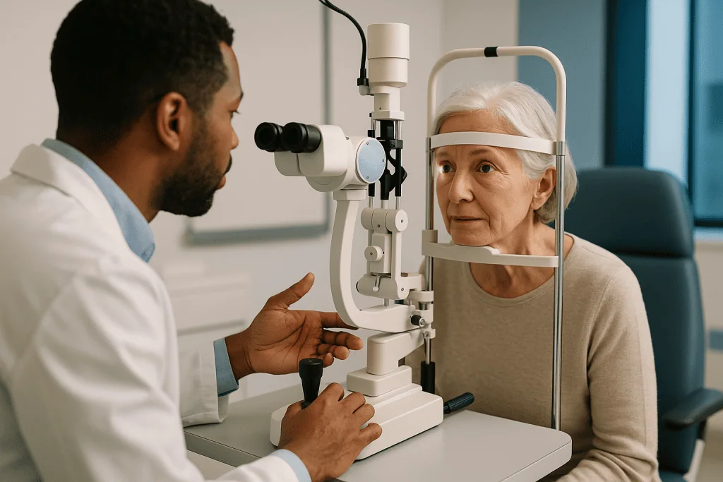
Selective laser trabeculoplasty, or SLT, is the workhorse for open-angle glaucoma, the common form where the drain is accessible but underperforming. Low-energy pulses are applied to the meshwork, selectively absorbed by its pigmented cells, and the tissue responds by improving outflow over the following weeks. The Mayo Clinic describes laser trabeculoplasty as an option when eye drops are not enough or are poorly tolerated, and notes that it can take several weeks to see the full effect. Some guidelines now consider it a reasonable first treatment rather than a fallback. The pressure-lowering effect can fade over months to years, and the treatment can sometimes be repeated; drops may still be needed afterward. Whether to adjust any medicine remains a decision for the prescribing ophthalmologist.
Laser peripheral iridotomy is for a different anatomy. In angle-closure glaucoma, or eyes at risk of it, the iris bows forward and physically blocks the drain. A YAG laser makes a pinpoint hole in the peripheral iris so fluid can pass freely and the iris falls back. In an acute attack, with sudden pain, a red eye, halos and nausea, this is an emergency treatment; in narrow-angle eyes it is preventive.
Cyclophotocoagulation targets the ciliary body, the tissue that produces aqueous fluid, and reduces production. It is generally reserved for glaucoma that has resisted other treatments, because it is harder to titrate and carries more inflammation risk.
None of these lasers repair the optic nerve. They protect what remains by lowering pressure, which is why regular visual field testing continues after treatment.
YAG laser capsulotomy: the laser after cataract surgery
Cataract surgery removes the cloudy natural lens and replaces it with a clear artificial one, leaving the thin natural capsule that held the old lens in place as a support. In a proportion of people, cells left on that capsule grow across it over months or years, producing a haze behind the new lens. This is posterior capsule opacification, sometimes called a secondary cataract, although it is not a cataract at all.
The symptoms mimic the original cataract: gradual blur, glare from oncoming headlights, colors that look washed out. People often assume the surgery has failed. It has not; the implant is fine, and the fix is quick.
A YAG laser capsulotomy uses photodisruption to open a small central window in the cloudy capsule. The Mayo Clinic describes it as a painless outpatient procedure taking about five minutes. Drops dilate the pupil, a lens may be placed on the eye, and a series of clicks follows as the laser creates the opening. Light then passes cleanly through to the retina again.
Vision usually clears over the following day or two once the dilating drops wear off. You may notice new floaters for a while, which are fragments of capsule drifting in the vitreous; they typically settle or sink out of view. Pressure in the eye is often checked afterward because it can rise briefly, and some people are given a short course of anti-inflammatory drops at the clinician’s discretion.
The opening does not close again, so the procedure is rarely repeated. The uncommon risks are a temporary pressure rise, swelling of the macula, and, rarely, retinal detachment, which is why a retina check is part of follow-up if new flashes or a shadow appear.
Corneal lasers: PRK, LASIK and SMILE explained
All three refractive lasers do the same fundamental thing: they remove a precisely calculated sliver of corneal tissue so that the front surface of the eye focuses light onto the retina. For short-sightedness the cornea is flattened; for long-sightedness it is steepened in the center; for astigmatism it is made more evenly curved. The differences lie in how the surgeon reaches the tissue.
PRK, photorefractive keratectomy, is the oldest approach. The surgeon removes the thin surface skin of the cornea, the epithelium, then applies the excimer laser directly to the layer beneath. The epithelium regrows over several days. LASEK and epi-LASIK are variants that loosen and replace the surface layer rather than removing it.
LASIK, laser-assisted in situ keratomileusis, creates a hinged flap in the cornea with a femtosecond laser or a fine blade, lifts it, applies the excimer laser underneath, and lays the flap back down. Because the surface is preserved, comfort returns fast and vision is often usable the next day. The trade-off is a flap that, while it heals firmly, remains a plane in the cornea.
SMILE, small incision lenticule extraction, uses only a femtosecond laser to cut a disc of tissue inside the cornea, which is then drawn out through a small side incision. There is no flap and no surface removal. It is the newest of the three to reach mainstream use and is currently applied mainly to short-sightedness and some astigmatism; its range for long-sightedness is more limited.
The NHS notes that laser eye surgery is generally offered to adults whose prescription has been stable for a couple of years and that it is not suitable for every prescription; lens-based surgery may be considered where the cornea is too thin or the correction too large. Those limits are set by measurement, not by preference.
LASIK vs PRK: what is actually better?
This is the most searched question in refractive surgery, and the honest answer is that neither is universally better. They reach similar destinations by different roads, and the right road depends on your cornea and your life.
On final visual result, the evidence from comparative studies is broadly reassuring: for the same prescription, the two procedures produce similar outcomes once healing is complete. Where they differ is the journey. The Mayo Clinic describes LASIK vision as typically clearing within a day or so, with stability over two to three months. PRK asks for patience: the surface layer takes several days to regrow, during which the eye is sore, light-sensitive and blurry, and crisp vision may take weeks longer to settle.
So why would anyone choose PRK? Anatomy and occupation. A cornea that is thinner than average leaves less margin for a flap, and PRK preserves more structural tissue. People whose work or sport involves a real risk of a blow to the eye, such as contact athletes or some military roles, may be steered toward PRK because there is no flap to dislodge. Certain surface irregularities also favor it.
LASIK, by contrast, suits people who need to function quickly and whose measurements allow a flap. Dry eye symptoms are common after both but tend to be more noticeable in the early months after LASIK because more corneal nerves are cut; they usually improve over time, though not for everyone.
The NHS puts the choice plainly: the surgeon will recommend the procedure that suits your eyes, and complications and results are broadly comparable across the main techniques. If a clinic offers only one method regardless of measurements, that is worth noticing.
Types of eye laser treatment at a glance
Seeing the procedures side by side makes the pattern clear: the part of the eye treated tells you the purpose, the purpose tells you what “success” means, and that in turn tells you what recovery looks like.
| Laser procedure | Part of eye | What it aims to do | Typical setting and feel | What it does not do |
|---|---|---|---|---|
| Panretinal or focal photocoagulation | Retina | Shrink abnormal vessels, seal leaks, protect central vision | Clinic chair; dull ache or bright flashes; may need more than one session | Restore vision already lost |
| Retinal tear barricade | Retina | Weld a tear to prevent detachment | Clinic; brief; scar matures over about two weeks | Remove floaters |
| Selective laser trabeculoplasty (SLT) | Trabecular meshwork | Lower eye pressure by improving drainage | Clinic; mild discomfort; effect builds over weeks | Repair the optic nerve; always replace drops |
| Laser peripheral iridotomy | Iris | Open a channel to prevent or treat angle closure | Clinic; brief sting; sometimes urgent | Treat open-angle glaucoma |
| Cyclophotocoagulation | Ciliary body | Reduce fluid production in resistant glaucoma | Clinic or theater; more inflammation than SLT | Serve as a first-line option |
| YAG capsulotomy | Lens capsule | Clear haze after cataract surgery | Clinic; painless; minutes | Change your glasses prescription |
| PRK / LASEK | Corneal surface | Reshape cornea to reduce glasses dependence | Laser suite; sore for days; vision settles over weeks | Prevent age-related reading changes |
| LASIK | Cornea under a flap | Reshape cornea to reduce glasses dependence | Laser suite; comfortable within a day; stable in 2-3 months | Suit very thin corneas |
| SMILE | Cornea, no flap | Reshape cornea for short sight and astigmatism | Laser suite; quick comfort; flapless | Treat most long-sightedness at present |
Timelines in the table are typical ranges drawn from the Mayo Clinic and NHS resources listed in the references, not guarantees. Your surgeon’s estimate, based on your own measurements, always takes precedence.
Who is usually a candidate, and who is asked to wait?
For the sight-protecting lasers, candidacy is driven by disease rather than preference. Someone with proliferative diabetic retinopathy or a fresh retinal tear is usually offered laser promptly because delay carries risk. Someone with open-angle glaucoma may be offered SLT early or after drops, depending on pressure, nerve damage and how well drops are tolerated. Angle-closure risk usually leads to iridotomy without much debate. The main reason to defer these treatments is an eye that is too inflamed, bleeding too heavily, or too cloudy for the laser to reach its target; the team then treats the obstacle first.
Refractive laser is where the “wait” conversation happens most. The NHS describes typical candidates as adults over 18 with a prescription that has been stable for around two years, healthy corneas of adequate thickness, and no significant eye disease. People are commonly asked to postpone or reconsider if they are pregnant or breastfeeding, because hormonal shifts can temporarily change the prescription; if their glasses have changed recently; if they have uncontrolled dry eye, since surgery can worsen it; or if they have keratoconus, a condition where the cornea thins and bulges, because removing tissue could accelerate it. Autoimmune conditions, certain medicines that affect healing, and unrealistic expectations are also reasons a surgeon may hesitate.
Age cuts both ways. Younger than the early twenties, the eye may still be changing. Past the mid-forties, the natural lens begins to stiffen and reading vision drifts regardless of what the cornea does, so a person may be steered toward lens-based options or a frank discussion about reading glasses later.
For YAG capsulotomy, the question is simply whether the haze is affecting vision or interfering with examination of the retina. A faint film that causes no symptoms is often left alone.
What do the days and weeks after eye laser look like?
Recovery depends almost entirely on which tissue was treated, so it helps to know your category before you go home.
After retinal laser, expect a dazzled, dim or blurred eye for several hours from the bright flashes and dilating drops, sometimes with a headache-like ache. Peripheral vision may feel slightly reduced after heavy scatter treatment, and night vision can be affected, as the National Eye Institute notes. Most people return to ordinary activities the next day, avoiding heavy lifting if a tear was treated until the surgeon confirms the scar has set. Follow-up is usually within a few weeks, and further sessions are common in diabetic eye disease.
After SLT, the eye may be mildly achy and light-sensitive for a day or two, and a small pressure spike can occur in the first hours, which is why many clinics check pressure before you leave. The pressure-lowering effect builds over several weeks according to the Mayo Clinic, so you should continue any prescribed drops exactly as directed unless the ophthalmologist tells you otherwise. Iridotomy recovery is similar; a few people notice a faint line of light or glare afterward, related to the new opening.
After YAG capsulotomy, vision clears as dilation fades, typically within a day or two, and new floaters may drift for a few weeks.
After LASIK, the Mayo Clinic describes itching, grittiness, burning and watering for a few hours, blur that lifts within a day, and vision that continues to settle over two to three months. Protective shields at night, no eye rubbing, and a pause on swimming and eye makeup are standard early advice. PRK follows a slower curve: a bandage contact lens for several days, real discomfort while the surface heals, and vision that sharpens over weeks. Dry eye is common after both and is managed with lubricating drops as advised.
What's the downside? Laser eye surgery side effects and risks
Every effective treatment has a cost in risk, and a good explainer should say so plainly rather than bury it in reassurance.
For retinal photocoagulation, the intended scar is the main side effect. Scatter treatment can reduce peripheral vision, dim night vision and slightly affect color perception. Focal treatment near the macula carries a small risk of a blind spot if a burn lands closer to the center than intended. Rarely, the laser can trigger bleeding or, over years, a scar can enlarge. These risks are weighed against the far larger threat of untreated proliferative disease.
Glaucoma lasers are generally low-risk. SLT can cause a temporary pressure rise, inflammation and, uncommonly, a persistent pressure increase requiring treatment. Its effect can also wane, which is a limitation rather than a complication. Iridotomy may produce glare or a visual line, and cyclophotocoagulation carries a higher chance of inflammation and, rarely, excessively low pressure.
YAG capsulotomy risks include a brief pressure spike, macular swelling and, infrequently, retinal detachment.
Refractive surgery is where risk deserves the most candor, because the eye was healthy to begin with. The Mayo Clinic and NHS list dry eye that can persist for months and occasionally longer; glare, halos and double images especially at night; under- or over-correction that may need glasses or a second procedure; regression, where the prescription drifts back over years; flap problems specific to LASIK, including wrinkles or dislodgement after trauma; infection; and, very rarely, corneal ectasia, a progressive bulging that can seriously affect vision. Losing best-corrected vision, meaning worse sight even with glasses, is uncommon but real. None of these can be promised away, and any surgeon should walk you through them before you sign anything.
What are the alternatives to eye laser treatment?
Laser is rarely the only option, and knowing what sits beside it makes the conversation with your team more balanced.
In diabetic macular edema, anti-VEGF injections, drugs that block the vessel-growth signal, have largely become first-line, with steroid implants as another route and laser used in selected patterns or in combination. For proliferative retinopathy, anti-VEGF can also be used instead of or alongside scatter laser; each approach has different demands on follow-up, and the retina specialist will explain the trade-offs. Vitrectomy, a surgical removal of the vitreous gel, is reserved for bleeding or traction that laser cannot address. The single most powerful non-laser intervention remains blood glucose, blood pressure and lipid control, which the National Eye Institute emphasizes as the foundation of prevention.
In glaucoma, eye drops from several classes lower pressure either by reducing fluid production or improving outflow; they work as long as they are taken, and the prescribing clinician manages any changes. Minimally invasive glaucoma surgeries insert tiny stents or open channels, often at the time of cataract surgery. Trabeculectomy and tube shunts create a new drainage route for more advanced disease. Cataract removal itself can lower pressure in angle-closure eyes.
For refractive error, the obvious alternatives are glasses and contact lenses, which carry no surgical risk though contact lenses have their own infection risks. Implantable collamer lenses are placed inside the eye in front of the natural lens and can treat prescriptions too large or corneas too thin for laser. Refractive lens exchange replaces the natural lens and is more often considered in older adults who are approaching cataract age.
For posterior capsule opacification, there is no meaningful alternative to YAG capsulotomy other than watchful waiting when symptoms are mild.
What people often get wrong about eye lasers
Myth: “Laser means I won’t need glasses.” Only the corneal refractive lasers aim at that. Retinal, glaucoma and YAG lasers do not change your prescription, and even refractive surgery does not stop the age-related loss of near focus that arrives in the mid-forties. Reading glasses may still be in your future.
Myth: “If my glaucoma laser worked, I can stop my drops.” Sometimes the ophthalmologist does reduce medication after SLT, and sometimes the laser is not enough on its own. The National Eye Institute notes that many people still need drops after laser. Changing them without the prescriber’s instruction is one of the more common ways glaucoma quietly progresses.
Myth: “Retinal laser damaged my night vision, so it failed.” Reduced night and side vision after scatter laser is an expected effect, not a mistake. The treatment succeeded if the abnormal vessels regressed and central vision was protected.
Myth: “My cataract came back.” Cataracts do not regrow. Haze on the capsule is a separate, common event with its own quick fix.
Myth: “The newest laser is the best laser.” SMILE is newer than LASIK, which is newer than PRK, yet all three remain in use because each fits certain eyes. Evidence, not release dates, should guide the choice.
Myth: “Laser eye surgery is painless and permanent.” PRK is genuinely uncomfortable for days. Corneal reshaping is permanent, but eyes keep aging, and some prescriptions drift back over years, which the NHS flags as a recognized possibility.
Myth: “Bigger prescription, bigger benefit.” The reverse is closer to the truth: very high prescriptions are harder to correct fully, use more tissue and carry more risk of regression, which is why lens-based alternatives exist.
Questions to ask your care team before any eye laser
A ten-minute consultation goes further when you arrive with specific questions. These are the ones that tend to unlock the most useful answers, grouped by the kind of laser being proposed.
For any laser: Which part of my eye are you treating, and what problem is it meant to fix? What happens if I do nothing for now? What would you expect me to notice in the first week, and what would worry you? How will we know whether it has worked, and when will that be measured?
For retinal laser: Is this to protect vision or to improve it? Will I need more than one session? How will this affect my night driving or side vision? Are injections an option instead of or alongside laser, and why are you recommending this order?
For glaucoma laser: Is this instead of drops, in addition to them, or a step before surgery? How long might the effect last, and can it be repeated? Should I keep taking my current drops exactly as before afterward? How often will my pressure and visual field be checked?
For YAG capsulotomy: Is the haze definitely what is affecting my vision, or is something else contributing? Will you check my retina afterward?
For refractive laser: Which procedure fits my corneal thickness and prescription, and why? What are my realistic odds of needing glasses for some tasks afterward, based on my measurements rather than averages? How do you manage dry eye if it persists? What is your policy if I am under- or over-corrected? Who do I call at night if something feels wrong?
Write the answers down. The most reassuring sign in any of these conversations is a clinician who describes the limits of the treatment as readily as its benefits.
When to call your doctor after eye laser treatment
Most laser recoveries are uneventful, and mild grittiness, light sensitivity or a dull ache in the first day or two is expected. Some symptoms are different in kind, not degree, and should prompt a same-day call to the treating clinic or, out of hours, an emergency eye service.
Call urgently for a sudden shower of new floaters, flashes of light, or a curtain or shadow moving across part of your vision, at any point after any eye procedure. These can indicate a retinal tear or detachment, which is time-sensitive.
Call urgently for severe or rapidly worsening eye pain, especially with a red eye, nausea, headache or halos around lights. After glaucoma laser or YAG capsulotomy this can signal a sharp pressure rise; after any procedure it can indicate inflammation or infection.
Call urgently for a sudden marked drop in vision that is not explained by dilating drops wearing off, or vision that was improving and then abruptly worsens. After LASIK, this may indicate a flap problem, particularly if it follows rubbing or a knock to the eye.
Call urgently for increasing redness with thick discharge, a white or gray spot on the cornea, or pain that intensifies rather than eases over the first days after PRK or LASIK. Infection is rare but requires prompt treatment.
Call promptly, though not necessarily out of hours, if dry eye becomes disabling, if glare and halos are not improving over the weeks your surgeon described, or if your glasses prescription seems to be changing again.
Whatever the symptom, do not adjust prescribed drops, restart old ones or stop new ones on your own. Describe what you are experiencing and let the team decide the next step; they would far rather hear from you about a false alarm than miss a real one.
Frequently asked questions
What is better, LASIK or PRK?
Neither is better for everyone; comparative evidence shows similar final vision for the same prescription. LASIK recovers faster, often with usable vision the next day, but requires enough corneal thickness for a flap. PRK removes the surface layer instead, which means several sore days and slower sharpening, yet it preserves more tissue and avoids flap-related problems, which suits thinner corneas and people at risk of eye trauma. Your surgeon’s measurements decide.
What's the downside to laser eye surgery?
The main downsides are dry eye that can last months, night glare and halos, the possibility of under- or over-correction needing glasses or a second procedure, and gradual regression over years. Rarer risks include infection, flap complications after LASIK, and corneal ectasia, a progressive bulging that can seriously affect vision. Because the eye was healthy to begin with, these risks deserve honest discussion before you decide.
What's the worst vision LASIK can fix?
There is no single cutoff; the limit depends on corneal thickness and shape as much as the prescription. Higher prescriptions require removing more tissue, which raises the risk of regression and weakens the cornea, so surgeons set individual limits from scans. The NHS notes laser is not suitable for very high prescriptions, where implantable lenses or lens exchange are usually considered instead. Ask what your own measurements allow.
What is the newest form of laser eye surgery?
SMILE, small incision lenticule extraction, is the most recent refractive laser in mainstream use. It uses only a femtosecond laser to carve a thin disc inside the cornea, which is removed through a small incision, so there is no flap and no surface removal. It is currently used mainly for short-sightedness and astigmatism. Newer does not mean better for every eye; ask how the evidence compares for your prescription.
Does SLT laser for glaucoma replace eye drops?
Sometimes, but not always. Selective laser trabeculoplasty improves drainage and lowers pressure over several weeks, and some people can reduce their drops afterward under their ophthalmologist’s direction. Others need laser and drops together, and the effect can wear off over months to years, at which point it may be repeated. Never stop or change glaucoma drops on your own; the prescribing clinician decides based on your pressure readings.
Is a YAG laser capsulotomy ever needed twice?
Rarely. The laser opens a permanent window in the cloudy capsule behind the lens implant, and that opening does not close. Occasionally a surgeon enlarges the opening if the initial one was small or if floaters of capsule remain in the visual axis. If vision clouds again years later, the cause is usually something else, such as macular changes or a new glasses prescription, and needs its own examination.
Does retinal laser hurt?
It ranges from uncomfortable to briefly painful, depending on the amount of treatment and where it is aimed. Focal laser for a small tear is usually just bright and startling. Heavy scatter laser for diabetic retinopathy can produce a deep ache or sharp jabs, and some clinics numb the eye more thoroughly or split treatment across sessions. Afterward, expect dazzle, blur and a dull headache for several hours.
Can glaucoma laser reverse damage to the optic nerve?
No. Nerve fibers already lost to glaucoma do not regrow, and no laser or medicine currently restores them. Laser treatments such as SLT and iridotomy lower eye pressure to slow or halt further loss, which is why they are most valuable when started before damage is advanced. Regular visual field tests and nerve scans continue after laser to confirm the pressure achieved is protecting what remains.
Do the results of laser eye surgery wear off?
The corneal reshaping itself is permanent, but the eye keeps changing. Some prescriptions drift back over years, more commonly with higher initial corrections, and the natural lens begins stiffening in the mid-forties, bringing reading glasses regardless of earlier surgery. The NHS lists regression as a recognized possibility. An enhancement procedure is sometimes offered if enough corneal thickness remains, a decision the surgeon makes from fresh measurements.
Can I have laser eye surgery if I have diabetes or I am pregnant?
Pregnancy and breastfeeding usually mean waiting, because hormonal changes can temporarily shift the prescription and affect corneal healing. Diabetes is not an automatic bar to refractive surgery, but surgeons look for stable glucose control, a stable prescription and no active retinopathy, since healing and infection risk can be affected. People with diabetic eye disease may instead need retinal laser, which is a different procedure with different goals.
References
- NHS: Laser eye surgery and lens surgery
- NIH National Eye Institute: Diabetic retinopathy
- NIH National Eye Institute: Glaucoma surgery
This article is for general information only and is not a substitute for professional medical advice. Please consult a qualified doctor about your individual situation.

More from the Blog
Ocular Surface Disease Explained: How Tear Film, Eyelids and Cornea Fail Together
Ocular surface disease is an umbrella term for conditions in which the tear film, eyelids and cornea stop working as a unit, most often…

Eye Surgery in Older Adults With Heart Disease or Diabetes: How the Plan Is Adjusted
Eye surgery in older adults with heart disease or diabetes is usually still possible, but the plan changes. Most procedures, especially cataract surgery, are…

Why You Must Pause Contact Lenses Before a Myopia Laser Assessment (and for How Long)
Contact lenses press on the cornea and temporarily change its shape, so measurements taken too soon after removing them can be inaccurate and could…
Injections, Laser or Surgery for Macular Diseases: How the Retina Team Picks a Path
Macular disease treatment depends on what is happening in the macula. Leaking new blood vessels and swelling are usually treated with repeated anti-VEGF eye…

How Tear Duct Irrigation and Imaging Confirm Where the Blockage Sits
A blocked tear duct test locates where tears stop draining rather than simply confirming that they overflow. An eye specialist usually starts with a…
What Happens in the Operating Room During Cataract Surgery, Minute by Minute
During cataract surgery, a surgeon numbs the eye with drops or a small injection, makes an incision of a few millimeters at the edge…