

Skin Cancer Excision Explained: Margins, Closure Options and What Happens to the Sample
Key Takeaways
- The margin drawn on the skin before surgery is usually wider than the margin the pathologist later measures, because skin shrinks after removal and preservation.
- Melanoma is typically treated in two operations: a narrow diagnostic excision first, then a wide local excision whose margin is scaled to the tumor's Breslow thickness.
- Mohs surgery is not a more aggressive operation but a more precise one, examining the entire edge of each layer during the visit so the least possible skin is removed.
- Standard excision specimens are sliced at intervals, so a clear margin report describes the sections examined rather than every point along the edge.
- An involved or close margin is not an emergency; options include a targeted re-excision, Mohs surgery, radiotherapy, or observation, decided with the treating team.
- A new scar usually looks reddest around the second month and keeps fading and softening for a year or more, and protecting it from sun helps prevent permanent darkening.
A skin cancer excision procedure removes the visible tumor plus a rim of normal-looking skin, called the margin, usually under local anesthetic in an outpatient room. The wound is then closed with stitches, a skin flap, or a graft, or left to heal on its own. The removed tissue goes to a pathologist, who checks under a microscope whether the margins are clear; the care team uses that report to decide whether anything further is needed.
The spot on her temple was smaller than a pencil eraser. It had bled twice while she washed her face, scabbed over, and then quietly come back, which is exactly why the dermatologist wanted it out. Now she is sitting on paper-covered vinyl with a purple ink line drawn around it, wondering how something so small needs a sterile tray, a stack of gauze, and a consent form.
That gap between the size of the lesion and the seriousness of the setup is where most questions about a skin cancer excision procedure begin. Why cut out healthy skin too? How will they close it? Will it hurt afterward? And what, exactly, happens to the piece of you that leaves the room in a labeled jar?
This guide walks through each of those steps in plain language, using what mainstream medical evidence actually shows rather than what waiting-room folklore suggests. Every decision, though, belongs with you and the team holding the scalpel.
What is a skin cancer excision procedure, and what actually happens in the room?
An excision is surgery in its simplest form: a scalpel removes a defined piece of skin, and the surgeon repairs what is left. Most skin cancer excisions take place in an outpatient procedure room rather than an operating theater, and you go home the same day, according to the NHS and Mayo Clinic descriptions of treatment for both melanoma and non-melanoma skin cancer.
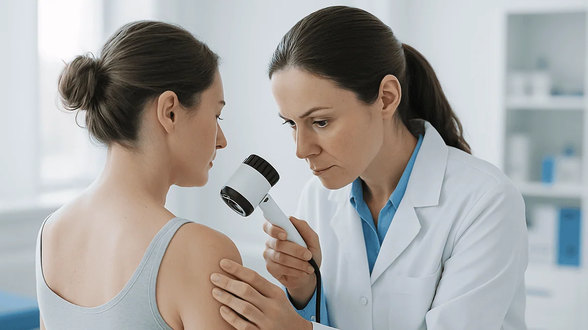
The sequence is remarkably consistent. First, the surgeon examines the lesion, sometimes with a handheld magnifier called a dermatoscope, and draws the visible edge in ink. A second line is drawn a set distance outside the first; that ring is the margin, meaning the band of normal-appearing skin removed alongside the tumor. Together they are usually shaped into an ellipse roughly three times as long as it is wide, because that geometry lets the two sides come together without puckered corners.
Next comes the local anesthetic, a numbing medicine injected just beneath the skin so the area loses sensation while you stay fully awake. Once the skin is numb, the surgeon cuts along the outer line down to the layer of fat beneath the skin, the subcutaneous fat, and lifts the tissue away in one piece. Small bleeding vessels are sealed with an electrocautery device, a pen-like tool that uses electrical current to stop bleeding.
The specimen is oriented, often with a single stitch or an ink mark, so the pathologist knows which edge faced which direction, then placed in preservative. Finally the wound is closed, dressed, and you receive written care instructions. The whole visit for a standard excision commonly lasts well under an hour, though a Mohs procedure, described later, can take several hours because tissue is examined while you wait, as Mayo Clinic notes.
Who is usually offered excision, and who is asked to wait or consider alternatives?
Excision is the workhorse treatment for the three most common skin cancers: basal cell carcinoma, squamous cell carcinoma, and melanoma. The NHS lists surgery as the main treatment for non-melanoma skin cancer and describes surgical removal as the first-line approach to melanoma. It suits lesions with a clear edge, a size the surrounding skin can accommodate, and a location where a straight scar is acceptable.
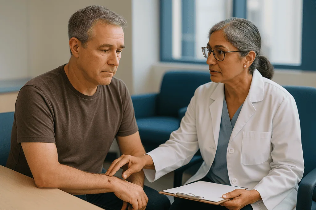
Some people are steered toward other options first. Very superficial basal cell carcinomas on the trunk may be offered scraping and cautery, freezing with liquid nitrogen, or prescription creams, which the NHS lists among alternatives for selected low-risk tumors. Cancers on the nose, eyelid, ear, or lip, where every millimeter of tissue matters, are often referred for Mohs surgery so the least possible skin is removed. Large or aggressive tumors, or those near important nerves, may be planned in a full operating theater with a plastic surgeon involved.
Waiting is sometimes the right call, too. An active skin infection near the site usually needs treatment first. If you take a blood-thinning medicine, the team will ask about it, but you should never stop or adjust it on your own; many excisions proceed safely with the medicine continued, and any change is the prescriber’s decision. People with implanted heart devices are asked to mention them, because certain cautery tools may need adjusting. For a small number of frail patients with slow-growing tumors, careful observation is a legitimate option the team may raise.
The point is that eligibility is not a checklist you can score at home. It comes from a conversation about the tumor’s type, its exact position, your health, and what matters to you about the outcome.
Skin excision margins explained: why healthy-looking skin is removed too
Cancer cells do not respect the border you can see. Beneath the surface, basal and squamous cell carcinomas send microscopic extensions outward and downward, sometimes several millimeters beyond the visible edge. The margin exists to capture those invisible roots in one clean sweep.
Two different margins matter, and mixing them up causes confusion. The clinical margin is the distance the surgeon measures and marks on the skin before cutting. The histological margin is what the pathologist later measures under the microscope: the actual distance between the last cancer cell and the cut edge of the specimen. Skin shrinks after removal and preservation, so the histological number is typically smaller than the clinical one. A report saying the margin was narrower than what was drawn is usually describing that shrinkage, not a surgical error.
How wide the clinical margin should be depends on the diagnosis. For many well-defined basal cell carcinomas, a few millimeters is standard practice. For melanoma, the margin is scaled to the tumor’s depth, measured in millimeters from the skin surface to the deepest cancer cell, a figure known as the Breslow thickness. Mayo Clinic explains that the amount of skin removed for melanoma depends on how thick the tumor is, with deeper melanomas warranting wider excisions, often a centimeter or more on each side.
Wider is not automatically better. Every extra millimeter means a longer scar, more tension on the closure, and on the face, a greater chance of pulling an eyelid or lip out of line. Surgeons therefore aim for the narrowest margin the evidence supports for that specific tumor. If the pathology later shows cancer at the edge, a second, targeted excision is planned rather than having taken excess skin from everyone up front.
What does wide local excision mean for skin cancer such as melanoma?
Melanoma is usually treated in two stages, and understanding why prevents a common shock. The first stage is a diagnostic excision: the suspicious mole is removed with only a narrow rim of skin so the pathologist can confirm melanoma and measure its Breslow thickness. The second stage, once the diagnosis and depth are known, is the wide local excision, a deliberately larger removal that takes a further margin of skin and the fat beneath, down to the fibrous layer over the muscle, from around the original scar.
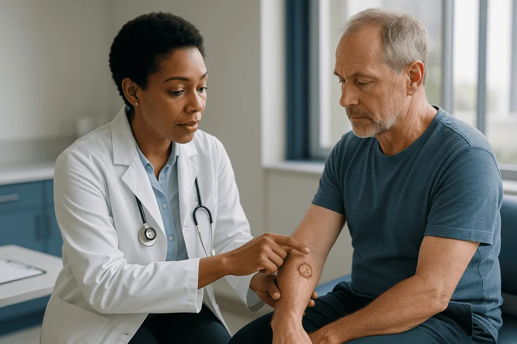
People are often startled to hear they need a second operation on a site that already looks healed. The reason is that the safe margin cannot be chosen until the depth is known, and the depth cannot be known until the mole is under a microscope. The NHS describes this sequence as standard care, with the wide excision performed after the initial biopsy result.
For melanomas beyond a certain thickness, the team may also propose a sentinel lymph node biopsy at the same time as the wide excision. Lymph nodes are small filters in the immune system; the sentinel node is the first one the skin at the melanoma site drains into. A tracer dye and a small amount of radioactive marker are injected near the scar, the node that picks them up is identified and removed, and it is examined for stray cancer cells. Mayo Clinic notes this test helps determine whether melanoma has begun to spread and shapes further planning.
Wide local excision is also used for some high-risk squamous cell carcinomas. The principle is identical: match the margin to the tumor’s behavior, remove it in one piece, and let the pathologist confirm the edges are clean before calling the surgical part finished.
Standard excision, Mohs surgery, or something else? A side-by-side comparison
Not every skin cancer is treated the same way, and the differences come down to how the margin is checked and how much tissue is spared. The table summarizes the main options that guideline-level sources such as the NHS and Mayo Clinic describe for skin cancer.
| Approach | How it works | Margin checked? | Typically considered for |
|---|---|---|---|
| Standard excision | Tumor plus a pre-set margin removed in one piece; wound closed the same day | Yes, by a pathologist days later | Most basal cell, squamous cell, and melanoma on trunk and limbs |
| Mohs micrographic surgery | Thin layers removed and examined under the microscope while you wait; repeated until edges are clear | Yes, same day, 100% of the edge examined | Face, ears, nose, hands; recurrent or ill-defined tumors |
| Curettage and electrocautery | Tumor scraped away and base sealed with heat | No formal margin | Selected small, low-risk basal or squamous cell carcinomas |
| Cryotherapy | Freezing with liquid nitrogen destroys the lesion | No | Some superficial, low-risk lesions and precancerous patches |
| Topical treatments and photodynamic therapy | Cream or light-activated medicine destroys abnormal cells | No | Superficial basal cell carcinoma and precancerous change in selected cases |
| Radiotherapy | Targeted radiation over several sessions | No | People unsuitable for surgery, or as an add-on after incomplete removal |
Mohs surgery deserves a closer look because it is often misunderstood as a stronger operation. It is not more aggressive; it is more precise. Mayo Clinic describes it as removing skin one thin layer at a time, with each layer mapped and checked under a microscope during the same visit, so the surgeon stops the moment the edges are clear. That precision spares tissue where tissue is scarce, which is why it is favored on the face. The trade-off is time, specialized training, and availability. For a small basal cell carcinoma on the back, a standard excision achieves the same goal with less fuss. Your team’s recommendation reflects that balance, not a ranking of better and worse.
How painful is skin excision, really?
The honest answer from patients and clinicians alike is that the sharpest moment is the first few seconds of the anesthetic injection. Local anesthetics such as lidocaine sting as they spread through the tissue, a sensation many people compare to a bee sting or a vaccination that lasts a little longer. Surgeons often buffer the solution or inject slowly to soften this. Within a minute or two the area goes numb, and from then on you should feel pressure, tugging, and the odd vibration from the cautery, but not pain. If you do feel a sharp sensation, say so; more anesthetic can be added.
Fear tends to hurt more than the procedure. Being awake and hearing the instruments is unsettling for some people, and it is reasonable to ask for a running commentary, or for silence, or to bring headphones. Some teams offer a mild oral sedative for anxious patients; whether that suits you is a decision for the treating clinician, not something to arrange yourself.
Afterward, the numbness wears off over a few hours and the site feels bruised and tight. Mayo Clinic’s guidance on Mohs surgery, which involves the same kind of wound, describes discomfort that is usually manageable with ordinary pain relief and that settles over the first days. Areas under tension, such as the shin or the back, ache more than loose skin on the cheek. Most people describe soreness rather than severe pain, and increasing pain after the first couple of days is a reason to call, not to push through.
Before taking any pain reliever, check with your team. Some common medicines increase bleeding tendency, and your surgeon may prefer you avoid them for a short period. That advice, like everything here, is theirs to give.
Closure options after skin cancer excision: stitches, flaps, grafts, or letting it heal
Once the tumor is out, the surgeon is left with a hole, and how it is filled shapes both the healing timeline and the final scar. There are four main strategies, and choosing among them is part craft, part geometry.
Primary closure is the simplest: the two sides of the elliptical wound are brought together and stitched in a straight line. Most excisions on the trunk, arms, and legs are closed this way. Surgeons frequently use two layers of stitches. Deep absorbable stitches, which dissolve on their own over weeks, hold the tension so the surface line can heal without being pulled apart; fine surface stitches, either absorbable or removed at a follow-up visit, align the skin edges neatly.
When the wound is too wide to close directly, or closing it would distort a nearby feature, a local flap may be used. A flap is a piece of neighboring skin, still attached by its own blood supply, that is lifted and rotated or advanced into the defect. The donor area then closes in a line hidden along a natural crease. Flaps are common on the nose, cheek, and forehead.
A skin graft is different: a thin sheet of skin is taken entirely free from another site, often behind the ear, the collarbone area, or the thigh, and laid into the wound, where it must grow a new blood supply over roughly a week. Grafts work well for shins and scalps but can look paler or shinier than the surrounding skin.
Healing by secondary intention means leaving the wound open, dressed, and letting the body fill it from the base and edges. It sounds alarming but produces surprisingly good results in concave areas such as the inner corner of the eye or the temple, at the cost of several weeks of dressing changes. For melanoma and some ill-defined tumors, surgeons may deliberately delay a flap or graft until the pathology confirms clear margins, so that no complex repair has to be undone.
What happens to the sample after it leaves the room?
The jar on the counter contains the most important part of the day. Before the specimen goes anywhere, the surgeon orients it, typically with a suture at one edge or ink of different colors on different sides, and records that on the request form so the pathologist can say not just whether cancer reaches an edge but which edge. It then drops into formalin, a preservative that fixes the tissue and stops it breaking down.
In the pathology laboratory, technicians measure and describe the specimen, then paint its outer surface with colored ink that survives processing, so under the microscope the true cut surface is unmistakable. The tissue is sliced at intervals along its length, a technique often called bread-loafing because the pieces resemble slices from a loaf. Each slice is dehydrated, embedded in a block of paraffin wax, shaved into sections thinner than a sheet of paper, mounted on glass slides, and stained with dyes that turn nuclei blue-purple and the surrounding tissue pink.
A pathologist, a doctor who specializes in diagnosing disease from tissue, then reads the slides. They confirm the type of cancer, note features that affect risk, and measure the distance from tumor to inked edge. One honest limitation is worth knowing: bread-loafing samples the specimen at intervals, so a standard excision report describes the margins on the sections examined rather than every micron of the edge. That is why Mohs surgery, which maps and examines the entire margin on frozen sections during the visit, is preferred where recurrence would be hard to manage.
Timing varies by laboratory. The NHS advises that results can take a few weeks to come back, and melanoma specimens sometimes need extra stains or a second opinion, which adds time. A slow report usually means thoroughness, not bad news.
Reading the pathology report: clear, close, and involved margins
Pathology reports are written for doctors, but three words on them carry most of the meaning for you. Clear, or negative, margins mean no cancer cells were seen at the inked edges of the tissue examined. Close margins mean cancer approached the edge within a small distance, sometimes fractions of a millimeter. Involved, or positive, margins mean cancer cells reached the cut edge, so some may remain in the skin.
Beyond margins, the report records the specific diagnosis and its subtype, because these behave differently. Basal cell carcinoma is described as nodular, superficial, infiltrative, or other patterns, and the infiltrative types are more likely to have subtle roots. Squamous cell carcinoma is graded by how abnormal the cells look and how deeply they invade. Any spread along nerves, called perineural invasion, is flagged because it raises the risk of the cancer returning. For melanoma, the report includes Breslow thickness, whether the surface has broken down, and how many cells are dividing, all of which feed into staging.
An involved or close margin is not a failure and not an emergency. Depending on the tumor type, location, and your circumstances, the options include a further excision aimed at the specific edge the pathologist identified, referral for Mohs surgery to clear the remaining tissue precisely, a course of radiotherapy, or in some cases of low-risk basal cell carcinoma, close observation, since not every remaining cell goes on to regrow. Complex decisions are usually taken at a multidisciplinary team meeting, a regular conference where surgeons, dermatologists, pathologists, and oncologists review cases together.
Ask for a copy of your report and ask your team to translate any word you do not recognize. Understanding those three margin terms will make every later conversation clearer.
Skin cancer excision recovery time: what the first days and weeks usually look like
Recovery unfolds in fairly predictable phases, though your surgeon’s written instructions override any general timeline.
In the first day or two, the wound is covered by a firm pressure dressing to limit bleeding and swelling. Mayo Clinic’s guidance for Mohs wounds, which heal in the same way as excisions, describes keeping the initial dressing dry and in place for the period your surgeon specifies before switching to daily care. Mild oozing onto the dressing is normal; steady bleeding that soaks through is not. Keeping the area elevated where possible, such as sleeping with an extra pillow after a facial excision, reduces throbbing.
Over the first week, you will likely be cleaning the site gently, applying a thin layer of ointment as instructed, and covering it with a fresh dressing. Bruising around the wound, particularly near the eye, can look dramatic and then fade through yellow and green. Stitches on the face are commonly removed sooner than those on the body, where the skin is thicker and under more tension; your team will give you a specific date, which is often within about one to two weeks, and absorbable stitches dissolve on their own over several weeks.
For roughly two to three weeks, most surgeons advise avoiding heavy lifting, vigorous exercise, and swimming, because tension on a healing line can widen the scar or open it. Legs heal more slowly than the face because circulation is poorer, and grafts need particular protection while they establish a blood supply.
The scar itself keeps changing for much longer than people expect. It typically looks its reddest and firmest around the first two months, then softens and fades over the following year, a maturation process Cleveland Clinic describes as continuing for many months. Sun protection on the new scar during that period helps prevent it from darkening permanently.
What are the risks and complications of a skin cancer excision procedure?
Excision is low-risk surgery, but no surgery is risk-free, and a neutral list is more useful than reassurance. Mayo Clinic and the NHS describe the same core complications for skin surgery, and they fall into a handful of groups.
Bleeding is the most common early problem, especially for people on blood-thinning medicines or those with wounds on the scalp, which bleeds generously. Most bleeding stops with firm, uninterrupted pressure; a collection of blood under the skin, called a hematoma, occasionally needs to be drained. Infection shows up as spreading redness, warmth, increasing pain, and sometimes discharge, and is more likely in wounds on the lower leg, in people with diabetes, or after long procedures. Wound dehiscence, meaning the stitched edges pull apart, happens most often when a wound under tension is stretched too soon.
Nerve injury is a specific concern in certain locations. A branch of the facial nerve runs shallowly across the temple, and cutting near it can weaken the eyebrow on that side. Numbness around any scar is expected and usually improves over months as small sensory nerves regrow, though a patch may remain permanently dulled. Flaps and grafts carry their own risks: a flap tip or a graft can lose its blood supply and partially die, leaving an area that heals slowly or needs revision.
Every excision leaves a scar. Most flatten and fade, but some people, particularly on the chest, shoulders, and in darker skin tones, form raised hypertrophic scars or keloids, which are overgrowths of scar tissue extending beyond the original wound. The final risk is the one the whole procedure aims to prevent: incomplete removal or later recurrence, which is why the pathology report and follow-up skin checks matter as much as the operation itself. Alternatives, from Mohs to radiotherapy, exist precisely so that the plan can be tailored when one of these risks weighs heavily.
What people often get wrong about skin cancer removal
Several persistent beliefs make excision seem either scarier or more trivial than it is. Correcting them in plain terms is worth a few minutes.
The first is that a spot which looks like a pimple must be a pimple. Basal cell carcinoma frequently appears as a small, pearly or flesh-colored bump that may bleed, crust, and seem to heal before returning, which is why it is so often dismissed for months. People searching for pictures of skin cancer that looks like a pimple are usually trying to reassure themselves, but no photograph can settle the question. The Mayo Clinic’s advice is simple: a blemish that has not resolved after several weeks, or that keeps bleeding, deserves a professional look rather than a comparison with images online.
The second is that cutting into a cancer spreads it. This fear is old and understandable, but there is no evidence from mainstream oncology that a properly performed excision seeds skin cancer into the body. Surgery remains the standard first treatment precisely because removal with a margin is effective.
The third is that non-melanoma skin cancers do not matter. Basal cell carcinoma rarely spreads, but left alone it destroys tissue locally, and the CDC notes that squamous cell carcinoma can spread to other parts of the body if not treated. Small lesions removed early need smaller margins and simpler closures.
The fourth is that clear margins mean the story is over. Clear margins mean that particular tumor is gone. The skin that grew one cancer has usually had decades of sun exposure and is capable of growing another, so follow-up skin checks and daily sun protection remain part of the plan. The fifth is that people with darker skin cannot get skin cancer. They can, and because diagnosis often comes later, the CDC emphasizes that everyone, regardless of skin tone, benefits from awareness and protection.
Questions to ask your care team before and after the procedure
A good consultation leaves you knowing not just what will happen but why this plan was chosen over the alternatives. Bring these questions written down; consultations are short and adrenaline erases memory.
- What type of skin cancer do you think this is, and has a biopsy confirmed it?
- How wide a margin are you planning, and what guides that choice for my tumor?
- Is standard excision the best option here, or would Mohs surgery, curettage, or a non-surgical treatment be reasonable alternatives for me?
- How do you expect to close the wound, and if that changes during the procedure, what are the possibilities?
- Roughly where will the scar sit, and how long will it be?
- Should I continue all my regular medicines, including anything that thins the blood, exactly as prescribed? Please confirm with whoever prescribes them.
- Will I be able to drive home, and should someone come with me?
- How do I care for the dressing, when can I shower, and when will stitches be removed or dissolve?
- What activities should I avoid, and for how long?
- When and how will I receive the pathology results?
- If the margins are not clear, what are the likely next steps?
- How often should I have skin checks afterward, and who does them?
After the procedure, add a second round: which specific signs should prompt me to call, and which number do I call outside office hours? Ask for a copy of the pathology report when it arrives and request that any unfamiliar term be explained. None of these questions is a challenge to your team’s judgment. They are how you become an informed participant in decisions that remain, rightly, with the people treating you.
When to call your doctor
Most excision wounds heal without drama, but a few signs mean you should not wait for your scheduled follow-up. Contact your surgical team the same day if you notice any of the following, and if you cannot reach them, use an urgent care service.
- Bleeding that does not stop after firm, continuous pressure for the length of time your surgeon specified, or that soaks through the dressing repeatedly
- Redness spreading outward from the wound, increasing warmth or swelling, thick or foul-smelling discharge, or a fever
- Pain that worsens after the first couple of days rather than easing, or pain the recommended relief does not touch
- Stitches that pull apart or a wound that gapes open
- A skin graft or flap that turns dark purple, black, or pale white
- A rapidly enlarging, tense, painful lump under the wound, which can indicate blood collecting
- New weakness or drooping in the face, or numbness that spreads rather than improves
- A rash, hives, or swelling suggesting a reaction to a dressing, ointment, or medicine
Call emergency services immediately for chest pain, difficulty breathing, fainting, or facial swelling that affects your airway; these are rare but need emergency care, not a message to the clinic.
Beyond the wound itself, seek medical advice for any new or changing spot on your skin at any time after treatment. The NHS advises that a sore that does not heal within a few weeks, a growth that bleeds or crusts repeatedly, or a mole that changes in size, shape, or color should be assessed by a clinician. Having had one skin cancer is the strongest predictor of another, and the earlier a new lesion is checked, the smaller the excision it is likely to need. Your treating team, not a search engine, is the right place to take that worry.
Frequently asked questions
How painful is a skin excision?
The sharpest moment is usually the anesthetic injection, which stings for a few seconds before the area goes numb. During the procedure you feel pressure and tugging but should not feel pain, and more anesthetic can be given if you do. Afterward the site feels bruised and tight for a few days, with soreness rather than severe pain in most people. Ask your team which pain relievers are appropriate before taking anything.
How long does it take to recover from skin cancer excision surgery?
Most people return to desk work within a day or two, while the wound itself needs one to two weeks before stitches are removed and about two to three weeks of avoiding heavy lifting, vigorous exercise, and swimming, depending on your surgeon’s instructions. Legs heal more slowly than the face. The scar continues to soften and fade for a year or more. Your written care plan sets the exact timeline.
Can skin cancer be treated successfully with excision?
For most basal cell, squamous cell, and early melanoma, surgical removal with clear margins is the standard and effective treatment, and the NHS describes surgery as the main approach for both non-melanoma and melanoma skin cancer. Outcomes depend on the cancer type, its depth, and whether it has spread, which is why the pathology report matters. Your team will explain what your specific result means and whether any further treatment or monitoring is advised.
Can skin cancer look like a pimple, and can I compare it with pictures online?
Yes, basal cell carcinoma often appears as a small pearly or flesh-colored bump that bleeds, crusts, and seems to heal before returning. Photographs online cannot confirm or rule out cancer, because benign and malignant spots overlap in appearance. A useful rule from mainstream guidance is that a blemish persisting for several weeks or bleeding repeatedly deserves a clinician’s examination, not a picture comparison.
What does wide local excision mean for skin cancer?
Wide local excision is a second, deliberately larger removal of skin and underlying fat around the scar from a diagnostic biopsy, most often for melanoma. The width of the margin is chosen after the pathologist has measured the tumor’s depth, because that depth predicts how far microscopic cells may extend. It is sometimes combined with a sentinel lymph node biopsy to check whether the melanoma has begun to spread.
Why do I need a second surgery if the mole was already removed?
Because the safe margin cannot be decided until the pathologist has confirmed the diagnosis and measured how deep the cancer went. The first procedure is diagnostic and uses only a narrow rim of skin; the second, wide local excision, takes the evidence-based margin for that specific depth. Doing it in two steps avoids removing excess skin from people whose lesions turn out to be benign or very thin.
How long does it take to get the pathology results after excision?
It varies by laboratory, but the NHS advises that results can take a few weeks. Tissue must be preserved, embedded in wax, sliced, stained, and read by a pathologist, and melanoma specimens sometimes need extra stains or a second opinion, which adds time. A slower report usually reflects thoroughness rather than bad news. Ask your team how and when you will be told.
What happens if the margins are not clear?
The report will say which edge is involved or close, and your team will discuss options, which may include a targeted re-excision of that edge, referral for Mohs surgery, a course of radiotherapy, or in some low-risk basal cell carcinomas, careful observation. Complex cases are often reviewed at a multidisciplinary team meeting. It is a common situation with a clear pathway, not a sign the operation failed.
What are the different skin cancer excision closure options?
The wound may be stitched directly in a straight line, closed with a flap of neighboring skin that keeps its own blood supply, covered with a skin graft taken from elsewhere, or left to heal from the base over several weeks. The choice depends on the wound’s size, location, skin laxity, and whether the surgeon prefers to wait for clear margins before a complex repair. Each option produces a different scar pattern.
Will I have a scar after skin cancer excision, and will it fade?
Every excision leaves a scar, typically a line roughly three times the width of the lesion for a standard closure. It usually looks reddest and firmest around the second month, then flattens and fades over a year or longer. Some people, especially on the chest, shoulders, or in darker skin tones, form raised scars. Sun protection on the healing scar helps prevent permanent darkening; ask your team about scar care.
References
This article is for general information only and is not a substitute for professional medical advice. Please consult a qualified doctor about your individual situation.

More from the Blog

Before a Mastectomy: Blood Thinners, Herbal Products, Smoking and the Days Leading Up
Preparing for a mastectomy usually means giving your surgical and anesthesia team a complete list of prescription medicines, over-the-counter drugs and herbal products, following…
Sperm Banking Before Testicular Cancer Treatment: Why Your Team Raises It Early
Sperm banking before testicular cancer treatment means freezing one or more semen samples, ideally before surgery, chemotherapy or radiotherapy begins, because these treatments can…
How Long Do International Patients Stay After Mohs Surgery Before Flying Home?
No published guideline sets a fixed waiting period for flying after Mohs surgery. In practice, surgeons often ask patients with a simple closure to…
When Immunotherapy Enters a Cancer Treatment Plan: First Line, After Surgery or Later
Immunotherapy is used at several points in a cancer treatment plan, not only late. Depending on the cancer type, its stage and biomarker test…
Why Head Cancer Follow-Up Keeps Examinations and Scans on a Schedule After Treatment
Head and neck cancer follow up scans and examinations stay on a schedule because most recurrences appear within the first few years after treatment,…
Ablation or Surgery for Liver Cancer? What Makes Each Option Suitable
Ablation and surgery are both used to treat liver cancer that has not spread beyond the liver. Ablation destroys a small tumor in place…