

What a Sentinel Lymph Node Biopsy Tells Your Team About Melanoma Spread

Key Takeaways
- A sentinel lymph node biopsy stages melanoma rather than treating it; the wide excision of the skin is the treatment step.
- The procedure is generally discussed for melanomas thicker than about 0.8 mm or with ulceration, and usually not for very thin, non-ulcerated stage IA tumors.
- In the MSLT-I trial, roughly one in six people with intermediate-thickness melanoma had a positive sentinel node, meaning most results come back clear.
- A positive node reclassifies the melanoma as stage III, a wide category in which deposit size and node count matter more than the stage label alone.
- The MSLT-II trial found that removing all remaining nodes after a positive sentinel node did not improve melanoma-specific survival and quadrupled the rate of lymphedema compared with ultrasound surveillance.
- Lymphedema after sentinel biopsy alone affected about 6 percent of MSLT-II participants, and seroma, numbness, and minor wound problems are the more common complications.
A sentinel lymph node biopsy for melanoma removes the first one or two lymph nodes that drain the skin where the melanoma grew, so a pathologist can check them for cancer cells. A negative result means no spread was found in those nodes; a positive result moves the melanoma to stage III and shapes follow-up, imaging, and treatment discussions with the care team.
The mole came off two weeks ago, and the pathology report said melanoma. Now the surgeon is describing a second, smaller operation: an injection near the scar, a scan, a shallow cut in the armpit, and the removal of a node or two that most people never knew they had. The question that hangs in the room is the one nobody quite says out loud. Has it gone anywhere yet?
A sentinel lymph node biopsy for melanoma exists to answer that question as precisely as current surgery can. It does not treat the melanoma; the wide excision does that. It gathers information, and the information changes what happens next.
This explainer walks through what the procedure involves, who is usually offered it, what the pathologist is actually looking for, and, most importantly, what a positive or negative result does and does not tell you about the road ahead.
What is a sentinel lymph node biopsy for melanoma?
Lymph nodes are small, bean-shaped filters scattered along the lymphatic vessels that drain fluid from every part of the skin. When melanoma cells leave the original tumor, the lymphatic channels are one of the first routes they take, and the first node those channels reach is called the sentinel node. It stands guard, in the old military sense, at the gate.
A sentinel lymph node biopsy for melanoma is a surgical procedure that identifies that gatekeeper node, removes it, and sends it for detailed examination. The logic is simple: if the sentinel node is clear, the nodes further along the chain are very likely clear too. If it contains melanoma cells, the team knows the disease has begun to travel and can plan accordingly.
The distinction between this and the original skin surgery matters. The wide local excision, in which the surgeon removes the melanoma with a rim of healthy skin around it, is the treatment step. The sentinel node biopsy is the staging step, staging being the process of working out how far a cancer has spread. Mayo Clinic describes it as a way to find out whether cancer has spread beyond the primary tumor without removing an entire group of nodes.
Both procedures are often done under the same anesthetic. The surgeon widens the scar from the first biopsy, then makes a separate small incision over the node basin, usually the armpit, groin, or neck, depending on where the melanoma sat. For a melanoma on the calf, the sentinel node is most often in the groin; for one on the shoulder blade, the armpit or the neck. Sometimes the tracer reveals drainage to two basins at once, and the surgeon samples both.
How the procedure actually works, step by step
Finding a single node the size of a lentil inside the fat of an armpit is not guesswork. The sentinel node is mapped, not searched for.
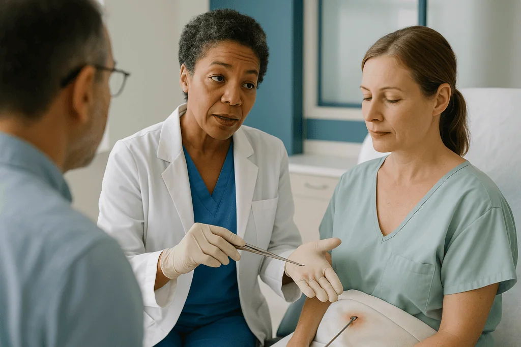
The morning of surgery, or occasionally the day before, a small amount of a weak radioactive tracer is injected into the skin around the melanoma scar. The injections sting briefly. Over the next hour or so the tracer travels along the same lymphatic channels a cancer cell would use and collects in the first node it meets. A scan called lymphoscintigraphy, essentially a camera that photographs where the radioactivity has pooled, shows the surgeon which basin to open and roughly how deep the node lies.
In the operating room, many surgeons add a second marker: a blue dye injected at the same site just before the incision. The dye turns the lymphatic vessels and the sentinel node a vivid blue, visible to the naked eye. The surgeon then passes a handheld gamma probe, a wand that clicks louder as it nears radioactivity, over the skin to pinpoint the node before cutting.
The node that is both blue and radioactive is removed, along with any other node that reads strongly on the probe. Mayo Clinic notes that anywhere from one to a few nodes may be taken. The incision is typically a few centimeters long and is closed with dissolvable stitches or fine sutures. The whole node portion of the operation often takes well under an hour, though the combined procedure with the wide excision runs longer.
Most people go home the same day. The blue dye may tint the urine green or blue for a day or two and can leave a faint blue mark at the injection site that fades over weeks; both are expected and harmless.
What stage of melanoma requires a lymph node biopsy, and who is asked to wait?
Not every melanoma warrants this second operation. The decision rests mainly on features in the original pathology report, and two features carry the most weight.
The first is Breslow thickness: how deep, in millimeters, the melanoma has grown from the skin surface. Thicker tumors have had more chance to reach the lymphatics. The second is ulceration, meaning the skin over the melanoma had broken down under the microscope, a sign associated with more aggressive behavior. Other details such as a high mitotic rate, the count of dividing cells, can tip the balance in borderline cases.
Very thin melanomas, those under roughly 0.8 mm without ulceration, are usually classed as stage IA, and guideline bodies generally do not recommend a sentinel node biopsy because the chance of finding spread is low enough that the operation’s risks outweigh its information. The NHS explains that a sentinel node biopsy is typically discussed when the melanoma is stage IB or higher, and Mayo Clinic frames the same threshold around thickness and ulceration.
At the other end, someone whose nodes are already enlarged on examination or ultrasound usually does not need a sentinel biopsy at all. A needle sample of the suspicious node answers the question more directly, and the team moves to imaging of the rest of the body.
Age, other health conditions, and personal preference all enter the conversation. An older adult with significant heart or lung disease might reasonably decide, with the team, that the information is not worth a general anesthetic. The melanoma lymph node biopsy stage threshold is a starting point for discussion, not a rule that bypasses the person in the chair.
Why not just scan the nodes, or remove them all?
People often ask why a scan cannot do the job. The honest answer is resolution. Sentinel node deposits are frequently microscopic, sometimes a cluster of a few dozen cells. CT scans, PET scans, and ultrasound detect nodes that are enlarged or metabolically busy; they cannot see a deposit smaller than a grain of sand. A normal scan is reassuring but does not exclude microscopic spread, which is exactly what the biopsy is designed to find.
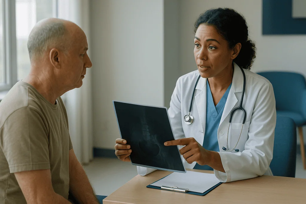
The opposite question, why not remove all the nodes to be safe, has a clearer answer from history. For much of the twentieth century, surgeons did remove the entire regional node basin in people with thicker melanomas, an operation called elective lymph node dissection. Most of those patients turned out to have no cancer in any node, yet many lived with permanent arm or leg swelling called lymphedema, a consequence of cutting the lymphatic drainage. Randomized trials failed to show a survival benefit for the group as a whole.
The sentinel node technique, developed in the early 1990s, offered a middle path: sample the one node most likely to be involved, spare the rest. The Multicenter Selective Lymphadenectomy Trial (MSLT-I), published in the New England Journal of Medicine, randomized more than 2,000 people to sentinel biopsy or observation and confirmed that the procedure accurately identified node status and gave earlier, more precise staging, though it did not improve melanoma-specific survival across the whole study population.
The realistic alternatives to a sentinel biopsy today are therefore not scans instead of surgery, but observation: regular clinical examination of the node basins, sometimes with scheduled ultrasound, accepting that any spread will be found later, when a node becomes palpable. Some people choose that path knowingly, and it is a legitimate option to discuss.
What the pathologist looks for, and why a negative result still has limits
Once removed, the node is not simply glanced at under a microscope. It is sliced into thin sections at several levels, stained with standard dyes, and then stained again with immunohistochemistry, a technique that uses antibodies to light up proteins found on melanoma cells but not on normal node tissue. This is how deposits of a handful of cells become visible.
The report that comes back a week or two later describes whether melanoma cells were present, how many nodes were examined, the size of the largest deposit measured in millimeters, and where in the node the cells sat. A deposit confined to the outer rim behaves differently from one spreading through the whole node or breaking through its capsule into the surrounding fat.
A negative result, meaning no melanoma cells found, is genuinely good news and the outcome for the large majority of people who have the procedure. In MSLT-I, roughly one in six people with intermediate-thickness melanoma had a positive sentinel node, which means five in six did not.
The limit is that no test is perfect. A small proportion of people with a negative sentinel node later develop melanoma in that same node basin, a so-called false negative. It can happen when the tracer maps to the wrong node, when cells bypassed the sentinel node, or when a tiny deposit was missed despite careful sectioning. This is why a negative result does not end follow-up. Skin checks and node examinations continue on a schedule set by the team, and any new lump near the scar or in the basin is taken seriously regardless of the earlier result.
What happens if a sentinel node biopsy is positive for melanoma?
A positive sentinel node melanoma result means cancer cells have reached at least one lymph node. The melanoma is reclassified as stage III, whatever its original thickness. That word lands hard, and it is worth slowing down over what it does and does not mean.
Stage III is a wide category. It includes someone with a 0.3 mm cluster of cells in a single node and someone with several bulky, matted nodes. The two situations carry very different outlooks, and the staging system subdivides stage III into groups partly on that basis. Your report’s details, the size of the deposit, the number of positive nodes, the presence of ulceration in the original melanoma, are what your team will weigh, not the stage label alone.
Practically, several things usually follow. The team typically orders whole-body imaging, often a CT or PET-CT scan, to check whether melanoma has traveled beyond the regional nodes. Blood tests may be added. The original tumor tissue is frequently sent for molecular testing to look for a mutation in a gene called BRAF, which drives cell growth in roughly half of melanomas and determines whether a particular class of targeted medicine is even an option.
Then comes the central conversation: what to do about the remaining nodes in that basin, and whether to offer additional treatment aimed at reducing the chance of the melanoma returning. Both questions have shifted substantially in the past decade, and the next sections take them in turn. The case is usually reviewed by a multidisciplinary team, a meeting of surgeons, oncologists, dermatologists, pathologists, and radiologists, before a recommendation reaches you.
How serious is melanoma in the lymph nodes? Understanding stage III
Melanoma that has reached a lymph node is more serious than melanoma confined to the skin, because it has proven it can travel. The nodes themselves are rarely the danger; the concern is that cells capable of reaching a node may also have reached the bloodstream and seeded distant organs, where they may remain undetectable for months or years.
That is the sober part. The other part is that stage III is, by definition, still regional disease. The melanoma has not been shown to have spread to distant organs, and the goal of treatment at this stage is to prevent that from happening or to catch it early if it does. People with a single microscopic deposit and a non-ulcerated primary sit at the more favorable end of the stage III spectrum; those with multiple involved nodes and an ulcerated primary sit at the other.
Outlook figures for stage III vary so widely across these subgroups that a single number would mislead more than inform. Your oncologist can quote ranges specific to your substage from the current staging manual and explain what they are based on. It is reasonable to ask for that, and equally reasonable to say you would rather not hear numbers yet.
Where the melanoma sat on the body, the tumor’s genetic profile, and your overall health all shape the picture. So does the fact that treatment for stage III melanoma has changed more in the last ten years than in the previous fifty. A positive sentinel node today opens the door to options that did not exist for people diagnosed a generation ago, and follow-up is more structured and more sensitive than it once was.
Completion dissection versus ultrasound surveillance: what the trials showed
Until recently, a positive sentinel node was almost automatically followed by a completion lymph node dissection, a second operation to remove all the remaining nodes in that basin. The reasoning was that if one node was involved, others might be, and taking them out would stop further spread.
The second Multicenter Selective Lymphadenectomy Trial (MSLT-II) tested that assumption directly. It randomized nearly 2,000 people with a positive sentinel node to either immediate completion dissection or active surveillance with regular ultrasound of the node basin, with surgery only if a node later became involved. The trial, published in the New England Journal of Medicine, found that immediate dissection did not improve melanoma-specific survival. It did give slightly better control of disease within the node basin and more staging information, at the price of a markedly higher rate of lymphedema.
| Approach after a positive sentinel node | What it involves | What MSLT-II found |
|---|---|---|
| Completion lymph node dissection | Second operation removing all remaining nodes in the basin | No improvement in melanoma-specific survival; better regional control; lymphedema in about 24 percent |
| Active surveillance | Clinical examination plus scheduled ultrasound of the basin; surgery only if a node becomes involved | Equivalent melanoma-specific survival; lymphedema in about 6 percent |
Since those results, most guideline bodies have moved toward surveillance as the usual path for people with low-volume sentinel node disease, reserving dissection for particular situations such as a large deposit, disease breaking out of the node, or circumstances where surveillance would be impractical. The choice still belongs to you and your team, and the trial’s findings are one input among several.
Treatments that may follow a positive result, by class
A positive sentinel node often opens a discussion about adjuvant therapy, meaning treatment given after surgery, when no cancer can be seen, to reduce the chance of it coming back. Two broad classes are used in stage III melanoma, and understanding how they work helps make sense of the conversation.
Immune checkpoint inhibitors are antibody medicines that release a brake on the immune system. Melanoma cells often exploit a signaling pathway, involving a protein called PD-1 on immune cells, to switch off the T cells that would otherwise attack them. Blocking that pathway lets the immune system recognize and destroy residual melanoma cells. These medicines are given by intravenous infusion, and their side effects come from the same mechanism: an unleashed immune system can also inflame healthy tissue, from the skin and gut to the thyroid and other glands, which is why regular blood monitoring accompanies treatment.
Targeted therapy applies only to melanomas carrying a BRAF mutation. The mutated BRAF protein sits in a growth-signaling chain and keeps it permanently switched on. BRAF inhibitors and MEK inhibitors, taken as tablets, block two consecutive steps in that chain. They tend to cause fever, fatigue, skin changes, and joint aches rather than immune inflammation.
Whether adjuvant treatment is offered depends on the substage, the deposit size, your other health conditions, and your own priorities about trading months of side effects for a reduced but not eliminated risk. Not everyone with a positive node is offered it, and not everyone who is offered it chooses it. The prescribing oncologist explains the specific medicines, their monitoring, and typical treatment length; nothing here substitutes for that conversation.
Sentinel node biopsy melanoma recovery: the days and weeks that follow
Recovery from the node portion of the surgery is usually gentler than people expect, though the combined wound from the wide excision can make the first days more uncomfortable than the armpit or groin incision itself.
The first two or three days bring soreness at the node site, bruising, and a feeling of tightness when raising the arm or walking if the groin was involved. Over-the-counter pain relief recommended by the team is usually sufficient; ask before taking anything, since some common pain relievers increase bleeding. The dressing typically stays on for several days, and showering is usually permitted once the team confirms the wound is sealed.
Many people return to desk work within a week, according to the NHS and Mayo Clinic guidance on this procedure, while jobs involving heavy lifting or repetitive use of the affected limb may need two to three weeks. Driving should wait until you can perform an emergency stop without pain and are no longer taking sedating medicines. Gentle movement of the shoulder or hip from the first day helps prevent stiffness; a few slow range-of-motion exercises, if the team provides them, are worth doing.
The pathology result generally arrives one to two weeks after surgery. That waiting period is often the hardest part of the whole experience, and it is normal to find concentration difficult. Ask at discharge exactly how and when the result will be communicated so you are not left checking a phone.
A firm ridge or small fluid pocket under the incision is common in the second and third weeks and usually settles without intervention. Numbness in a patch of skin near the scar can persist for months and sometimes permanently; it comes from small sensory nerves cut during surgery rather than from any problem with healing.
Sentinel node biopsy side effects and risks in plain terms
The procedure is low-risk by surgical standards, but low-risk is not no-risk, and honest counseling covers the following.
Seroma, a collection of lymphatic fluid under the incision, is the most frequent complication. It feels like a soft, sometimes wobbly swelling. Small seromas absorb on their own; larger ones may need to be drained with a needle in clinic, occasionally more than once. Wound infection occurs in a minority and shows as spreading redness, increasing pain, warmth, or discharge; it usually responds to a course of antibiotics chosen by the team.
Lymphedema, persistent swelling of the arm or leg from disrupted lymphatic drainage, is the complication people fear most. After sentinel biopsy alone it is uncommon; in the MSLT-II surveillance arm, where participants had only the sentinel procedure, about 6 percent developed it, compared with about 24 percent after full dissection. Groin biopsies carry a somewhat higher risk than armpit biopsies because the leg has further to drain against gravity.
Allergic reactions to the blue dye are rare but can be serious, ranging from hives to a drop in blood pressure during surgery; the anesthesia team monitors for this and treats it if it occurs. The radioactive tracer dose is very small and poses no meaningful risk to you or those around you.
Nerve irritation causing numbness or tingling in nearby skin is common and usually improves over months. Rarely, in the neck, a nerve supplying a shoulder muscle can be affected, causing weakness. Bleeding requiring a return to the operating room is unusual.
Finally, there is the risk that comes with any general anesthetic, which the anesthesiologist assesses individually and which rises with age and other health conditions. For most people it is small, but it is a legitimate part of the decision.
What people often get wrong about sentinel node biopsy for melanoma
Misunderstandings cluster around a few themes, and clearing them up tends to lower anxiety more than any reassurance.
The first is that the biopsy itself treats the cancer. It does not. Removing one or two nodes that may or may not contain cells is not a therapeutic step; the value lies in the information. Some people, hearing that MSLT-I found no overall survival benefit from the procedure, conclude it is pointless. The trial’s own authors argued the opposite: the procedure identifies the minority who need further care and spares the majority unnecessary surgery.
The second is that a negative result means the melanoma is gone for good. A clear sentinel node is strongly reassuring but does not eliminate the possibility of later recurrence, whether through a false negative or through cells that traveled by the bloodstream rather than the lymphatics. Follow-up continues for years for this reason.
The third is that surgery can spread the cancer. The idea that cutting into a node releases cells has no support in trial data; MSLT-I compared surgery with observation in thousands of people and found no such effect.
The fourth is that a positive node means the melanoma is everywhere. Stage III is regional disease. Whole-body imaging after a positive result frequently shows nothing beyond the node basin.
The fifth is that a positive node automatically leads to removing all the nodes. As MSLT-II showed, surveillance is now a standard option for many people.
A last, quieter error: believing the outcome was determined by how quickly the biopsy happened. Within the typical planning window of a few weeks, timing does not change what the node contains. The cells were either there or they were not.
Questions to ask your care team before and after the procedure
Consultations move quickly, and the best questions are the ones written down in advance. These are the ones that tend to matter most, grouped by the moment they arise.
Before surgery, ask what features of your pathology report led to the recommendation, and what the team estimates the chance of a positive node to be in your case. Ask which basin or basins the surgeon expects to sample, whether blue dye will be used, and what the plan is if the tracer maps somewhere unexpected. Ask what would happen if you chose observation instead, and how that follow-up would be organized. If you have lymphedema risk factors such as prior surgery, obesity, or a groin site, ask how those affect the decision.
Around the result, ask exactly when it is expected and who will call. If it is negative, ask what your follow-up schedule will be, how often you will be examined, and what changes you should report. If it is positive, ask for the specific details: how many nodes were positive, the size of the largest deposit in millimeters, whether the deposit extended beyond the node, and which stage III substage this places you in.
Looking ahead after a positive result, ask whether the team recommends completion dissection or ultrasound surveillance and why, what imaging will be done, whether BRAF testing has been ordered, and whether adjuvant treatment is being considered. Ask what the treatment would involve week to week, what monitoring it requires, and what happens if you decline. Ask how the multidisciplinary team meeting works and whether you can hear its reasoning.
One question serves every stage: what would make you change this recommendation? The answer reveals how firm the plan is and where your own preferences carry weight.
When to call your doctor
Most recoveries are uneventful, but certain signs need a same-day call to the surgical team or, out of hours, an urgent care or emergency assessment.
Seek care promptly if the incision develops spreading redness, increasing rather than easing pain, warmth, foul-smelling discharge, or if the wound edges open. A temperature of 38 °C (100.4 °F) or higher after surgery warrants a call, as does shaking or chills. Rapidly enlarging swelling at the site, particularly if it feels tense or is accompanied by bruising that spreads, may indicate bleeding and should be assessed the same day.
Go to an emergency department without delay if you experience sudden shortness of breath, chest pain, or a swollen, painful calf, which can signal a blood clot after any surgery. Treat facial swelling, hives, or difficulty breathing in the hours after the blue dye as an emergency.
In the weeks and months beyond, contact the team about any new lump in the operated node basin or along the path between the scar and the basin, any new dark spot or nodule near the original melanoma site, persistent unexplained swelling of the arm or leg on that side, or general symptoms such as unexplained weight loss, persistent headaches, or new bone pain. These do not necessarily mean recurrence, but each deserves examination rather than watchful hope.
Distress is also a reason to reach out. The wait for pathology, and the adjustment to a stage III diagnosis, can be harder than the surgery. Nurse specialists, psycho-oncology services, and patient support organizations exist precisely for this, and asking to be connected with them is part of good care, not an admission of weakness.
Frequently asked questions
What happens if a sentinel node biopsy is positive for melanoma?
A positive result means melanoma cells were found in at least one node, and the cancer is restaged as stage III. The team usually orders whole-body imaging, tests the tumor for a BRAF mutation, and discusses two decisions: whether to monitor the remaining nodes with ultrasound or remove them surgically, and whether to offer adjuvant treatment such as immune checkpoint inhibitors or targeted therapy to lower the chance of recurrence.
What stage of melanoma requires a lymph node biopsy?
Guideline bodies generally recommend discussing a sentinel node biopsy from stage IB upward, meaning melanomas thicker than about 0.8 mm or any thickness with ulceration. Very thin stage IA melanomas usually do not warrant it because the chance of nodal spread is low. People whose nodes are already enlarged typically have a needle biopsy of the suspicious node instead, since the sentinel technique is designed for clinically normal nodes.
How serious is melanoma in the lymph nodes?
Melanoma in a lymph node is stage III disease, which is more serious than skin-confined melanoma because it shows the cancer can spread, yet it remains regional rather than distant disease. Outlook varies widely within stage III depending on the size of the deposit, the number of involved nodes, and whether the original melanoma was ulcerated. Your oncologist can give substage-specific ranges from the current staging manual.
How long does sentinel node biopsy melanoma recovery take?
Most people go home the same day and return to light or desk-based work within about a week, according to NHS and Mayo Clinic guidance, with heavier physical work usually resuming after two to three weeks. Soreness, bruising, and tightness at the incision ease over the first several days. Numbness in nearby skin can persist for months. The pathology result typically takes one to two weeks.
What are the main sentinel node biopsy side effects?
The most common problems are seroma, a fluid collection under the incision that may need draining, wound infection, bruising, and patchy numbness from small nerves cut during surgery. Lymphedema, lasting swelling of the arm or leg, affected about 6 percent of people who had sentinel biopsy alone in the MSLT-II trial. Rare risks include allergic reaction to the blue dye and the general risks of anesthesia.
Can a sentinel node biopsy miss melanoma spread?
Yes, a small proportion of negative results are false negatives, where melanoma later appears in the same node basin. This can happen when the tracer maps to the wrong node, when cells bypass the sentinel node, or when a tiny deposit escapes detection despite multiple sections and special stains. Because of this, follow-up examinations continue after a negative result, and any new lump should be reported.
Does a positive sentinel node mean all the lymph nodes must be removed?
Not usually anymore. The MSLT-II trial found that immediately removing all remaining nodes did not improve melanoma-specific survival compared with active surveillance using regular ultrasound, and it caused far more lymphedema. Most guideline bodies now favor surveillance for people with low-volume disease in the sentinel node, reserving completion dissection for situations such as large deposits or disease extending outside the node.
What is the difference between a sentinel node biopsy and a lymph node dissection?
A sentinel node biopsy removes only the first one or few nodes draining the melanoma site, identified with a tracer, to check for spread. A lymph node dissection removes all the nodes in a regional basin, such as the entire armpit or groin group. The biopsy is a staging test with a low complication rate; dissection is a larger treatment operation with a substantially higher risk of lymphedema.
Is the radioactive tracer used in sentinel node mapping dangerous?
No, the amount of radioactivity injected is very small and decays within hours, posing no meaningful risk to you, family members, or children. The blue dye sometimes used alongside it is also safe for most people, though it can tint urine green or blue for a day or two and leave a temporary blue mark at the injection site. Serious allergic reactions to the dye are rare and are managed by the anesthesia team.
Can I choose not to have a sentinel node biopsy for melanoma?
Yes, it is an optional staging procedure, and declining it is a legitimate choice after an informed discussion. The alternative is observation, with regular clinical examination of the node basins and sometimes scheduled ultrasound, accepting that any spread will be detected later when a node becomes palpable. The trade-off is less precise staging and potentially later access to adjuvant treatment options that depend on knowing node status.
This article is for general information only and is not a substitute for professional medical advice. Please consult a qualified doctor about your individual situation.

More from the Blog

When Targeted Therapy Stops Working: Resistance, Repeat Biopsies and the Next Options
When targeted therapy stops working, it usually means the cancer has found a way around the drug, a process called acquired resistance. The care…
Why Cystoscopy Surveillance Continues for Years After Bladder Cancer Treatment
Cystoscopy surveillance after bladder cancer continues for years because the disease has one of the highest recurrence rates of any solid tumor, and new…

Radiation Precautions After Radioiodine Treatment: Distance, Sleeping Alone and Hygiene
Radiation precautions after radioactive iodine usually mean keeping extra distance from other people, sleeping alone, and taking care with urine, saliva and sweat for…

How Acute Lymphocytic Leukemia Treatment Runs in Phases, From Induction to Maintenance
Acute lymphoblastic leukemia is treated in sequential phases rather than one course. Induction, usually about four weeks, aims to clear leukemia cells from the…

What Sarcoma Follow-Up Involves: Scheduled Imaging, Wound Review and Function Checks
Sarcoma follow-up combines scheduled imaging, wound checks and function assessments. Guideline consensus suggests chest imaging by CT or X-ray, plus MRI or ultrasound of…

Flying Home After Radioiodine Treatment: Airport Detectors, Timing and What to Carry
Flying after radioactive iodine treatment is usually possible once your treating team confirms your radiation level has fallen enough for close contact with other…