

What Are the Risks of Fat Transfer? Cysts, Fat Necrosis and Uneven Volume Explained

Key Takeaways
- Transplanted fat survives only where it lies within a fraction of a millimeter of existing blood vessels, which is why surgeons inject it in many thin threads rather than one pool.
- Fat necrosis is grafted fat that died and was walled off with scar; it usually feels like a firm, mobile nodule and often shrinks over months, but any new breast lump still needs a clinical check.
- Oil cysts and calcifications after breast grafting are typically benign but can require extra imaging, so every future mammography service should be told about the procedure.
- Fat embolism, mainly a risk of buttock grafting, happens when fat enters torn gluteal veins and travels to the lungs; injection above the muscle with a moving blunt cannula is the described safeguard.
- National patient guidance describes swelling after liposuction as taking up to around six months to settle, which is roughly when final grafted volume can be judged.
- Surviving fat is permanent tissue that gains and shrinks with your weight, an advantage over implants that are not lifetime devices, but a drawback when a face or breast is overfilled.
Fat transfer risks fall into two groups. At the liposuction donor site: bruising, numbness, fluid collections, contour dents and infection. At the recipient site: uneven volume as some fat is reabsorbed, fat necrosis (firm lumps where grafted fat has died), oil cysts and calcifications that can complicate breast imaging, asymmetry and overfilling. Rare but serious risks include infection and fat embolism, mainly after buttock grafting. Results are not fully predictable.
The consent form is three pages long, and one word keeps pulling the eye back: necrosis. It sits between “bruising” and “asymmetry” as if it belonged there, and the person holding the pen is not sure it does. The consultation covered volume, natural feel, the appeal of using your own tissue. The paperwork, as paperwork tends to, covers everything that can go sideways.
That gap between the conversation and the form is where most anxiety about fat transfer risks lives. The procedure sounds almost gentle: take fat from a place you have too much, move it to a place you would like more. No implant, no foreign material. Yet transplanted fat is living tissue that has to survive a move, and not all of it does.
What follows is an honest map of what can happen, why it happens, how common it tends to be, and what the days and years afterward usually look like, so the word on page two loses its sting and becomes something you can ask about.
What actually happens during fat transfer, step by step
Fat transfer, also called fat grafting or lipofilling, is really two procedures joined by a preparation step. First comes liposuction, the removal of fat through a thin hollow tube called a cannula, usually from the abdomen, flanks or thighs. The surgeon injects a fluid containing local anesthetic and a blood-vessel-narrowing medicine into the area, then moves the cannula back and forth to loosen and suction out fat (MedlinePlus).
Next, the harvested fat is processed. The aim is to separate living fat cells from blood, oil released by ruptured cells and the injected fluid. Surgeons do this by letting the mixture settle, spinning it in a centrifuge, rinsing it or passing it through filters. Each method has trade-offs, and no single technique has been shown in guideline-level evidence to be clearly superior.
Finally, the purified fat is injected into the recipient site: breast, buttock, face or hand. This is the part that decides almost everything about outcome. Fat cells have no blood supply of their own once moved. They survive only if placed within a fraction of a millimeter of the recipient’s existing blood vessels, which can then grow into the graft over the following days. So surgeons deposit the fat in many tiny threads across several layers, rather than one pool, using dozens or hundreds of passes.
Picture planting seedlings. Spread them thinly through good soil and most take root. Dump them in a heap and the ones in the middle starve. A large fraction of the complications described below, from lumps to lost volume, trace back to that simple biology of blood supply.
Fat transfer risks at a glance: what is common and what is rare
Because fat transfer has two surgical sites, its risks are best sorted by where they happen and how urgent they are. The table below summarizes what patient information from mainstream medical sources typically lists for liposuction and fat grafting (Mayo Clinic; NHS).
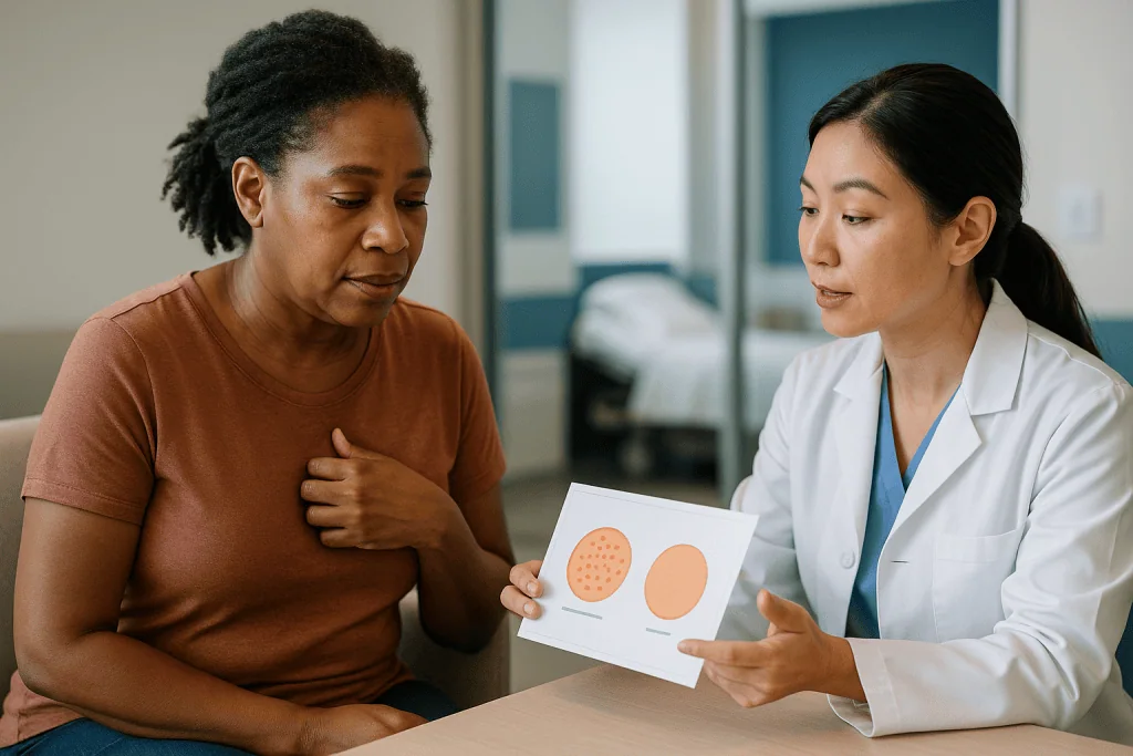
| Risk | Where | What it is | Usual course |
|---|---|---|---|
| Bruising, swelling | Both sites | Expected after any tissue disruption | Settles over weeks; final shape judged after months |
| Volume loss or unevenness | Recipient | Part of the graft is reabsorbed, not always evenly | Becomes clear over several months; may prompt a repeat session |
| Fat necrosis | Recipient | Grafted fat dies and forms a firm lump | Often softens or shrinks; sometimes needs imaging or removal |
| Oil cysts, calcifications | Recipient (breast) | Liquefied fat walled off, or calcium in scar | Usually harmless; may need extra imaging to characterize |
| Seroma | Donor | Fluid collection under skin | Often reabsorbs; may be drained |
| Contour dents, numbness | Donor | Uneven fat removal, nerve irritation | Numbness usually eases; dents may persist |
| Infection | Either | Bacteria in wound or graft | Needs prompt medical assessment |
| Fat embolism, blood clots | Systemic | Fat or clot entering blood vessels, lodging in lungs | Rare but life-threatening; emergency |
Two patterns are worth noticing. The common problems are aesthetic and usually settle or can be revised. The rare problems are medical emergencies, and they cluster around specific situations: large-volume grafting into the buttocks, injections near facial arteries, and anyone with a raised clotting risk. Knowing which category a symptom belongs to is most of what a patient needs.
Why does some of the fat disappear? Uneven volume explained
Fat that fails to gain a blood supply in the first days does not simply vanish; the body breaks it down and clears it over weeks to months. The NHS notes plainly that with fat transfer to the breast, some of the injected fat may be reabsorbed, so results are less predictable than with an implant and more than one procedure may be needed (NHS).
Reabsorption itself is expected. The problem is that it is rarely uniform. Fat placed near well-perfused muscle may survive well while fat in a tighter, scarred or previously radiated area struggles. The result can be a breast that is fuller on one side, a cheek that dips where it once matched, or a buttock with a subtle shelf where two zones of graft met. Surgeons often overfill slightly in anticipation, but that is a judgment call, and judgment sometimes lands short or long.
Several factors push the odds around:
- Recipient tissue quality: good blood supply and loose, unscarred tissue favor survival.
- Volume per session: the more fat pushed into a fixed space, the more sits too far from vessels.
- Handling: fat exposed to air, heat or rough suction for long periods survives less well.
- Pressure after surgery: sitting on freshly grafted buttocks or sleeping face-down on grafted cheeks compresses tiny vessels before they mature.
- Weight change: surviving fat behaves like the fat it came from, gaining and shrinking with the rest of you.
This is why a reputable surgeon will decline to promise a size or a percentage, and why the honest answer to “how much will stay?” is a range shaped by your tissue, not a number from a brochure.
What is fat necrosis after fat transfer, and why does it form a lump?
Fat necrosis is the death of fat tissue, in this context grafted fat that never connected to a blood supply. The word sounds alarming, but the process is closer to a bruise that heals badly than to anything dangerous. Dead fat cells release their oil, the surrounding tissue reacts with inflammation, and the body walls the area off with scar. What you feel is a firm, sometimes tender nodule, often round and mobile, occasionally with a faint dimpling of the skin over it.
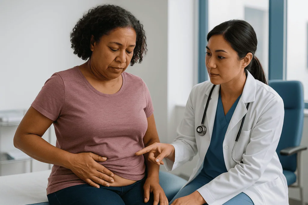
Why some grafts and not others? Volume and placement, chiefly. A large deposit in one spot leaves a core no vessel can reach. Grafting into a tight pocket, or into tissue scarred by earlier surgery or radiotherapy, does the same. Smoking narrows small vessels and starves the graft. So does infection, which can convert a healthy graft into a necrotic one.
The natural history is usually reassuring. Many nodules soften and shrink over months as the body clears the debris. Some persist as small, stable firm areas that never change. A minority enlarge, become painful, or develop into an oil cyst or a calcified mass that the person can feel.
In the breast, a new lump always deserves assessment, regardless of how likely fat necrosis is. Medical guidance is consistent that breast lumps should be checked, because a clinical exam and imaging, not the patient’s history alone, distinguish a benign lump from something that needs treatment (MedlinePlus). Ultrasound, mammography or MRI can often identify fat necrosis by its typical appearance. When imaging is unclear, a needle biopsy settles the question. Treatment, if any, ranges from watching and waiting to draining a cyst or surgically removing a bothersome nodule; that call rests with your surgeon and breast team.
Oil cysts and calcifications: what they mean on a mammogram
An oil cyst is a thin-walled sac of liquefied fat, formed when a pocket of grafted fat breaks down and the body seals it rather than absorbing it. On a mammogram it has a characteristic look: a round, dark, fat-density lesion, sometimes rimmed with calcium like an eggshell. Radiologists recognize this pattern readily, and it is widely regarded as benign.
Calcifications are deposits of calcium that form in scar tissue or dying fat. Most after fat grafting are coarse, rounded or rim-like, patterns radiologists associate with benign processes. The concern patients often hear about is that fine, clustered calcifications can also be a sign of early breast cancer. The two usually look different to an experienced reader, but not always at first glance. That is why fat grafting to the breast can lead to additional imaging views, a follow-up scan in a few months, or occasionally a biopsy to be certain.
None of this means fat transfer causes cancer. The evidence base, reflected in patient information from national health services, does not show that fat grafting increases breast cancer risk (NHS). What it can do is add noise to the pictures your future screening depends on.
Practical steps reduce that noise. Ask for baseline breast imaging before surgery if your team thinks it is appropriate for your age and history, so any later changes have a comparison. Tell every future mammography service that you have had fat grafting, including the year, so the radiologist reads with the right context. Keep imaging reports where you can find them. A surgeon who works regularly with a breast radiology team is well placed to explain how your local screening program handles grafted breasts.
Facial fat grafting risks: can it go wrong?
Yes, and the ways it goes wrong in the face differ from the body. Volumes are small, the margin for error is millimeters, and the tissue is crowded with important structures. Most problems are cosmetic; a few are serious.
Overfilling is the most common complaint. Surgeons add a little extra to allow for reabsorption, but if more survives than expected, the cheeks or under-eye area can look puffy or heavy. Unlike dissolvable fillers, surviving fat is permanent tissue, so correction means surgical removal or liposuction of a delicate area. Asymmetry follows the same logic: uneven survival between the two sides, which may not be obvious until swelling fully settles.
Lumps and visible irregularities are a particular worry in the thin skin below the eyes and along the jawline, where even a small clump of fat shows. Prolonged swelling around the eyes can last longer than people expect. Bruising can track down into the neck. Nerve irritation may cause temporary numbness or, rarely, weakness in a facial muscle.
The serious risk is vascular injury. Fat injected into or against an artery can block it. Because the arteries around the nose, between the brows and beneath the eyes connect to the vessels supplying the eye and, indirectly, the brain, blockage can cause skin death in the injected zone, sudden vision loss or stroke. These events are rare, but they are well described in the literature on facial injections of all kinds, and they are the reason experienced injectors use blunt cannulas, small volumes, slow technique and constant awareness of anatomy.
Fat also changes with the face over time. Weight gain can enlarge grafted cheeks disproportionately. Aging shifts tissue downward, and grafted fat travels with it. A result that suits a face at one weight or age may need revisiting later.
Fat grafting complications in the buttocks: the fat embolism question
Buttock fat grafting carries the one complication of fat transfer that has caused deaths in otherwise healthy people: fat embolism. An embolism is a blockage of a blood vessel by material carried in the bloodstream. Mayo Clinic lists fat embolism among liposuction and fat-grafting risks, noting that loosened fat can enter a blood vessel and travel to the lungs or brain, a medical emergency (Mayo Clinic).
The gluteal region is uniquely hazardous because large veins run within and beneath the gluteal muscles. If a cannula tears one of those veins and fat is injected into or near the tear, fat can be drawn into the circulation, reach the heart and lodge in the lungs, sometimes within minutes. Grafting only into the layer above the muscle, staying away from the deep pelvic vessels, using larger blunt cannulas and injecting only while the cannula is moving are the technical safeguards most often described. Patients cannot check technique themselves, which is why asking directly how and where a surgeon places fat is reasonable.
Two other risks matter here. Deep vein thrombosis, a clot in a leg vein, is a concern after any longer operation, and buttock grafting often means long hours face-down followed by days of limited sitting. Signs include one-sided calf pain, swelling or warmth. A clot that travels to the lungs, a pulmonary embolism, causes sudden breathlessness or chest pain and is an emergency.
Finally, this is the procedure where large volumes of fat are moved, so the ordinary complications scale up: more seroma at donor sites, more swelling, more chance of uneven survival, and pressure-related graft loss when sitting or lying on the area too early. The recovery instructions about cushions and positioning are not fussiness; they protect both the graft and, in a sense, the person carrying it.
Risks at the donor site: the liposuction half of the story
Patients tend to focus on the destination and forget the departure. Liposuction has its own list of complications, and most of the recovery discomfort after fat transfer comes from the donor area (MedlinePlus; NHS).
Bruising and swelling are universal. The suctioned space fills with fluid and blood, and the skin over it becomes tender and often numb because small sensory nerves are disrupted. Numbness usually recovers over weeks to months as nerves regrow, but patchy altered sensation can persist.
Contour irregularities are the donor-site complaint people notice most. Removing fat unevenly, or removing too much from one spot, leaves dents, ridges or waviness once swelling settles. Skin with poor elasticity may sag rather than retract. These are cosmetic, but they can be difficult to correct and sometimes require, ironically, fat grafting into the dent.
A seroma is a pocket of clear fluid that collects in the emptied space. Small ones reabsorb; larger ones may need needle drainage, occasionally more than once. Compression garments are used partly to limit this.
Less common problems include infection at the small entry incisions, skin discoloration, and, with larger-volume liposuction, shifts in body fluid that can strain the heart and kidneys. Local anesthetic in the injected fluid is dosed carefully by the team for that reason, and there are recognized upper limits on how much fat can safely be removed in a single session. Anyone considering large-volume harvest should expect a frank conversation about that ceiling.
The surgeon’s honest answer to “where will the fat come from?” therefore matters twice: it decides what tissue you can spare, and it decides where you will feel it for the next month.
Who is fat transfer usually for, and who is usually asked to wait?
Fat transfer suits people who want a modest, natural-feeling increase in volume, who have enough spare fat to harvest, and who accept that results are partly unpredictable and may need topping up. It is often chosen for breast reconstruction after cancer surgery, for smoothing the edges of implants, for restoring facial volume lost with age, and for buttock and hand contouring. The candidate who does best is at a stable weight, does not smoke, has realistic expectations and can follow positioning restrictions afterward.
Can you be too thin for fat transfer? Yes, in practical terms. Every graft requires a donor supply, and a person with little subcutaneous fat may not have enough to achieve a meaningful change, or harvesting it may leave visible dents. Some surgeons will decline; others will suggest a smaller goal or a different approach. It is a legitimate anatomical limit, not a judgment.
People commonly asked to wait or to reconsider include those who:
- smoke or use nicotine products, because nicotine narrows the small vessels a graft depends on;
- are actively losing or gaining weight, since surviving fat will change with the body;
- plan pregnancy or breastfeeding soon, when breast tissue will change substantially;
- have poorly controlled diabetes, bleeding disorders or a history of blood clots;
- have active infection anywhere, or a recent surgery still healing;
- expect a large size change in one session, which fat transfer rarely delivers.
Age is not a cutoff. Search engines are full of “best age for breast fat transfer,” and there is no such number in medical guidance. What matters is that breast development is complete, that health is stable, and that screening arrangements are in place for people old enough to need them. Someone with a strong family history of breast cancer should expect their team to discuss imaging and, possibly, involve a breast specialist before agreeing to graft (NHS).
What the days and weeks after fat transfer usually look like
The first three days are the sore, swollen, uncertain stretch. Donor sites ache in a deep, bruised way; recipient sites feel tight and look larger than the final result will be. Compression garments over the liposuctioned areas are worn most of the day. Small incisions may leak tinged fluid for a day or two. Walking around the house is encouraged from the first day, because movement lowers clot risk; strenuous activity is not.
Through the first two weeks, bruising blooms and fades, often traveling downward with gravity. Most people return to desk work within a week or two after liposuction, according to national patient guidance, with heavier activity resuming later (NHS). Grafted areas must be protected from pressure: no sleeping on a grafted face, no sitting directly on grafted buttocks except with a special cushion, no tight bras over grafted breasts. The tiny new vessels feeding the graft are fragile in this window.
From weeks three to eight, swelling recedes noticeably and the shape begins to look like yours. This is also when uneven reabsorption starts to show, and when firm spots may be felt. Many of those soften. Numbness at the donor site lingers.
Both Mayo Clinic and the NHS describe swelling after liposuction as taking several months, up to around six, to settle fully (Mayo Clinic). Surgeons generally judge the final grafted volume in a similar timeframe, once the body has cleared the fat that did not survive. Decisions about a second session, if wanted, usually wait until that point.
Everyone’s timeline shifts with volume grafted, site and health. Your team’s written instructions, not this outline, are the schedule to follow.
Fat transfer breast augmentation long term: what happens after 10 years?
People searching “fat transfer breast augmentation after 10 years” are asking a fair question, and the honest answer has two parts: what surviving fat does, and what the long-term evidence can and cannot say.
Fat that has gained a blood supply becomes ordinary breast fat. It does not expire, harden or need replacement. It behaves like the tissue it came from, which means it grows with weight gain and shrinks with weight loss, sometimes more dramatically than native breast fat because it retains the character of abdominal or thigh fat. Pregnancy, breastfeeding and menopause all remodel the breast, and grafted fat is remodeled with it. Aging brings the same descent and softening that any breast experiences.
Fat necrosis and calcifications may appear years after surgery, or may be found for the first time on a routine mammogram. Their appearance is usually benign, but as discussed above, they can trigger additional imaging. Telling every screening service about the history matters as much in year ten as in year one.
On cancer risk, national guidance does not show that fat grafting increases the chance of breast cancer, and there is no recognized need for special surveillance beyond standard screening for the person’s age and risk, though many teams recommend a baseline scan (NHS). Long-term studies are limited and mostly observational, so the strongest statement the evidence supports is absence of a demonstrated signal, not proof of zero effect.
The comparison people often have in mind is with implants. The NHS is explicit that breast implants are not lifetime devices and will probably need replacing at some point, with their own long-term concerns such as capsular contracture and rupture. Fat carries none of those device-specific issues but offers less volume and less predictability. Neither is universally better; they are different bargains, and the right one depends on goals, anatomy and tolerance for uncertainty.
What people often get wrong about fat transfer risks
“It’s my own fat, so it’s risk-free.” Autologous means no rejection and no foreign material. It does not mean no surgery. Fat transfer involves anesthesia, two operated sites and living tissue that may not survive. The most serious deaths in cosmetic surgery in recent years have involved fat grafting, not implants.
“Whatever is there at week two is what I keep.” The opposite. Week two is peak swelling plus fat that has not yet been cleared. Final volume is judged months later, once the body has removed what did not take.
“A lump means the surgery gave me cancer.” Most lumps after fat grafting are fat necrosis or oil cysts, and current evidence does not link fat grafting to breast cancer. Every new breast lump still needs a professional check, because history alone cannot tell the difference (MedlinePlus).
“More fat in one session means a bigger result.” Past a point, extra fat starves. Overpacking raises the rate of necrosis, cysts and lumps without adding lasting volume. Staged sessions exist for a biological reason.
“It doubles as weight loss.” Liposuction removes a limited amount of fat from specific areas and has no effect on metabolic health. Weight can shift to other regions afterward.
“Fat and fillers are the same thing.” Injectable fillers dissolve over months and can be reversed with an enzyme in many cases. Surviving fat is permanent tissue. That is an advantage when the result is right and a real problem when it is not.
“Fat grafts can’t be damaged after surgery.” In the early weeks, pressure, smoking and infection can all kill a graft that was healthy on the operating table. Aftercare is part of the treatment, not an optional add-on.
Questions to ask your care team before fat transfer
A good consultation should leave you able to describe your own risks in your own words. These questions tend to draw out the answers that matter, and a surgeon comfortable with them is a reassuring sign in itself.
- Do I have enough donor fat for the change I want, and where exactly will it be taken from?
- How much of the grafted fat do you expect to survive in my case, and what makes you think so? Which of my characteristics lower the odds?
- How many sessions might I realistically need, and how far apart?
- For breast grafting: should I have baseline imaging first? How will fat necrosis or calcifications be handled if they show up on a future mammogram?
- For buttock grafting: in which tissue layer do you place the fat, and what technique do you use to avoid injecting into or near the deep veins?
- For facial grafting: how do you reduce the risk of injecting into an artery, and what is your plan if signs of a vascular injury appear?
- What will the donor site look and feel like at one month, and what happens if it heals with a dent?
- What is your plan for clot prevention during and after surgery, given my history?
- What symptoms should make me call the same day, and who answers that call at night or on a weekend?
- How do weight change, pregnancy or menopause affect this result over the years?
- If I am unhappy with asymmetry or a lump, what are the revision options, and how long do you wait before considering them?
Write the answers down or ask permission to record them. The two-week post-operative brain, sore and impatient, will not remember what the pre-operative one was told.
When to call your doctor after fat transfer
Most recovery symptoms are expected and unpleasant rather than dangerous: bruising, swelling, tightness, tenderness at the donor site, small amounts of fluid from incisions in the first day or two. A short list of signs, however, should prompt a same-day call to your surgical team or emergency care, because they may indicate infection, a clot, a fat embolism or a threatened graft (Mayo Clinic; NHS).
Seek emergency help immediately for:
- sudden shortness of breath, chest pain, rapid heartbeat or coughing up blood;
- confusion, drowsiness, difficulty speaking, weakness on one side or a seizure;
- after facial grafting: sudden vision change, severe eye pain, or skin that turns white, mottled or dark over the injected area;
- fainting, or lips and fingertips turning blue.
Call your surgical team the same day for:
- fever, chills or feeling systemically unwell;
- redness that spreads beyond the incision, warmth, increasing pain, or pus or foul-smelling drainage;
- pain that worsens rather than eases after the first few days, or pain out of proportion in one area;
- a rapidly enlarging, tense swelling at the donor or recipient site;
- calf pain, swelling or warmth in one leg;
- a wound that opens or bleeds persistently.
Over the longer term, any new breast lump, a lump that grows, changes in skin texture or nipple discharge should be assessed by a clinician, even years later and even though fat necrosis is the most likely explanation. Persistent hard nodules in the face or body that bother you are also worth a review; the team can image them and discuss whether watching, draining or removing is appropriate. Every one of these judgments belongs to the people who operated on you and know your anatomy, so when in doubt, call.
Frequently asked questions
What happens to fat transfer breast augmentation after 10 years?
Fat that survived the first few months becomes ordinary breast fat and stays, changing size with weight, pregnancy and menopause like the rest of the breast. Fat necrosis, oil cysts or calcifications may appear on later mammograms and are usually benign, though they can prompt extra imaging. Current evidence does not show an increased breast cancer risk, but long-term data are limited and mostly observational, so routine screening appropriate to your age and risk remains important.
Can facial fat grafting go wrong?
Yes. The most common problems are overfilling, asymmetry from uneven fat survival, visible lumps under thin skin near the eyes, and swelling that lasts longer than expected. Because surviving fat is permanent, correction often means surgery rather than dissolving a filler. Rarely, fat injected into an artery around the nose or eyes can cause skin death, vision loss or stroke, which is why technique, small volumes and anatomical knowledge matter so much.
Can you be too thin for fat transfer?
In practical terms, yes. Every graft needs a donor supply, and a person with little subcutaneous fat may not have enough to produce a meaningful change, or harvesting it could leave visible dents. A surgeon may suggest a smaller goal, staged sessions or an alternative approach. This is an anatomical limit, not a judgment, and the honest conversation is about what your body can spare rather than what you weigh.
What is the best age for breast fat transfer?
There is no best age in medical guidance. What matters is that breast development is complete, weight is stable, overall health is good, and screening arrangements are in place for people old enough to need them. Younger adults planning pregnancy may be advised to wait because breast tissue will change. Older adults should discuss baseline imaging and family history with their team so future mammograms can be read with the right context.
What are the most common fat grafting complications?
The most common are aesthetic rather than dangerous: uneven volume as some fat is reabsorbed, firm lumps of fat necrosis, prolonged swelling and bruising, and contour dents or numbness at the liposuction donor site. Seromas, or fluid collections, are also fairly common after liposuction. Serious complications such as infection, blood clots and fat embolism are uncommon but require urgent care, which is why knowing red-flag symptoms matters.
Does fat necrosis after fat transfer go away on its own?
Often, yes. Many nodules soften and shrink over months as the body clears the dead fat and inflammation settles. Some remain as small, stable, firm areas indefinitely, and a minority grow, become painful or turn into oil cysts or calcified masses. A clinician should assess any new breast lump with examination and imaging; treatment ranges from watching to draining or surgical removal, decided by your surgical team.
Can fat transfer lumps be mistaken for breast cancer?
They can complicate imaging. Fat necrosis, oil cysts and calcifications usually have recognizable benign patterns on mammograms and ultrasound, but occasionally the appearance overlaps with suspicious findings, leading to extra views, a follow-up scan or a biopsy. Telling every screening service that you have had fat grafting helps the radiologist interpret the pictures correctly. Current evidence does not show that fat grafting causes breast cancer.
How long does swelling last after fat transfer?
Swelling is worst in the first week, improves noticeably over several weeks, and national patient guidance describes it taking up to around six months to settle completely after liposuction. Grafted areas follow a similar arc, and surgeons generally judge final volume only after that period, once the body has cleared fat that did not survive. Your own timeline depends on the volume moved, the site and your health.
Does smoking affect fat transfer results?
Nicotine narrows the tiny blood vessels a graft depends on in its first days, which raises the risk of fat necrosis, poor volume retention and wound-healing problems at both sites. Many surgeons ask people to stop all nicotine products for a period before and after surgery, and some decline to operate on active smokers. Your team can advise on timing and support; the decision about surgery sits with them.
Can uneven volume after fat transfer be fixed?
Usually, though not always perfectly. Options include a second grafting session to fill areas where less fat survived, gentle liposuction to reduce an overfilled zone, or removal of a firm nodule. Surgeons typically wait several months until swelling has settled and surviving fat is stable before judging what needs correction. Each revision carries the same risks as the original procedure, which is worth weighing with your team.
References
- MedlinePlus: Liposuction
- NHS: Liposuction
- NHS: Breast enlargement (including fat transfer)
- MedlinePlus: Breast lump
This article is for general information only and is not a substitute for professional medical advice. Please consult a qualified doctor about your individual situation.

More from the Blog
When an Arm Lift Needs Revision: Widened Scars, Residual Laxity and a Second Procedure
An arm lift revision is a second operation, or sometimes a smaller office procedure, that addresses problems left after a brachioplasty: scars that have…
How Does J Plasma Skin Tightening Work? Helium Plasma and Radiofrequency Explained Simply
J Plasma is a common name for helium plasma skin tightening, a procedure in which radiofrequency energy ionizes helium gas into a plasma that…

Fat Grafting Recovery: Protecting the Grafted Area While the Donor Site Heals
Fat grafting recovery runs on two tracks at once: the grafted area needs protection from pressure, heat and movement while transplanted fat cells form…

BBL Safety Explained: Why Fat Placement Depth Matters and How Surgeons Reduce the Risk
A Brazilian butt lift (BBL) is safest when fat is placed only in the subcutaneous layer above the gluteal muscle. Fat injected into or…
Laser Liposuction vs Traditional Liposuction: Incisions, Downtime and Skin Response Compared
Laser liposuction and traditional liposuction both remove fat through small skin incisions with a thin tube called a cannula. The laser version first heats…
Loose Skin After Weight Loss: What Improves on Its Own, and When Surgery Is the Answer
Mild skin laxity after weight loss often improves gradually over 12 to 24 months as collagen remodels, especially in younger people who lost a…