

What Does CT Pulmonary Angiography Show? How a Pulmonary Embolism Is Confirmed Before Treatment
Key Takeaways
- CT pulmonary angiography shows a clot as a dark gap inside a contrast-filled pulmonary artery, which radiologists call a filling defect.
- Guidelines steer most low-risk people through a Wells score and D-dimer blood test before any scan, so a normal D-dimer can avoid CTPA altogether.
- A CTPA can show strain on the right ventricle by comparing its width to the left, a finding that feeds directly into how embolism risk is categorized.
- Catheter pulmonary angiography threads a tube through the heart into the lung arteries and is now reserved mainly for cases where treatment through that catheter is planned.
- Shellfish allergy is not a reason to avoid iodine contrast; a previous reaction to contrast itself is what the team needs to know about.
- The NHS states that anticoagulant treatment after a confirmed embolism usually continues for at least three months, with the duration set by the prescribing clinician.
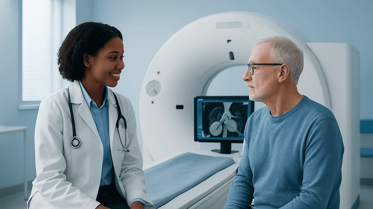
CT pulmonary angiography (CTPA) is a chest CT scan timed to a rapid injection of iodine-based contrast so the pulmonary arteries light up. It shows whether a blood clot is blocking those arteries, its size and location, and gives clues about strain on the right side of the heart. It is the usual imaging test used to confirm or rule out pulmonary embolism before treatment is decided.
It is just after midnight in a busy emergency department. A woman in her fifties who drove nine hours to see her grandchildren sits on the edge of a trolley, one hand pressed to her chest. She is not in agony. She is simply short of breath in a way she cannot explain, and her pulse is quicker than the monitor would like. The doctor listens, asks about the long drive and a swollen calf, and then says something that sounds far more alarming than it is meant to: “We’d like to do a CT pulmonary angiography.”
Most people have never heard those three words together. They know what a CT scan is. They may know what a blood clot is. What they want to know, in that moment, is what the machine is actually looking for and what happens if it finds it.
This explainer answers that question the way a radiologist might if given twenty unhurried minutes: what the scan shows, why it is ordered, who is steered toward a different test, and what the days afterward usually hold.
What does CT pulmonary angiography actually show?
A CT pulmonary angiography scan is a computed tomography study of the chest taken at the precise moment a fast injection of contrast dye fills the pulmonary arteries, the vessels carrying blood from the heart to the lungs. Contrast is an iodine-based liquid that blocks X-rays, so any vessel it fills appears bright white on the images. A clot does not take up contrast. It shows as a dark gap inside a bright vessel, which radiologists call a filling defect.
That single visual trick answers the question the emergency team is asking: is there a pulmonary embolism, a clot that has traveled to the lungs, or not? According to the Mayo Clinic, CTPA is now the imaging test most often used to make that call, because it is fast, widely available around the clock and shows the arteries directly rather than by inference.
Modern scanners slice the chest into images thinner than a coin, so the report can describe where a clot sits (in a main artery, a branch to one lobe, or a small peripheral vessel), how much of the vessel it fills, and whether it appears fresh or older and partly scarred into the wall.
The same images capture a great deal besides. The scan shows the heart chambers, the aorta, the lung tissue, the pleura that lines the lungs, and the bones of the chest wall. That is why a CTPA report often lists findings that have nothing to do with clots: a patch of pneumonia, fluid around a lung, an enlarged heart, or a nodule that needs follow-up. Later in this article we look at what those extras mean and which ones matter.
What CTPA cannot do is tell you where the clot came from. Most pulmonary emboli begin as deep vein thrombosis in a leg or pelvic vein, and the chest scan does not image those veins unless a separate leg study is added.
How a CTPA scan for pulmonary embolism works, step by step
The experience is shorter than most people expect. The NHS notes that a CT scan usually takes around 10 to 20 minutes from lying down to sitting up, and the part that acquires the pictures lasts a matter of seconds.
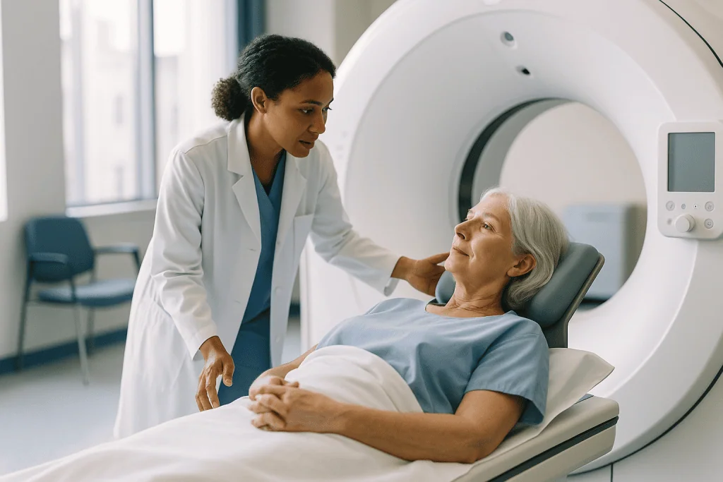
First, a nurse or technologist places a small plastic cannula, a thin flexible tube, into a vein in the arm, usually near the elbow. A larger vein is preferred because the contrast must be pushed in quickly to reach the lungs as a dense bolus rather than a dilute trickle. The cannula is then connected to a mechanical injector.
You lie on your back on a narrow table with your arms above your head. The table slides into a ring-shaped opening, not a tunnel; the scanner is open at both ends and the staff can see and speak to you throughout.
The injector starts. Many people feel a warm flush spreading through the chest and pelvis, a metallic taste, or a sudden feeling that they have wet themselves. All three are common, harmless and pass within a minute. The scanner tracks the dye as it arrives in the heart, and the moment it reaches the pulmonary arteries the machine begins imaging. You will be asked to hold your breath for a few seconds, because a moving lung blurs small vessels.
Afterward the cannula stays in for a short while in case a reaction develops, then comes out. You can normally eat, drink and move about immediately unless the team says otherwise. Drinking fluids over the rest of the day helps the kidneys clear the contrast.
A radiologist, a doctor who specializes in reading scans, reviews the images. In an emergency setting a preliminary read is often available within the hour, though the formal report can take longer.
Why doctors usually check a risk score and a blood test first
A CTPA is rarely the first move. Guidelines from bodies such as NICE and the European Society of Cardiology ask clinicians to estimate how likely a clot is before choosing a test, because a scan that involves contrast and radiation should be reserved for people in whom it will change management.
The Wells score is the structured checklist most emergency departments use. It gives points for things like clinical signs of a leg clot, a fast heart rate, recent surgery or immobility, previous clots, coughing up blood, active cancer and the clinician’s own judgment that embolism is the most likely explanation. The total sorts people into “unlikely” or “likely” groups.
For the unlikely group, a D-dimer blood test comes next. D-dimer is a fragment released when the body breaks down a clot. A normal result in a low-risk person makes embolism improbable enough that the Mayo Clinic and NHS both describe it as a reasonable point to stop and look for another cause. A raised D-dimer does not confirm a clot; pregnancy, infection, recent surgery, cancer and simple aging all raise it. What it does is justify moving on to imaging.
For the likely group, clinicians frequently skip D-dimer and go straight to CTPA, because a normal blood test would not be reassuring enough to change the plan.
This layering has an unglamorous benefit. Every imaging test produces some false positives, and the fewer low-probability people are scanned, the fewer will be told they have a clot when a tiny artifact was misread. Equally, no test catches everything. The risk score, the blood test and the scan are meant to be read together, and the treating team weighs all three rather than treating any single result as the final word.
CT angiogram vs pulmonary angiogram: what is the difference?
The names overlap, and the confusion is understandable. “Angiogram” simply means a picture of blood vessels. The word before it tells you which vessels and which method.

A CT angiogram is a broad category: any CT scan timed to contrast so that a particular set of arteries lights up. A coronary CT angiogram targets the arteries on the surface of the heart. A CT aortogram targets the aorta. A CT pulmonary angiogram, the subject of this article, targets the arteries of the lungs. The technique is the same; the timing and the region scanned differ. When a doctor says “CT angiogram” in the context of possible embolism, they mean CTPA.
A conventional pulmonary angiogram, sometimes called catheter or invasive pulmonary angiography, is a different procedure. As MedlinePlus describes it, a long thin tube called a catheter is threaded from a vein in the groin or arm, through the right side of the heart and into the pulmonary artery. Contrast is then injected directly and X-ray movies are recorded. It is done in a specialized suite, requires local anesthetic and often sedation, and carries the procedural risks of any catheter passed through the heart.
For decades the catheter study was the reference standard for diagnosing embolism. It has largely been replaced by CTPA for diagnosis because the CT version is non-invasive, faster and shows lung tissue and heart as well as vessels. Catheter angiography still has a role, chiefly when a team is planning to treat a large clot through the catheter itself, or when CT images are inconclusive and the answer matters urgently.
A third relative, magnetic resonance angiography, uses magnets rather than X-rays. It avoids radiation but, according to the Mayo Clinic, is less widely available and less sensitive for small clots, so it is used selectively.
Who is usually sent for CT pulmonary angiography, and who is asked to wait
CTPA is the default imaging test for most adults with a plausible story for pulmonary embolism who can lie flat, hold their breath briefly and receive iodine contrast safely. That covers the great majority of people in an emergency department. Several groups, however, are routinely steered toward alternatives or asked to pause while the team gathers more information.
Kidney function matters. Iodine contrast is cleared by the kidneys, and in people whose kidneys are already impaired it can cause a temporary further decline. Clinicians will often check a recent blood test for kidney function before injecting. If it is significantly reduced, they may choose a ventilation-perfusion (V/Q) scan instead. A V/Q scan is a nuclear medicine test that compares where air reaches in the lungs with where blood flows, looking for mismatched areas that suggest a blocked vessel.
A previous serious allergic reaction to iodine contrast is another reason to reconsider. Mild past reactions can sometimes be managed with pretreatment decided by the team; severe ones usually mean a different test.
Pregnancy calls for a specific conversation. Both CTPA and V/Q involve some radiation, and the choice between them balances the dose to the fetus against the dose to the breast tissue and the quality of the answer. Guidelines from NICE and the ESC support imaging when embolism is genuinely suspected, because untreated embolism poses a greater risk to mother and baby than either scan, but the sequence is individualized.
People who are too unstable to lie flat or leave the resuscitation bay may have a bedside echocardiogram, an ultrasound of the heart, first. It cannot show the clot directly but can show a right heart under strain, which in the right context may be enough to start treatment while imaging is arranged.
None of these decisions belongs to the patient alone or to a protocol alone. They are made by the treating team with the person in front of them.
What not to do before a CT angiogram
Preparation for CTPA is lighter than for many hospital tests, and in an emergency there may be no time to prepare at all. When the scan is scheduled, a handful of instructions are common.
Do not assume you must fast for hours. Johns Hopkins Medicine notes that for a contrast CT of the chest you may be asked not to eat or drink for a few hours beforehand, chiefly to reduce nausea if a reaction occurs. The imaging department gives the exact instruction, and it varies. Water is usually fine unless told otherwise.
Do not stay silent about kidney disease, diabetes, thyroid disease, asthma or any previous reaction to contrast or to shellfish-related myths (the shellfish link is not real, but the team will still want to hear about any allergy history). Each of these can change how the contrast is handled.
Do not stop or skip a prescribed medicine on your own initiative. Some people are told to hold a particular oral diabetes medicine around the time of contrast, and others on kidney-related medicines receive specific guidance. Those decisions rest with the prescribing clinician, who knows your full picture. Bring a list of everything you take, including supplements.
Do not wear metal near the chest if you can help it. Necklaces, underwire, zippers and metallic threads create streaks on the image. You will likely be offered a gown.
Do not forget to say if you might be pregnant or are breastfeeding. Pregnancy changes the imaging plan; breastfeeding generally does not need to be interrupted after iodine contrast, but the team will confirm current guidance.
Finally, do not dress for a long wait in the dark. Warm layers help, because scanner rooms are kept cool to protect the equipment, and a cold, tense body finds breath-holding harder.
Can CTPA show heart problems?
Yes, within limits, and this is one of the scan’s quiet strengths. Because the heart sits in the middle of the imaged volume and fills with contrast during the study, CTPA gives a reasonable look at its size, shape and chambers even though that is not its main purpose.
The finding that matters most in embolism is right ventricular strain. When a large clot blocks the pulmonary arteries, the right ventricle, the chamber that pumps blood to the lungs, has to push against sudden resistance. It stretches and enlarges. Radiologists measure the width of the right ventricle against the left and report the ratio; a right ventricle wider than the left is a marker that the clot is placing real load on the heart. The ESC and other guideline groups use this finding, together with blood tests and blood pressure, to sort embolism into risk categories that shape how aggressively it is treated. The AHA describes this same emphasis on right heart function in its patient material on venous thromboembolism.
The scan can also reveal an enlarged heart overall, fluid in the sac around the heart, calcium in the coronary arteries, valve calcification and abnormalities of the aorta such as widening or, rarely, a tear. Any of these may be mentioned in the report as an incidental finding.
What CTPA does not do well is assess the coronary arteries themselves or how the heart muscle is moving. Those need a dedicated coronary CT angiogram, an echocardiogram or other heart-specific tests. A CTPA report that says “heart unremarkable” means nothing alarming was seen on a study designed for the lungs, not that a cardiology workup has been completed.
If a heart finding does appear, the treating team decides whether it needs follow-up, and how urgently.
Reading your CTPA report: what the findings usually mean
Radiology reports are written for other doctors, and the vocabulary can feel like a wall. The table below translates the phrases people most often ask about. It is a glossary, not a diagnosis; only your treating team can tell you how a finding applies to you.
| Report phrase | Plain meaning | Why it matters |
|---|---|---|
| No filling defect / no evidence of PE | Contrast fills all visible pulmonary arteries; no clot seen | Embolism is unlikely; the team looks for other causes of symptoms |
| Filling defect in a lobar or segmental artery | A clot is blocking a branch supplying a lobe or a segment of a lobe | Confirms embolism; treatment is usually started |
| Subsegmental defect | A small clot in one of the finest visible branches | Real but small; guidelines note these can be harder to confirm and are weighed individually |
| Saddle embolus | A clot straddling the point where the main pulmonary artery splits | Indicates a large clot burden; the team assesses heart strain closely |
| RV/LV ratio increased | The right ventricle is wider than the left | Suggests the heart is under strain; influences risk category |
| Mosaic attenuation / webs | Patchy lung density or thin bands inside vessels | Can suggest older, chronic clots rather than a fresh one |
| Suboptimal contrast opacification | The dye did not fill the arteries densely enough | Technical limitation; the report may say small clots cannot be excluded |
| Incidental pulmonary nodule | A small spot in lung tissue unrelated to clots | Usually benign; often needs a follow-up scan on a schedule the team sets |
Two things are worth holding onto. A negative scan is genuinely reassuring in most people, and the Mayo Clinic lists CTPA among the tests that can rule embolism in or out. Yet a report that describes a technically limited study is not a clean negative, and a careful team will say so and decide whether repeat imaging or a V/Q scan is warranted.
What the contrast dye does, and what can go wrong
Contrast is the reason a CTPA works and the reason it carries risks a plain CT does not. Understanding both sides helps the consent conversation feel less like a formality.
The dye is an iodine-based compound that dissolves in blood and absorbs X-rays. Injected quickly, it travels from the arm vein to the right heart, through the pulmonary arteries and on around the body within a few heartbeats. Its concentration in the lung arteries peaks briefly, and the scanner is programmed to capture exactly that window. The body does nothing with the dye except excrete it; the kidneys filter most of it out over the following day, which is why drinking fluids afterward is encouraged.
The most common side effects are the sensations described earlier: warmth, a metallic taste and a false urge to urinate. Nausea and a brief headache occur in a minority. None of these is an allergy.
Allergic-type reactions do happen. The NHS describes them as uncommon and usually mild, taking the form of a rash, itching or hives that settle with or without medication. Severe reactions affecting breathing or blood pressure are rare, which is why staff keep the cannula in place and watch you briefly afterward, and why every CT department is equipped to treat them immediately.
Kidney effects are the other concern. In people with normal kidney function the risk of contrast harming the kidneys is small. In those with existing kidney disease, dehydration or diabetes the risk is higher, and the team may check blood tests, give fluids or choose a different scan. Anyone told they have reduced kidney function should make sure the imaging staff know before the injection.
Occasionally the dye leaks from the vein into the surrounding tissue at the injection site, causing swelling and discomfort in the arm. This is unpleasant but usually settles over a few days with elevation and observation, and the team will tell you what to watch for.
How much radiation is involved in a CT pulmonary angiography, honestly
Radiation is the question people are often embarrassed to ask and clinicians sometimes skate over. The honest answer has two parts: the dose is real, and in the situation where CTPA is being considered it is almost always justified.
CT uses X-rays, and a scan of the chest delivers considerably more than a single plain chest X-ray. The NHS puts it in everyday terms: the exposure from a CT scan is roughly equivalent to somewhere between a few months and a few years of the natural background radiation everyone receives from the environment, depending on the type of scan. CTPA sits within that range. Modern scanners use dose-reduction software that adjusts the beam to body size, and departments follow protocols designed to keep exposure as low as is consistent with a readable image.
What does that dose mean for cancer risk? The NHS is candid that any exposure carries a very small increase in lifetime risk, and that this risk is weighed against the benefit of the information. For a person in whom an untreated clot could be life-threatening within hours, that balance is not close.
Where the balance deserves more thought is in younger people, especially women, because breast tissue lies in the beam, and in anyone likely to need repeated scans over years. This is part of why guidelines push clinicians toward risk scores and D-dimer testing first, and why V/Q scanning is sometimes preferred in pregnancy or in someone who has already had several chest CTs.
It is reasonable to ask the team two things: whether the test will change what happens next, and whether a lower-radiation alternative would answer the question as well. A good clinician welcomes both questions. The answer, more often than not, will be that CTPA is the right test, but you will have heard the reasoning rather than simply the instruction.
When the scan is negative but you still feel unwell
A clear CTPA can feel like an anticlimax. You came in frightened, you were injected and scanned, and you are told there is no clot. Relief is appropriate. Confusion about what is causing the breathlessness is also appropriate, and a good team will not send you home with the first half of that sentence alone.
The symptoms that prompt a CTPA are shared with many other conditions. The same scan that clears the arteries frequently shows the actual culprit: an area of pneumonia, a collapsed segment of lung, fluid around a lung, or a heart that is larger than it should be. In those cases the report redirects the workup rather than ending it.
Sometimes the scan is entirely normal. Anxiety and panic can produce breathlessness, a racing heart and chest tightness that feel physically identical to more dangerous causes. Asthma, acid reflux, musculoskeletal chest wall pain, anemia and thyroid disorders are all on the list. A normal CTPA means the team can pursue these with less urgency, but it does not mean nothing is wrong.
Two caveats matter. First, a technically limited scan, one where the contrast did not fill the arteries well or breathing blurred the images, is not a true negative, and the team may repeat imaging or add a V/Q scan or leg ultrasound. Second, a negative chest scan does not exclude a clot in the leg veins. If a swollen or painful calf is part of the story, a leg ultrasound is often arranged.
The Mayo Clinic frames diagnosis as a sequence of tests interpreted together, and that is the right way to think about a negative result: one strong piece of evidence in a larger picture, not a verdict on whether your symptoms are real.
What the following days and weeks usually look like
If the scan confirms a pulmonary embolism, what happens next depends on how large the clot burden is and how the heart and circulation are coping. The team will already have placed you in a risk category using the scan, blood pressure, heart rate and blood tests, and that category drives the plan.
Most people diagnosed with embolism are started on an anticoagulant, a medicine that slows the blood’s ability to form new clots. It does not dissolve the existing clot; the body’s own systems do that over weeks to months. The medicine’s job is to stop the clot growing and prevent new ones forming while that happens. Some anticoagulants are given by injection at first and others by mouth from the outset; the choice, the duration and any changes are decided by the prescribing clinician based on your situation.
People in a lower-risk category are increasingly managed as outpatients after a short observation period, according to NHS guidance, provided they can get back to hospital quickly if needed. Those with heart strain or unstable blood pressure are admitted, and a small number receive clot-dissolving medicine or a catheter procedure to remove the clot directly.
The NHS notes that anticoagulant treatment usually continues for at least three months, and longer for people whose clot had no obvious temporary trigger or who have ongoing risk factors. Follow-up appointments review symptoms, bleeding risk and whether the underlying cause has been identified.
Breathlessness typically improves over days to weeks as the clot shrinks, but many people describe fatigue and reduced exercise tolerance lasting longer. A minority develop persistent symptoms that warrant reassessment, sometimes including repeat imaging to look for chronic clot. Report any new or worsening breathlessness to your team rather than waiting for the scheduled visit.
What people often get wrong about CTPA
Myths gather around any test people meet only in a crisis. Several are worth setting straight.
“The scan dissolves or treats the clot.” It does not. CTPA is purely diagnostic. Treatment, whether anticoagulant medicine, clot-dissolving drugs or a catheter procedure, is a separate decision made afterward.
“A CT angiogram of the chest checks my heart arteries too.” As discussed above, a CTPA shows the heart’s size and the right ventricle’s shape but is not designed to image the coronary arteries. That requires a differently timed coronary CT angiogram.
“If I’m allergic to shellfish I can’t have contrast.” This persistent belief has no evidence behind it. Shellfish allergy is a reaction to muscle proteins, not iodine. What matters is a previous reaction to contrast itself, or a history of severe allergies generally, and the team will ask.
“A negative scan means I’m fine.” It means embolism is unlikely. It does not explain your symptoms or exclude a clot in the legs.
“Small clots on the scan don’t matter.” Small subsegmental clots are a genuinely debated area. Guidelines acknowledge that their significance varies with the person’s risk factors and that they can sometimes be over-called. That is a reason for careful clinical judgment, not for dismissing the finding or for demanding treatment.
“The radiation is dangerous enough that I should refuse.” The dose is real, but the NHS and other bodies are consistent: when embolism is a serious possibility, the harm of missing it far outweighs the small long-term risk of the scan.
“CTPA and a lung V/Q scan are interchangeable.” They answer the same question by different routes and each has situations where it is preferred. The choice reflects kidney function, pregnancy, prior lung disease and availability, and the team explains their reasoning.
Questions to ask your care team
The minutes before a scan, and the conversation after the result, are the moments when asking one good question can change how well you understand what follows. These are the questions patients and families most often wish they had raised.
- Why this test rather than a D-dimer or a V/Q scan for me specifically, and would the result change the plan either way?
- Is my kidney function known, and does it affect whether I should receive contrast?
- I have had a reaction to contrast, or I have significant allergies. How will that be handled?
- I might be pregnant, or I am breastfeeding. Does that change the choice of scan or anything I need to do afterward?
- Should I keep taking all my usual medicines before and after the scan, and who makes that decision?
- If the scan is positive, what determines whether I stay in hospital or go home, and how will I know which category I am in?
- If the scan is negative, what is the working explanation for my symptoms and what happens next?
- The report mentions an incidental finding. Does it need follow-up, on what timeline, and who is responsible for arranging it?
- How long is treatment likely to continue, and what will the review at the end of that period involve?
- What symptoms should bring me straight back, and what number do I call?
Write the answers down, or ask whether a family member can be present. Emergency departments are noisy and frightening, and even calm people forget most of what they hear under stress. There is nothing unusual about asking a clinician to repeat an explanation or to write down the name of a finding so you can look it up later from a trustworthy source.
Remember too that the team expects these questions. A patient who understands why a test was chosen is more likely to return promptly if something changes, which is exactly what the team wants.
When to call your doctor
Two moments in this story carry red flags: before any scan has been done, when a clot is only a possibility, and after a diagnosis, when treatment brings its own risks.
Before diagnosis, seek emergency care without delay if you develop sudden breathlessness that is out of proportion to what you are doing, sharp chest pain that worsens when you breathe in, coughing up blood, a racing or irregular heartbeat with lightheadedness, or fainting. The Johns Hopkins Medicine patient material stresses that pulmonary embolism can be fatal within hours if untreated, which is why these symptoms warrant an ambulance rather than a wait-and-see approach. A painful, swollen or warm calf, especially after surgery, immobility or a long journey, should also prompt same-day medical contact.
After the scan, contact the imaging department or your doctor the same day if the arm used for injection becomes increasingly swollen, painful, numb or discolored, or if you develop a rash, itching, wheeze or facial swelling in the hours afterward. Severe breathing difficulty or a feeling of the throat closing is an emergency.
After a diagnosis and the start of anticoagulant treatment, call urgently if you notice bleeding that does not stop, blood in urine or stool, black stools, vomiting blood, a severe or unusual headache, sudden weakness, confusion or a fall that strikes the head. Also call if breathlessness returns or worsens, if chest pain comes back, or if a leg becomes newly swollen, because these can signal a new clot despite treatment.
For everything less dramatic, including persistent fatigue, worries about the medicine or a question about an incidental finding on the report, the routine route to your care team is the right one. Do not stop, skip or adjust any prescribed medicine because of a symptom without speaking to the clinician who prescribed it; that decision is theirs to make with you.
Frequently asked questions
Can CTPA show heart problems?
Partly. A CTPA captures the heart’s size and chambers because they fill with contrast during the scan, so it can reveal right ventricular enlargement from a large clot, an enlarged heart overall, fluid around the heart or aortic abnormalities. It is not designed to image the coronary arteries or assess how the heart muscle moves; those require a coronary CT angiogram or an echocardiogram. Any heart finding is reviewed by your treating team.
What is the difference between a CT angiogram and a pulmonary angiogram?
A CT angiogram is a non-invasive CT scan timed to intravenous contrast; when aimed at the lung arteries it is called a CT pulmonary angiogram. A conventional pulmonary angiogram is an invasive procedure in which a catheter is threaded through the heart into the pulmonary artery and contrast is injected directly. CT has largely replaced the catheter test for diagnosis, while catheter angiography is kept mainly for cases where the clot will be treated through the same catheter.
What not to do before a CT angiogram?
Do not stop or skip prescribed medicines on your own; the team decides whether anything should be held. Do not withhold information about kidney disease, diabetes, thyroid disease, allergies, previous contrast reactions, pregnancy or breastfeeding. Follow the department’s eating and drinking instructions rather than assuming a long fast is needed, and avoid metal near the chest. In an emergency, none of this should delay the scan; staff will ask what they need to know.
How much does a CT pulmonary angiogram cost?
This article does not publish prices, because the amount varies with the health system, the setting in which the scan is done, insurance coverage and whether it is part of an emergency admission or a scheduled outpatient test. The imaging department, hospital billing office or your insurer are the appropriate sources for a figure that applies to you. In an emergency, the clinical decision to scan is made on medical grounds first.
How long does a CTPA scan for pulmonary embolism take?
The NHS notes that a CT scan usually takes around 10 to 20 minutes in total, including positioning and cannula placement, while the image acquisition itself lasts only seconds during a single breath-hold. Expect some extra time afterward for observation while the cannula remains in place. In an emergency setting a preliminary read is often available within about an hour, though the formal radiology report can take longer depending on the department.
Is CT pulmonary angiography safe in pregnancy?
It can be used in pregnancy when a pulmonary embolism is genuinely suspected, because guidelines from NICE and the ESC consider the risk of a missed clot greater than the small radiation risk of imaging. The choice between CTPA and a ventilation-perfusion scan is individualized, weighing the dose to the fetus and to breast tissue and the quality of the answer. Tell the team if you might be pregnant so the plan can be adjusted.
What does a filling defect on CTPA mean?
A filling defect is an area inside a pulmonary artery that does not fill with contrast and appears dark against the bright vessel. In the context of suspected embolism it usually represents a clot. The report will describe its location, from the main artery down to small subsegmental branches, and how much of the vessel it occupies. Occasionally artifacts from breathing or poor contrast timing mimic a defect, which is why radiologists review images in several planes.
Why did I need a D-dimer test before the CT scan?
Because a normal D-dimer in a person judged low-risk by a Wells score makes pulmonary embolism unlikely enough that imaging can often be avoided, sparing contrast and radiation. D-dimer is a fragment released when clots break down, so a raised level supports moving on to CTPA but does not confirm a clot; pregnancy, infection, surgery and age all raise it. In high-risk people clinicians frequently skip the blood test and scan directly.
Can a CTPA miss a pulmonary embolism?
Rarely, yes. Very small clots in the finest branches can be below the scanner’s resolution, and a technically limited study, where contrast filling was poor or breathing blurred the images, may not exclude embolism reliably. The report will usually state when this is the case, and the team may repeat imaging, order a V/Q scan or a leg ultrasound, or rely on the overall clinical picture. A clean negative in a good-quality scan is strongly reassuring.
What happens after a CTPA confirms a pulmonary embolism?
The team places you in a risk category using the scan findings, blood pressure, heart rate and blood tests, and that category guides whether you are treated in hospital or observed briefly and managed at home. Most people begin an anticoagulant, a medicine that prevents clots from growing while the body slowly dissolves the existing one. The NHS states treatment usually lasts at least three months; the prescribing clinician decides the medicine and duration.
References
This article is for general information only and is not a substitute for professional medical advice. Please consult a qualified doctor about your individual situation.

More from the Blog

Life After a Pulmonary Embolism: Compression Stockings, Travel and Staying Active Safely
Life after a pulmonary embolism usually means several months of blood-thinning medicine, a gradual return to walking and everyday activity, and a plan for…

Percutaneous Closure Step by Step: How a Catheter Seals an Atrial Septal Defect or PFO
Percutaneous ASD closure is a catheter procedure: a cardiologist threads a thin tube from a groin vein to the heart, measures the hole between…

Echocardiography in Pregnancy and Childhood: Why Heart Ultrasound Is Used Across Ages
An echocardiogram in pregnancy and children is a painless heart ultrasound that uses sound waves, not radiation, to show how the heart is built…

Robotic Cardiac Surgery Recovery: Intensive Care, Early Walking and the Weeks Back Home
Robotic cardiac surgery recovery time is usually shorter than after open-chest surgery because the breastbone is left intact. Many people spend the first night…

Small Dense LDL: The Particle Size Debate Explained
Small dense LDL refers to the smaller, heavier low-density lipoprotein particles that appear to enter artery walls more easily and oxidize faster than larger…

Echocardiogram Preparation: Why Most Heart Ultrasounds Need Nothing Special Beforehand
For a standard transthoracic echocardiogram, you generally need no special preparation: eat and drink normally, take your usual medicines unless told otherwise, and wear…