

What Does Long-Term Eye Follow-Up After ROP Treatment Involve for Premature Babies?

Key Takeaways
- Retinal blood vessels normally finish growing around a baby's due date, so a child born at 26 weeks has a retina that is still being built long after discharge.
- After treatment, retina exams typically continue every one to two weeks until the vessels mature, and anti-VEGF injection extends that window because the disease can reactivate months later.
- In the ETROP trial, about one in four eyes treated early still had an unfavorable visual acuity result at age six despite good structural results, which is why follow-up tracks function, not just anatomy.
- Nearsightedness after ROP usually comes from a steeper cornea and thicker lens rather than a long eyeball, and it can be measured and corrected with glasses within the first year.
- Amblyopia responds best when found in the first few years of life, so the preschool years are the busiest period for vision testing after the retina has settled.
- Adults who had ROP carry a lifetime higher risk of retinal detachment and glaucoma, so flashes, new floaters or a spreading shadow in vision need same-day care at any age.
Long-term eye follow-up after ROP treatment means scheduled checks that begin within days of laser or injection, continue every one to two weeks until the retina's blood vessels finish growing, and then shift to periodic exams through childhood and adulthood. Eye doctors watch for reactivation, nearsightedness, misaligned eyes, lazy eye, glaucoma and late retinal detachment. The exact schedule is set by the treating ophthalmologist.
The discharge folder from a neonatal unit is thick. Feeding plans, hearing results, a car seat test. Somewhere near the back, often circled by a nurse, sits one line that parents tend to photograph with their phones: eye clinic, two weeks, do not miss.
Families who have already sat through retinopathy of prematurity (ROP) treatment sometimes read that line with a sinking feeling. Wasn’t the laser or the injection supposed to be the end of it? In truth, treatment is the beginning of a relationship with eye care that can last a lifetime. ROP long-term vision depends not only on what happened to the retina in the first months, but on how carefully the growing eye is watched for years afterward.
This explainer walks through what those visits involve, why the schedule looks the way it does, and which later problems the eye team is quietly checking for at every stage.
What is retinopathy of prematurity, and why doesn't treatment end the story?
The retina is the thin, light-sensing lining at the back of the eye. Its blood vessels grow outward from the optic nerve during pregnancy and normally finish reaching the retina’s edge around the time a baby is due. A baby born at 26 weeks arrives with a retina that is only partly supplied, and that unfinished growth is where ROP starts (MedlinePlus).
Outside the womb, vessel growth often stalls. Weeks later it can restart in a disorganized way, producing fragile new vessels and scar tissue that may tug on the retina. Doctors describe how far the disease has progressed using stages 1 through 5, where stage 1 is a faint line between supplied and unsupplied retina and stage 5 is a fully detached retina. They also note the zone, meaning how close to the center of the retina the changes sit, and whether “plus disease” (dilated, twisted vessels signaling active disease) is present (Cleveland Clinic).
Most ROP is mild and settles on its own without treatment. When it reaches a threshold where the risk of detachment is high, treatment with laser or an injection into the eye is offered to switch off the abnormal growth signal (NEI).
Here is the part that surprises families. Treatment addresses the active disease, not the fact that the eye itself is still immature. The eyeball keeps growing in length and shape for years, the retina’s periphery may stay thinner than usual, and the brain’s visual pathways are still wiring themselves. Each of those processes can go off course later. Follow-up exists to catch them while they are still manageable.
What does ROP long-term vision follow-up actually involve?
In infancy, a follow-up visit looks much like the screening exam parents may remember from the neonatal unit. Dilating drops are placed in each eye to widen the pupil. After they take effect, the ophthalmologist uses a small lid holder to keep the eye open, a head-mounted light called an indirect ophthalmoscope, and a handheld lens to view the retina. A blunt instrument may gently press the outside of the eye so the far edges of the retina roll into view. The looking part usually lasts a few minutes per eye, though the whole appointment, with waiting for the drops, takes longer (Johns Hopkins Medicine).
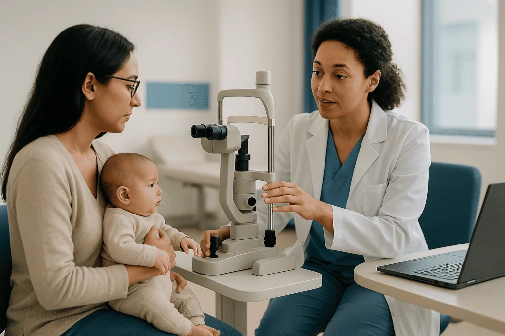
Babies protest, and that is expected. Swaddling, a pacifier and a parent’s voice help; the team will tell you which comfort steps they use. Some units photograph the retina with a wide-field camera so changes can be compared between visits.
As the child grows, the content of the visit changes. The retina still gets a dilated look, but the eye team adds measurements that matter for how a child actually sees:
- Vision testing, first by watching fixation behavior, later with matching pictures or letters.
- Cycloplegic refraction, where drops temporarily relax the focusing muscle so the true glasses prescription can be measured without the child straining.
- Alignment checks, covering one eye at a time to see whether the other drifts.
- Eye pressure measurement in children with scarred or structurally altered eyes.
Think of it as two tracks running side by side. One track asks whether the retina is anatomically safe. The other asks whether the visual system is developing well enough to use that retina. Both deserve attention, and a reassuring answer on one does not guarantee the other.
Who needs long-term follow-up, and who is usually asked to wait and watch?
Screening for ROP is generally recommended for babies born before about 30 weeks of gestation or weighing less than 1,500 grams (roughly 3 pounds 5 ounces) at birth, with the first exam typically a few weeks after birth depending on how early the baby arrived (MedlinePlus). Not every screened baby ends up in long-term follow-up. The intensity depends on what the exams found.
Babies who received laser or injection treatment sit at the top of the follow-up pyramid. Their eyes have been structurally altered by disease and by treatment, and the professional consensus is that they need ongoing monitoring through childhood and, for retinal health, into adult life (NEI).
A second group had ROP that regressed on its own. These children are usually followed until the retinal vessels have matured, then handed to a routine pediatric eye schedule with an eye exam in the toddler years and again before school. Nearsightedness and misalignment are still more common in this group than in children born at term, so “regressed” should not be read as “finished” (Cleveland Clinic).
The third group never developed ROP but were very premature. Prematurity itself raises the odds of vision problems, so a check in early childhood is widely advised even when the neonatal exams were clear (Mayo Clinic).
Who is asked to wait? Mostly it is a question of timing rather than exclusion. A baby who is still too unstable for a long exam may have the visit shortened or deferred by a few days, and a child who is otherwise well with stable findings may have the interval between visits stretched. Those calls belong to the treating ophthalmologist and neonatal team, who know the specific findings for that eye.
ROP eye exam schedule: what the first days and weeks after treatment usually look like
The first week after treatment is mostly about comfort and watching for early problems. After laser, the eyelids are often puffy and the eyes red for a few days. After an injection, a small red spot on the white of the eye is common. Any drops prescribed by the team should be used exactly as instructed, and questions about them go to the prescribing clinician, not to the internet.

Follow-up exams then resume on a tight cycle. During active disease, ophthalmologists typically examine the retina every one to two weeks, and that rhythm continues after treatment until the abnormal vessels have regressed and the normal vessels have grown out toward the retinal edge (Cleveland Clinic). The doctor is looking for several specific things at each visit:
- Whether the ridge of abnormal tissue is flattening and the twisted “plus” vessels are calming down.
- Whether any part of the retina shows new traction or lifting.
- Whether, after injection, vessel growth has resumed and is heading in the right direction.
Sometimes the retina does not respond fully, or responds in one area but not another. In that situation the team may discuss repeating treatment or adding a different type. Hearing the word “retreatment” is unsettling, but it is a planned contingency, not a sign that something went wrong.
Families often ask when the visits will finally space out. The honest answer is: when the retina shows the team that the vessels have matured, which varies from baby to baby and cannot be promised in advance. Many infants are still in two-weekly or monthly checks at the time they would originally have been due, and some, particularly after injection, are followed closely for several months beyond that. A written schedule from the clinic, pinned somewhere visible at home, is one of the simplest protective measures a family can take.
Why does follow-up differ after laser versus anti-VEGF injection?
Two main treatments exist for ROP, and they work by different mechanisms, which is why their follow-up timelines diverge (NEI).
Laser photocoagulation treats the part of the retina that has no blood supply yet. Those unsupplied cells are the source of the chemical distress signal driving abnormal growth. By treating that tissue, the laser removes the signal. The effect is permanent; the treated peripheral retina does not need to be revisited by vessels because it no longer sends the signal. Once regression is confirmed, reactivation is unusual. The trade-off is that the treated periphery is sacrificed, which is linked to the higher rates of nearsightedness seen after laser.
Anti-VEGF injections take a different route. VEGF, or vascular endothelial growth factor, is the protein that tells vessels to grow. A medicine in this class, injected into the gel of the eye, blocks that protein for a period of weeks. The abnormal vessels shrink, but the peripheral retina is preserved and normal vessels are expected to keep growing slowly toward the edge. Because the drug wears off while the retina is still unfinished, the disease can reactivate weeks or months later. Follow-up after injection therefore runs longer and more cautiously, often well past the original due date, until the vessels have actually reached the periphery or the team is satisfied the risk window has closed.
Questions remain about whether small amounts of injected anti-VEGF entering the bloodstream could affect development elsewhere in the body. Research is ongoing and the evidence is not settled; families should expect their team to describe this uncertainty plainly rather than dismiss it. Which treatment is chosen, and whether the two are ever combined, depends on the zone and stage of disease, the baby’s overall condition and the judgment of the treating ophthalmologist.
Retinopathy of prematurity follow up through the first year: what gets checked
Once the retina has settled, the pace changes but the stakes do not. The first year is when the visual brain is most plastic, so this is the window in which the eye team moves from “is the retina safe” to “is this child learning to see.”
At these visits, expect the ophthalmologist to confirm that vessel growth has finished and that any scar tissue is stable. Measurements that start to matter include the baby’s refractive error (the glasses prescription), whether the two eyes are similar to each other, and whether the eyes line up and move together. Parents are asked practical questions: does your baby fix on your face, follow a toy across the room, reach for things accurately, blink at bright light?
Corrected age matters here. A baby born three months early who is six months from birth behaves visually more like a three-month-old. Teams judge milestones against corrected age so that normal prematurity is not mistaken for a problem, and vice versa.
This is also the year when follow-up is most likely to fall apart. Families are juggling neonatal clinics, developmental assessments, feeding specialists and ordinary life with a baby who may still have medical equipment at home. ROP visits can feel like the least urgent item because the baby looks fine. Clinics counter this by tying eye visits to other appointments where possible and by sending reminders. If a visit is missed, the right response is a phone call to rebook, not waiting for the next scheduled one.
Where available, a neonatal follow-up program usually coordinates with the eye clinic so that findings about hearing, movement and cognition are read alongside vision results. A child who is not meeting visual milestones despite healthy-looking eyes needs that joined-up view (Mayo Clinic).
ROP and nearsightedness: why glasses are so common
Nearsightedness, or myopia, means the eye focuses images in front of the retina rather than on it, so distant objects blur. It is one of the most frequent long-term consequences of ROP and is listed among the later problems that children with a history of the disease are more likely to develop (NEI).
The mechanism is different from ordinary childhood myopia. In most nearsighted school-age children, the eyeball has simply grown too long. In children who had ROP, the front of the eye is often the culprit: the lens tends to be thicker and more curved, and the cornea steeper, so light is bent too strongly. The eye itself may actually be shorter than average. This pattern is more pronounced after laser treatment and after more severe disease, and it can appear early, sometimes within the first year, which is why refraction is part of infant visits rather than something saved for school.
A related issue is anisometropia, a meaningful difference in prescription between the two eyes. When one eye delivers a sharper image than the other, the developing brain quietly favors it, and the blurrier eye can slip into amblyopia (described in the next section). Catching an uneven prescription early is one of the main reasons eye doctors measure refraction so carefully in this group.
Glasses on a baby look startling. They are also one of the simplest, lowest-risk interventions in all of pediatric eye care. Soft frames with a strap, worn during waking hours as the team advises, give the brain a clear image to learn from. Prescriptions in this age group change quickly, so repeat measurements every few months are normal and do not mean the earlier prescription was wrong.
Strabismus and amblyopia: problems that appear after the retina settles
Strabismus is the medical term for eyes that do not point in the same direction; one may turn in, out, up or down. Amblyopia, often called lazy eye, means reduced vision in an eye that is structurally capable of seeing better, because the brain has learned to ignore its input during early childhood. Both are more common after ROP and after prematurity in general (Cleveland Clinic).
Several threads tie them to ROP. Scarring in the retina can drag the sharpest central area slightly out of position, so an eye that looks misaligned is actually pointing its best retina at the target. Uneven prescriptions push the brain toward one eye. Premature brains are also more prone to the coordination problems that underlie misalignment regardless of the retina.
Why the urgency? Visual development has a window. The brain’s ability to recover vision in a neglected eye is strongest in the first few years and fades through childhood, so the earlier amblyopia is identified, the more responsive it tends to be to treatment. That is the evidence-based reason follow-up visits cluster in the preschool years rather than being postponed until a child can read an eye chart.
Detection in a toddler relies on simple tools. The examiner covers one eye and watches whether the child objects more when one particular eye is covered, a sign that the uncovered eye sees poorly. Alignment is checked with a light reflection on the corneas and with cover testing. Management, decided by the eye team, may involve glasses, patching the stronger eye for set periods, or in some cases surgery on the eye muscles to improve alignment. Patching schedules are individualized and adjusted at each visit, so a plan that worked for another family’s child is not a template.
Can ROP cause blindness later? Retinal detachment, glaucoma and the adult years
The direct answer is that severe vision loss is uncommon in children whose ROP was treated and followed, but the risk of serious eye problems does not disappear with childhood. People who had ROP as infants carry a higher lifetime risk of retinal detachment, glaucoma and other conditions, which is why many ophthalmologists recommend continued periodic dilated examinations into adulthood (NEI).
Retinal detachment later in life is the concern that most shapes adult follow-up. The peripheral retina in these eyes may be thinner, with areas of weakness or old scarring where the gel inside the eye is unusually adherent. During adolescence and adulthood, as that gel naturally liquefies and pulls away, it can tear the retina at those weak spots. A tear that is spotted early can often be sealed; a detachment that progresses is a surgical emergency. Flashes of light, a sudden shower of floaters, or a shadow spreading across the vision are the signs that should never wait for the next routine visit.
Glaucoma, meaning damage to the optic nerve usually associated with raised pressure inside the eye, can develop years after ROP, particularly in eyes with significant scarring that narrows the drainage angle at the front of the eye. Because early glaucoma causes no symptoms, pressure checks are built into adult follow-up rather than triggered by complaints. Cataract, a clouding of the lens, is also seen earlier than usual in some eyes with a history of advanced disease.
None of this is a reason to live fearfully. It is a reason to keep a relationship with an eye clinic, to know the warning signs, and to make sensible choices about eye protection in contact sports. The frequency of adult checks is set individually, based on how the retina looks and what treatment was done.
What the evidence shows about ROP long-term vision outcomes
Parents reasonably want numbers, and the most useful ones come from long-term trials rather than single-center reports. The Early Treatment for Retinopathy of Prematurity (ETROP) study followed children treated as infants to age six. In eyes treated at an earlier, high-risk stage, 25.1 percent had an unfavorable visual acuity outcome, compared with 32.8 percent in eyes managed with the older, later-treatment approach; unfavorable structural outcomes were 9.0 percent versus 15.2 percent (ETROP Cooperative Group, PubMed).
Read those figures two ways. Earlier treatment clearly helped, and it changed practice. At the same time, roughly one in four treated eyes still did not reach a good acuity result even when the retina looked structurally sound. That gap is the single strongest argument for long-term follow-up: anatomy and function are not the same thing, and the problems that erode function, such as high myopia, amblyopia and misalignment, are the ones follow-up can modify.
A second layer of the evidence concerns the brain. Premature babies, especially those who were very unwell, are at increased risk of cerebral visual impairment, a condition in which the eyes may be healthy but the brain’s visual processing areas were injured. A child may see detail on a chart yet struggle to find a toy on a patterned carpet or recognize faces in a crowd. Standard eye exams can miss this, so neonatal follow-up programs increasingly include functional vision assessments and refer to early intervention services when needed (Mayo Clinic).
What the evidence cannot do is predict an individual child’s result. Trial percentages describe groups. The treating team’s assessment of a specific pair of eyes, revisited over time, is the only forecast that applies to your child, and even that comes with honest uncertainty.
At a glance: the typical ROP follow-up map from neonatal unit to adulthood
Schedules are individual and set by the treating ophthalmologist. The table below shows the general shape of follow-up described by major patient-information sources, so families know what kind of visit to expect at each stage (Johns Hopkins Medicine; NEI).
| Stage | What is usually checked | Why it matters |
|---|---|---|
| Days to weeks after treatment | Dilated retina exam every one to two weeks; eyelid and surface healing | Confirms regression; detects need for retreatment |
| Until vessels mature (longer after injection) | Dilated exams at intervals set by findings | Reactivation can occur months later after anti-VEGF |
| First year (corrected age) | Refraction, fixation and following, alignment, retinal stability | Early myopia and uneven prescriptions set up amblyopia |
| Toddler and preschool | Vision testing with pictures, cover testing, glasses updates, patching if advised | Peak window for treating amblyopia and strabismus |
| School age | Letter-chart acuity, refraction, dilated retina, functional vision in class | Learning depends on corrected vision; CVI may surface |
| Teen and adult years | Periodic dilated exam, eye pressure, retinal periphery | Lifetime risk of detachment and glaucoma |
Two patterns stand out. Visits are frequent while the retina is still changing and thin out once it is stable, but they never truly stop. And the purpose of the visit shifts from protecting the retina to protecting the way the child uses it, before returning, in adulthood, to the retina again.
If your child’s schedule looks different from this, that is not a warning sign. A retina that was treated in zone I, or an eye that needed a second procedure, will reasonably be seen more often than an eye with mild peripheral disease that regressed quickly. Ask the team to explain the reasoning behind your child’s intervals; most are glad to.
What people often get wrong about ROP follow-up
“The laser fixed it, so the eyes are fine now.” Treatment stops active disease. It does not change the fact that the eye is premature and still growing, nor does it remove the later risks of myopia, misalignment, glaucoma and detachment (NEI). Follow-up is part of the treatment, not an optional extra.
“My baby clearly sees well, so we can skip a visit.” Babies compensate beautifully. A child with one weak eye uses the strong one and looks completely normal at home. The problems follow-up is designed to catch are, by their nature, invisible to parents until late.
“Glasses mean the treatment failed.” Nearsightedness after ROP is an expected consequence of how these eyes grow, not a verdict on the procedure. A toddler in glasses is a sign the system worked: the prescription was found early enough to matter.
“Only babies who had severe ROP need eye checks.” Even children whose ROP regressed without treatment, and very premature children who never had ROP, have higher rates of refractive error and strabismus than term-born children and benefit from an early childhood eye exam (Mayo Clinic).
“The oxygen in the neonatal unit caused this, so someone made a mistake.” Oxygen management is one factor among many, and modern units balance it against the brain and lungs’ need for oxygen. Prematurity itself is the dominant cause (MedlinePlus).
“A normal eye exam means normal vision.” Not always. Cerebral visual impairment lives in the brain, not the eye. If a child with healthy-looking eyes struggles visually in cluttered or busy environments, that observation belongs in the clinic notes, not dismissed as clumsiness.
“Once they’re a teenager, they can stop going.” Adolescence is exactly when late retinal problems begin to appear, and when appointments are most often dropped.
Questions to ask your care team about ROP follow-up
A good follow-up relationship is a conversation, and parents who arrive with questions tend to leave with a clearer plan. Nothing on this list is a test of the team; it is a way of making sure the reasoning behind the schedule is shared with the people who have to carry it out at home.
- Which zone and stage did my child’s ROP reach, and was there plus disease? Knowing this helps you understand why the intervals are what they are.
- Which treatment was used, and does that change how long we need close monitoring?
- How will we know when the retinal vessels have finished growing, and what happens to the schedule then?
- What signs at home would make you want to see us sooner than planned?
- When will my child’s glasses prescription first be measured, and how often will it be rechecked?
- Who is checking for amblyopia and misalignment, and at what ages?
- Is anyone assessing how my child uses vision day to day, not just what the eye chart shows?
- How do the eye visits fit with the neonatal follow-up program, and do the two teams share notes?
- If we move or change insurance, what records should travel with us?
- At what age should my child start seeing an adult eye specialist, and what should that specialist be told?
Write the answers down, or ask whether the clinic can give them in writing. Families often describe a moment years later, in an unfamiliar emergency department or a new school’s health office, when a one-page summary of the child’s eye history turned out to be the most valuable document they owned. Ask for one before you need it (Johns Hopkins Medicine).
When to call your doctor
Most of ROP follow-up is planned and unhurried. A few situations should prompt a call to the eye clinic or, out of hours, urgent care, rather than waiting for the next scheduled visit (MedlinePlus; NEI).
In the days after laser or injection, contact the team the same day if the eye becomes increasingly red or swollen rather than settling, if there is thick discharge, if the eyelid will not open, or if the baby seems to be in persistent pain or is unusually irritable and feverish. A tiny red spot after an injection is expected; a cloudy or white appearance of the front of the eye is not.
In infancy and early childhood, call if you notice a white or gray reflection in the pupil in photographs or in dim light, an eye that looks larger or cloudier than the other, constant tearing with light sensitivity, eyes that shake or wander rhythmically, an eye turn that appears or worsens, or a baby who stops fixing on faces or following objects after previously doing so.
At any age, flashes of light, a sudden burst of new floaters, a shadow or curtain spreading across part of the vision, or a sudden drop in vision in one eye are emergency symptoms of a possible retinal tear or detachment. These need same-day assessment. Older children should be taught these signs in plain words so they can report them.
Finally, a missed appointment is itself a reason to call. Follow-up intervals in ROP are set because the eye can change in that window. Rebooking promptly matters more than apologizing for the gap. Every decision about what the symptoms mean and what to do next rests with the treating ophthalmologist and pediatric team, who know your child’s eyes.
Frequently asked questions
How long does retinopathy of prematurity follow up continue after treatment?
It continues in some form for life. Close exams run every one to two weeks until the retinal vessels mature, then spread out through childhood for vision, glasses and alignment checks, and continue periodically into adulthood for retinal detachment and glaucoma risk. The treating ophthalmologist sets the intervals based on the zone, stage and treatment type.
What is a typical ROP eye exam schedule in the first year?
After regression is confirmed, visits usually move from weekly or two-weekly to monthly and then to every few months, judged against corrected age. Each visit checks that vessel growth has finished, measures the glasses prescription, and watches fixation and eye alignment. Babies treated with injection are often followed more closely for longer because of late reactivation.
Can ROP cause blindness later in life even after successful treatment?
Severe vision loss is uncommon when ROP is treated and followed, but people who had ROP have a higher lifetime risk of retinal detachment, glaucoma and cataract. Periodic dilated eye exams in adulthood, together with knowing the warning signs of a retinal tear, are the recommended safeguards. No one can promise an individual outcome.
Why is my baby nearsighted after ROP, and will it get worse?
ROP and nearsightedness are linked because these eyes often develop a steeper cornea and thicker lens, bending light too strongly. Myopia tends to be more pronounced after laser and after severe disease. Prescriptions commonly change in early childhood, which is why refraction is repeated at visits. Glasses correct the blur and protect against amblyopia.
Does a normal eye exam mean my premature child's vision is normal?
Not necessarily. Prematurity can injure the brain’s visual processing areas, causing cerebral visual impairment, in which the eyes look healthy but the child struggles with cluttered scenes, faces or moving objects. If you see that pattern at home, tell both the eye clinic and the neonatal follow-up team so a functional vision assessment can be arranged.
What are the signs of amblyopia or lazy eye in a toddler who had ROP?
Often there are none a parent can see, because the child relies on the stronger eye. Clues include objecting strongly when one specific eye is covered, an eye that drifts, or head tilting. Eye doctors detect it with cover testing and age-appropriate vision tests, which is why preschool follow-up visits matter even for a child who seems to see well.
Why does follow-up last longer after anti-VEGF injection than after laser?
Laser permanently treats the unsupplied retina that drives abnormal growth, so once regression is confirmed, reactivation is unusual. An injected anti-VEGF medicine blocks the growth signal only for weeks, while the retina keeps developing slowly. Disease can therefore reactivate months later, so monitoring continues well past the original due date.
Will my child need glasses forever after ROP?
Many children with a history of ROP wear glasses long term, because the refractive changes are structural rather than temporary. Prescriptions often shift during growth and are rechecked regularly. Whether other options are appropriate in later years is a decision for the eye specialist, based on the stability of the prescription and the health of the retina.
Is it safe for a child who had ROP to play contact sports?
Most children can take part in ordinary activity, but eyes with a history of ROP have a higher risk of retinal detachment, and a direct blow can trigger a tear. Ask the ophthalmologist about protective eyewear and whether any specific sports deserve caution, since advice depends on how the retina looks in that individual child.
What should an adult who had ROP as a baby tell a new eye doctor?
Share that you were born prematurely, had ROP, which treatment you received and in which eye, and any history of glasses, patching or eye surgery. This history prompts the examiner to look carefully at the retinal periphery, check eye pressure and plan periodic dilated exams. A one-page summary from the childhood clinic is ideal.
References
- Retinopathy of prematurity – MedlinePlus Medical Encyclopedia
- Retinopathy of Prematurity – National Eye Institute (NIH)
This article is for general information only and is not a substitute for professional medical advice. Please consult a qualified doctor about your individual situation.

More from the Blog

How Long Does Whooping Cough Recovery Take? Why the Cough Outlasts Treatment Week by Week
Whooping cough typically lasts six to ten weeks, and sometimes longer. The mild cold-like first stage runs one to two weeks, the coughing-fit stage…

Growth Spurts and Height Prediction: What Decides How Tall a Child Will Be
Growth spurts are short periods when a child grows noticeably faster than usual, most dramatically in the first year of life and again during…

Nosebleeds in Kids: Why They Happen and How to Stop Them Properly
Nosebleeds in kids are common and usually harmless. Most start from small blood vessels at the front of the nasal septum that are irritated…
Snoring and Sleep Apnea in Children: Why It Affects Behavior and Growth
Sleep apnea in children is a condition in which the airway repeatedly narrows or closes during sleep, most often because of enlarged tonsils and…

Wilms Tumor Survival: What It Means, What to Expect and When to See a Specialist
Most children diagnosed with Wilms tumor survive it. Across all stages, roughly 9 in 10 children are alive five years after diagnosis, and for…

How Progress Is Measured in Pediatric Physical Therapy: Milestones, Function and Honest Pacing
Progress in pediatric physical therapy is measured less by the calendar than by function: whether a child can do more of what matters at…