

What Follow-Up After Lung Cancer Treatment Involves: Scans, Visits and Side-Effect Care
Key Takeaways
- Most lung cancer recurrences that occur appear within the first two years after treatment, which is why CT surveillance is front-loaded into that period and then spaced to annual.
- Studies comparing intensive imaging with standard schedules have not shown a consistent survival benefit from scanning more often, so the usual six-monthly-then-annual pattern is a reasoned compromise rather than a minimum.
- No blood test reliably detects lung cancer recurrence, so bloods in follow-up monitor treatment effects such as thyroid or liver function, not the cancer itself.
- Fleischner Society guidelines suggest an incidental solid nodule under 6 millimeters in a low-risk adult needs no routine follow-up, while one over 8 millimeters warrants a 3-month CT, PET-CT or tissue sampling.
- Chest CT does not image the brain, so new headaches, weakness or confusion after lung cancer need to be reported even when the last chest scan was stable.
- Radiation fibrosis can keep changing on scans for a year or more after treatment, and radiologists tell it apart from recurrence by comparing the whole series of images rather than a single scan.
Follow-up after lung cancer treatment usually combines scheduled clinic visits, a chest CT scan at set intervals, and ongoing care for side effects such as breathlessness, fatigue or nerve symptoms. Scans are typically more frequent in the first two to three years, when recurrence risk is highest, then spaced out. The exact schedule depends on stage, treatment type and overall health, and is set by the treating team.
The last radiotherapy session ends, or the surgical drain comes out, and a nurse hands over a card with a date three months away. For many people this is the strangest moment of the whole experience. Treatment had a rhythm: appointments, blood tests, a team who saw you weekly. Now the calendar is almost empty, and the only fixed point is a scan.
That is why lung cancer follow up CT scans carry so much weight. They are the tool the team uses to look for recurrence, to check how the lungs are healing, and to catch late effects of treatment. They are also, for most patients, the single biggest source of anxiety in the months after therapy.
This explainer sets out what surveillance actually involves, how often imaging is usually done, what the evidence says about scanning more or less, and what happens in the clinic between scans, where side-effect care does most of its work.
What lung cancer follow up CT scans are actually looking for
A follow-up CT after lung cancer treatment has three jobs, and it helps to know which one matters most at each stage.
The first is to look for recurrence. Lung cancer can return at the original site, in nearby lymph nodes, elsewhere in the lung, or in distant organs such as the liver, adrenal glands, bones or brain. A chest CT covers the lungs, mediastinum (the central chest compartment that holds the heart, windpipe and major lymph nodes) and usually the upper abdomen, so it captures most of the places where early recurrence is detected. According to the National Cancer Institute, the aim of follow-up care is to watch for a return of the cancer and to manage health problems caused by treatment.
The second job is to look for a new primary cancer. People who have had one lung cancer, particularly those with a long smoking history, carry a higher-than-average chance of developing a second, unrelated lung tumor over the following years. A surveillance scan is effectively also a screening scan for that second cancer.
The third job is to document healing. After surgery, the remaining lung expands into the space left behind. After radiation, the treated zone often develops scarring, called fibrosis, that can look alarming on an image but behaves predictably over time. Radiologists compare each new scan with the previous ones precisely so they can tell slow, expected change from something that is growing.
Most surveillance scans are performed without PET (positron emission tomography, a scan that highlights metabolically active tissue). PET-CT is generally reserved for clarifying a finding that CT alone cannot settle, because treated lung and inflamed tissue can light up on PET and produce false alarms, as the Mayo Clinic notes in its discussion of lung nodule assessment.
How a surveillance visit usually works, step by step
The pattern varies between hospitals, but a typical cycle runs like this.
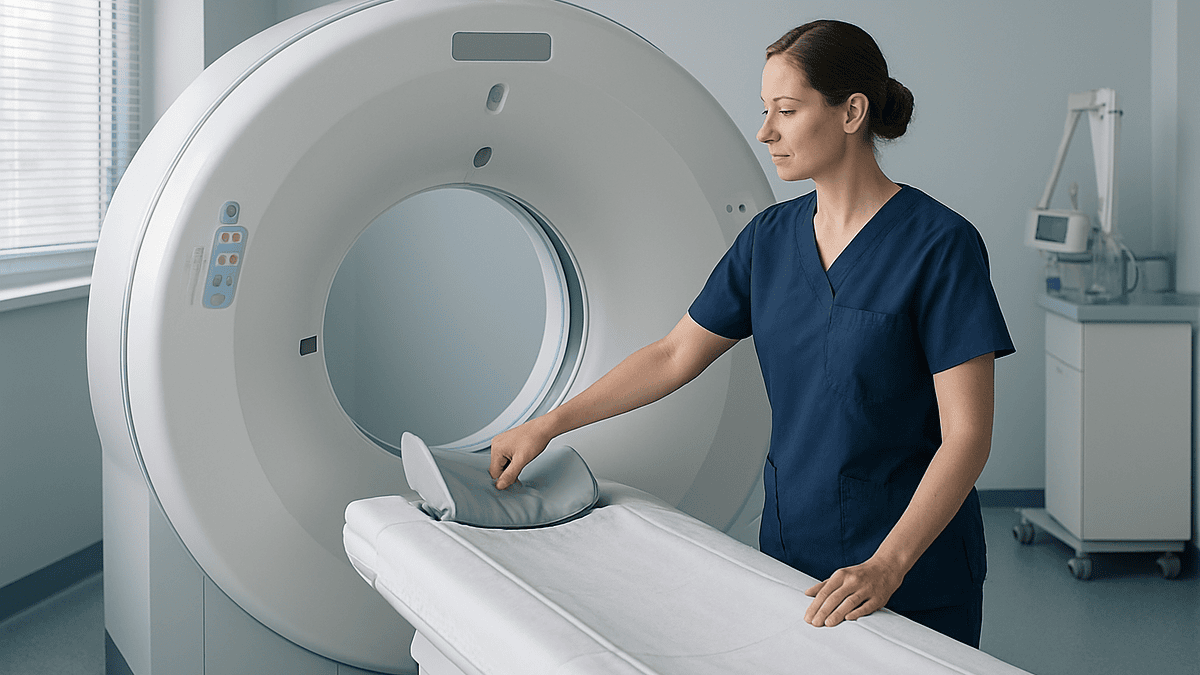
The CT is booked a few days to a week before the clinic appointment so the radiologist has time to read it and compare it with earlier images. For a plain chest CT you lie on a table that moves through a ring-shaped scanner; the scan itself takes a few minutes, and MedlinePlus describes the whole appointment as usually lasting under half an hour. Some surveillance scans use intravenous contrast dye to sharpen the view of blood vessels and lymph nodes; many use a low-dose protocol without contrast, which reduces radiation exposure and is enough to track known nodules.
Blood tests are not routinely part of lung cancer surveillance in the way they are for some other cancers, because there is no reliable blood marker that tracks lung cancer. Bloods are more often ordered to monitor treatment effects: kidney function after contrast, thyroid tests during or after immunotherapy, or liver tests on some targeted therapies.
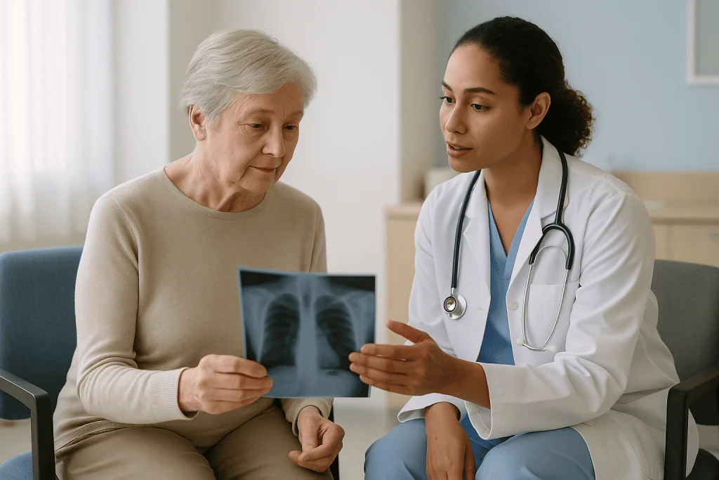
At the clinic visit, the oncologist, surgeon or specialist nurse goes through the report with you, asks about symptoms, listens to the chest, checks weight and oxygen saturation, and reviews any ongoing treatment. A good visit spends as much time on how you are functioning, breathlessness on stairs, sleep, appetite, mood, as on the scan itself.
Increasingly, some of these visits are nurse-led or by telephone, with the in-person review reserved for scan results or new symptoms. NHS guidance on living with lung cancer describes regular check-ups in the months and years after treatment, with the frequency decided by the care team. If a visit is by phone, ask who you contact between appointments if something changes.
How often are lung cancer follow up CT scans done?
This is the question every patient asks, and the honest answer has two parts: a common pattern, and a reminder that the pattern is a starting point, not a rule.
For people treated with the intent of removing all detectable disease, by surgery, radical radiotherapy or a combination, professional guideline groups in the United States and the United Kingdom broadly converge on a chest CT roughly every six months for the first two to three years, then once a year. The front-loading reflects the biology: most recurrences that are going to happen appear within the first two years after treatment, so that is when imaging has the most to find. The National Cancer Institute’s survivorship guidance explains that follow-up schedules are individualized by cancer type, stage and treatment, which is why your own interval may be shorter or longer than this.
For people on ongoing systemic treatment for advanced disease, scanning follows a different logic. Scans are timed to assess whether the treatment is working, often every two to four months in the early phase of a new drug, then less frequently once things are stable. Here the scan is not surveillance after treatment; it is part of treatment.
How long surveillance continues is less settled. Many teams keep annual imaging going for at least five years, and some continue beyond that because the risk of a second lung cancer does not fall to zero. Others switch to a model where the person is discharged with clear instructions and rapid access back to the clinic. Neither approach has been shown to be clearly superior, and the choice usually weighs age, lung function, other health conditions and the person’s own wishes.
Whatever the schedule, it belongs to you and your team together. Ask what interval has been chosen and why.
Who is usually offered scan-based follow-up, and who is asked to scan less
Not everyone who has had lung cancer treatment ends up on the same surveillance track, and the differences are deliberate.
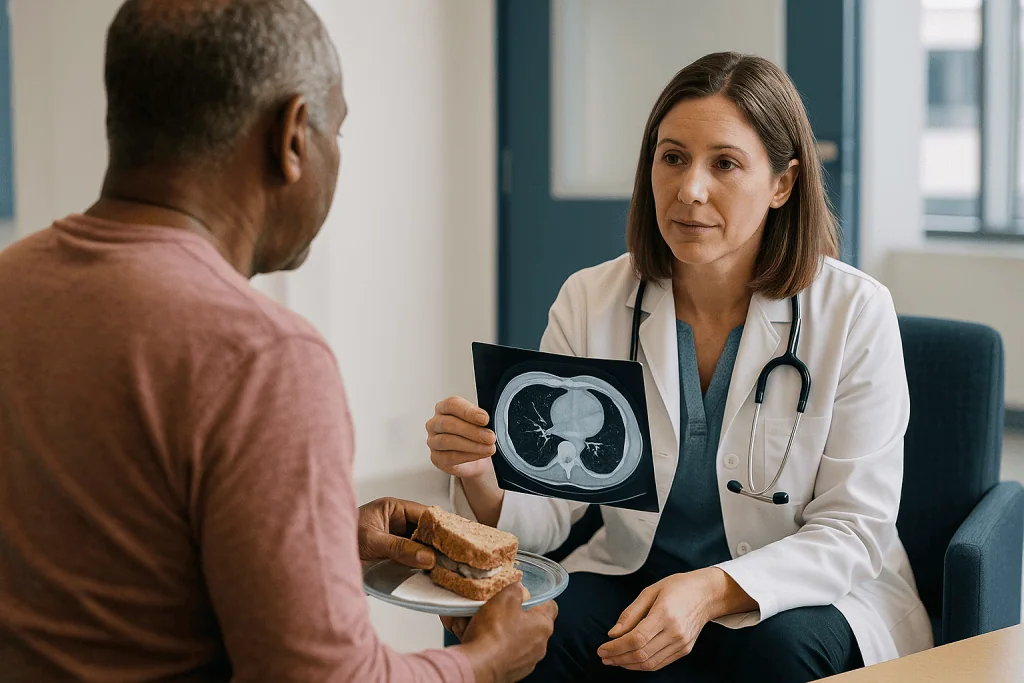
Regular CT surveillance is most clearly useful for people who could and would have further treatment if a recurrence or a new cancer were found. That generally means someone who has completed surgery or radical radiotherapy, whose lung function and general fitness would allow another operation, more radiotherapy or systemic therapy, and who wants that option. For this group, finding a small, isolated recurrence early can open doors that a later, symptom-driven diagnosis might not.
The team may suggest a lighter imaging schedule, or no routine scans, in a few situations:
- When frailty or other serious illnesses mean further aggressive treatment would not be offered, so a scan finding would not change care.
- When the person has decided they do not want further treatment and prefers to be seen only if symptoms arise.
- Late in surveillance, once several years of stable scans have passed and the yield of each additional scan falls.
None of these is a withdrawal of care. Clinic visits, symptom review and side-effect management continue; what changes is whether a CT is ordered in the absence of symptoms. The Cleveland Clinic’s overview of lung cancer emphasizes that follow-up is tailored to the individual rather than fixed.
There is also a middle ground. Some people with a small, slow-growing finding that was never treated, for example a ground-glass nodule (a hazy patch on CT that does not block the view of lung behind it) being watched rather than removed, sit on a nodule-surveillance pathway rather than a cancer-treatment pathway. Their intervals come from nodule guidelines, covered later in this article.
If you are unsure which track you are on, that is a fair question for the next visit.
Does scanning more often help? What the evidence shows
It feels intuitive that more scans must be safer. The evidence is more cautious, and understanding why can take some of the pressure off the calendar.
Studies that have compared intensive surveillance, meaning more frequent CT or routine PET-CT, with standard schedules have not consistently shown that people live longer when scanned more often. The pattern across the literature indexed in PubMed is that frequent imaging finds recurrences a little earlier, but earlier detection translates into better survival only when there is an effective treatment for what is found and the person is well enough to receive it. For isolated, surgically removable recurrences or second primaries, that condition is often met. For widespread recurrence, finding it two months sooner may change little except the length of time spent knowing.
Frequent scanning also has costs that are not financial. Each CT carries a small radiation dose; each one has a chance of showing an indeterminate finding that leads to a further scan, a PET, or a biopsy that turns out benign. MedlinePlus notes that the radiation from a single CT is low but cumulative exposure is worth tracking. The emotional cost is real too: the weeks before a scan are, for many survivors, the hardest of the year.
So the guideline consensus of six-monthly scans for a few years, then annually, is a compromise supported by observational evidence rather than a precise optimum. It concentrates imaging in the window when recurrence is most likely and when early detection is most likely to be actionable.
None of this means you should accept fewer scans than your team recommends. It means the schedule is a reasoned judgement, and that an extra scan requested for reassurance alone does not carry proven benefit.
Would lung cancer show up on a CT scan, and how often is it missed?
Two of the most searched questions about lung cancer imaging deserve direct answers.
Would lung cancer show up on a CT scan? Usually, yes. CT is the most sensitive routinely available imaging test for the lungs, able to show nodules a few millimeters across that a plain chest X-ray would miss entirely. The CDC’s screening guidance rests on exactly this capability: low-dose CT finds early cancers that X-ray does not. In follow-up, the question is slightly different, because the radiologist is comparing against a baseline. A new or growing shadow stands out more clearly when there is a previous scan to set it against.
How often is lung cancer missed on a CT? There is no single reliable percentage, and any article quoting one with confidence is oversimplifying. What the evidence does show is that misses happen in a minority of cases and tend to fall into recognizable groups: very small nodules under a few millimeters; lesions sitting against the heart, diaphragm or chest wall where contrast with surrounding tissue is low; central tumors inside or alongside airways; and changes hidden within post-radiation scarring or around a surgical staple line. Reader error also contributes, which is why many centers use double reading or computer-aided detection for surveillance scans.
A missed finding on one scan is often picked up on the next, which is one of the arguments for keeping to a regular interval rather than relying on symptoms. It is also why your symptoms matter even when the last scan was clear: a persistent new cough, hoarseness or pain between scans is worth reporting and may prompt an earlier image.
Finally, CT does not see everything. Brain metastases, for example, require MRI, which some teams include in follow-up for higher-risk cancers.
Follow up CT scan for a lung nodule: what the recommendations say
Many people reading about lung cancer follow-up are actually in an earlier situation: a scan done for another reason found a lung nodule, and they have been told to come back for another CT. The logic here is published and specific.
A lung nodule is a small, roughly round spot in the lung, under three centimeters across. Most are benign scars, healed infections or small clusters of lymph tissue. The Cleveland Clinic notes that the great majority of incidentally found nodules are not cancer. The challenge is distinguishing the few that are, without biopsying everyone.
The Fleischner Society guidelines, published in the journal Radiology and indexed on PubMed, are the most widely used framework. For a single solid nodule in an adult without a known cancer, they suggest, in broad terms:
- Under 6 millimeters: no routine follow-up in people at low risk; an optional CT at about 12 months in people at higher risk.
- 6 to 8 millimeters: a repeat CT at 6 to 12 months, and if stable, often another at 18 to 24 months.
- Over 8 millimeters: a CT at about 3 months, a PET-CT, or tissue sampling, depending on how suspicious the nodule looks.
Ground-glass and part-solid nodules follow a slower, longer track because the cancers they represent tend to grow very gradually; surveillance may extend to five years.
Two points are easily missed. First, these intervals apply to incidental nodules in people without a cancer diagnosis; once lung cancer has been treated, new nodules are usually assessed on a tighter schedule set by the oncology team. Second, growth over time is the key signal. A nodule that is unchanged across two years is very unlikely to be a solid-type cancer, which is why the recommended pattern ends rather than continuing indefinitely.
Reading your CT report: nodules, scarring, ground-glass and "stable"
Most patients now see their radiology report online before anyone has explained it. The vocabulary can be frightening when it does not need to be, so here is a plain-language guide to words that recur in lung cancer surveillance reports.
Stable is the word you want. It means the radiologist compared this scan with previous ones and found no meaningful change in size or appearance. Reports often say “stable post-treatment changes” or “no new or enlarging nodules.”
Post-radiation fibrosis or radiation pneumonitis describes the treated zone. Pneumonitis is inflammation that can appear within weeks to months of radiotherapy; fibrosis is the scarring that follows and can keep evolving for a year or more. The Mayo Clinic lists these among expected effects of lung radiotherapy. Scarring can look dense and irregular, which is why comparison with the planning scan matters.
Ground-glass opacity is a hazy area through which lung vessels are still visible. It can represent inflammation, infection, bleeding, or a very slow-growing early cancer. Context and change over time decide which.
Indeterminate nodule means the radiologist cannot say what it is from this scan alone. It is the most common reason for an extra follow-up image, and most such nodules turn out benign.
Lymphadenopathy means enlarged lymph nodes. After surgery or infection, nodes can swell reactively; growth over successive scans is more concerning than size on a single image.
Pleural effusion is fluid between the lung and chest wall. Small effusions are common after surgery and radiotherapy and often resolve.
If a word in your report worries you, write it down and ask. Radiologists describe everything they see; the clinical team decides what it means. A report that reads “abnormal” to a lay reader is often a straightforward description of a treated lung.
Side-effect care: what the clinic manages between scans
Scans get the attention, but most of the practical benefit of follow-up comes from managing the after-effects of treatment. Each therapy leaves a different footprint.
After surgery. Removing a lobe or a whole lung reduces breathing reserve. Breathlessness on exertion, chest wall pain along the incision, and numbness or sharp sensations where intercostal nerves were disturbed are common and can persist for months. The NHS notes that recovery after lung surgery often takes several weeks to a few months. Pulmonary rehabilitation, a supervised exercise and education program, is the intervention with the strongest evidence for improving breathlessness and stamina.
After radiotherapy. Fatigue, a dry cough, difficulty swallowing if the esophagus was in the field, and skin changes are typical early effects. Pneumonitis can emerge weeks to months later and presents with new cough, breathlessness or fever; it is treatable but needs prompt review, which is why teams ask you to report these symptoms rather than wait for the next scan.
After chemotherapy. Peripheral neuropathy, tingling or numbness in hands and feet, may improve slowly over months or persist. Hearing changes, kidney effects and lingering fatigue are also reviewed at visits.
During or after immunotherapy. Drugs that release the immune system’s brakes can cause inflammation in almost any organ: thyroid, bowel, skin, liver, lungs, joints. Some of these appear after treatment has finished. The team monitors blood tests and asks about new symptoms for this reason.
On targeted therapy. People with specific tumor mutations may stay on a daily oral drug for years. Rashes, diarrhea, nail changes and liver effects are managed over the long term, and decisions about continuing, pausing or changing medicine always rest with the prescribing oncologist.
Across all groups, mood, sleep, weight and return to work are legitimate agenda items, not afterthoughts.
Lung cancer surveillance after treatment: the days around each scan
Surveillance has a rhythm, and knowing it in advance helps people plan their lives around it rather than the other way round.
The week before a scan is usually the hardest. Survivors describe a familiar tightening of attention: every twinge becomes a possible sign, sleep gets lighter, concentration drifts. Clinicians sometimes call this scan anxiety; it is common enough that many cancer centers mention it explicitly in survivorship material, and the National Cancer Institute’s follow-up care guidance acknowledges that fear of recurrence is one of the most frequent concerns after treatment. Practical steps that people find useful include scheduling the scan and results visit as close together as possible, so the waiting window is short; arranging something absorbing for the day after the scan; and telling one trusted person what the date is.
On scan day, a non-contrast chest CT needs no fasting. A contrast scan usually requires a short period without food and a recent kidney function test, and you will be asked about allergies and whether you take certain medicines, which the imaging department will advise on individually.
Results commonly arrive within a few days to two weeks depending on the hospital’s process. If the scan is stable, the visit is often brief and the next interval is confirmed. If something needs clarifying, the team may order a PET-CT, an earlier repeat CT in a few months to check for growth, or in some cases a biopsy. An earlier repeat scan is a measured decision, not a verdict; it is the standard way to tell a slowly resolving area of inflammation from a growing lesion.
Between scans, the useful habit is simple: note new or persistent symptoms with dates and bring the list to the next visit, or call sooner if the symptom is one of the red flags described below.
A typical surveillance pathway at a glance
The table below summarizes a common pattern for someone treated with the aim of removing all detectable disease. It is a description of what guidelines broadly suggest, not a schedule for any individual; the treating team adjusts every element.
| Period after treatment | Imaging commonly used | Clinic focus | Why this pattern |
|---|---|---|---|
| First 3 months | Baseline chest CT after surgery or radiotherapy | Wound and breathing recovery, pain, early radiation effects, rehabilitation referral | Establishes the new normal anatomy that later scans are compared against |
| Months 3 to 24 | Chest CT roughly every 6 months | Recurrence symptoms, pneumonitis, fatigue, neuropathy, mood, return to activity | Most recurrences that occur appear in this window |
| Years 2 to 5 | Chest CT every 6 to 12 months, moving to annual | Late effects, second primary cancer risk, smoking cessation support, heart and lung health | Recurrence risk falls; second cancer risk persists |
| Beyond 5 years | Annual low-dose CT in many centers, or symptom-led review | General health, ongoing screening, long-term side effects | Balances continued second-cancer risk against radiation and anxiety burden |
Three caveats keep this table honest. People on ongoing systemic therapy for advanced disease follow a treatment-response schedule rather than this one. Those with higher-risk histology or known brain involvement may have periodic MRI added. And anyone who has been asked to scan less often because further treatment would not be appropriate should still expect regular clinical contact and side-effect care; the scan is only one part of follow-up.
The intervals cited here reflect the broad consensus described by the National Cancer Institute and national cancer services such as the NHS, which both stress that schedules are individualized.
What happens if a scan shows something new?
Sooner or later, many survivors get a report that is not simply “stable.” Knowing the usual next steps makes that moment less disorienting.
The first question the team asks is whether the finding is definitely new or growing, or whether it might be a previous area now seen at a slightly different angle. Comparison with every prior scan, not just the last one, often settles this. Radiologists are trained to spot slow change across a series that would be invisible between two consecutive images.
If the finding is genuinely new or enlarging, there are three broad routes. A short-interval repeat CT, typically within a few months, is appropriate when the change could plausibly be inflammation or infection that should settle. A PET-CT is used when the team wants to know whether the area is metabolically active and whether anything similar is present elsewhere in the body; the Mayo Clinic describes PET as useful for distinguishing active disease from scar, with the caveat that infection and inflammation can also light up. A biopsy, by needle through the chest wall, by bronchoscopy, or occasionally by surgery, is the only way to confirm what tissue is present, and it is chosen when the result would change management.
If recurrence is confirmed, the next conversation is about options, and it is often a wider conversation than people expect. An isolated recurrence may be treated with further surgery or focused radiotherapy. More widespread recurrence is usually approached with systemic treatment, and the tumor may be retested for mutations or immune markers because these can change over time and open different treatment paths. Every option carries risks and alternatives, including the option of symptom-focused care, and the choice rests with you and your team.
If the finding turns out to be benign, which happens often, the team will usually return you to the planned surveillance schedule, sometimes with an added interim scan for reassurance about that specific area.
What people often get wrong about lung cancer follow-up
Some beliefs circulate so widely that they are worth correcting directly.
“A clear scan means I am clear.” A stable CT is reassuring, but it is a snapshot of structures large enough to see. Microscopic disease is invisible to any scan, which is why surveillance continues after a clear result and why symptoms between scans still matter.
“More scans would catch it sooner, so I should push for them.” As described earlier, intensive imaging has not been shown to improve survival beyond standard schedules, and each additional scan adds radiation, false alarms and anxiety. Asking for a scan when you have a new symptom is sensible; asking for one purely for reassurance between scheduled dates carries no proven benefit.
“A blood test could replace the CT.” There is currently no blood marker reliable enough to monitor lung cancer, which is why guidelines do not include routine tumor markers in follow-up. Blood tests in surveillance exist to monitor treatment effects, not to detect recurrence.
“If I had no symptoms before diagnosis, symptoms will not help now.” Lung cancer is often silent at first diagnosis, but recurrence is frequently announced by symptoms, especially pain, hoarseness, headache or new neurological changes that CT of the chest would not capture. Reporting them promptly is part of surveillance.
“Scarring on the scan means the radiation is still damaging my lung.” Fibrosis is the lung’s settled response to treatment. It can evolve on imaging for a year or more, which is expected, and it is distinguished from recurrence precisely by comparison over time.
“Once I am discharged from follow-up, no one is watching.” Discharge usually comes with a direct route back to the team and, in many centers, continued annual low-dose CT through a screening program. The Cleveland Clinic and NHS both describe follow-up as a long-term arrangement rather than a fixed endpoint.
Questions to ask your care team
Visits are short and the agenda fills quickly. Taking a written list changes the conversation. These are questions that experienced patients wish they had asked earlier.
- What is my surveillance schedule, and what is the reasoning behind this interval for my stage and treatment?
- Will my scans be low-dose or standard, with or without contrast, and why?
- Who reads my scans, and will they always be compared with my earlier images?
- How and when will I get results, and who do I call if I have not heard by then?
- Which symptoms should prompt me to contact you before the next scheduled visit, and what number do I use?
- What does my latest report mean in plain language, especially any words like “indeterminate” or “post-treatment change”?
- Am I a candidate for pulmonary rehabilitation, and how do I access it?
- Which side effects of my treatment might appear late, and what would they look like?
- If I am on a long-term targeted or immune therapy, what monitoring does it need and what signs would make you reconsider it?
- Do I need any imaging outside the chest, such as brain MRI, and if not, why not?
- How long is surveillance planned to continue, and what happens when it ends?
- Is there smoking cessation, dietary or psychological support attached to this clinic?
- Would you be comfortable with me seeking a second opinion on my scan report or treatment plan, and how would that work?
A final question worth asking at every visit is simple: has anything changed in my situation that alters the plan? Guidelines evolve, new treatments arrive, and your own health shifts. Follow-up works best as a continuing conversation rather than a fixed protocol. The National Cancer Institute’s survivorship guidance recommends keeping a personal record of your treatment and follow-up plan so that any clinician you see, including a family physician, can understand your history quickly.
When to call your doctor
Surveillance scans are scheduled, but symptoms do not wait for appointments. Contact your cancer team, or seek urgent care, if you notice any of the following between visits.
- Coughing up blood, even a small amount, or a cough that is new or has changed and persisted for more than two to three weeks.
- Breathlessness that is new or clearly worse than your usual post-treatment level, especially if it comes with fever or chest pain; after radiotherapy this can indicate pneumonitis, which the Mayo Clinic notes needs prompt assessment.
- Chest pain that is new, persistent or different from incision or rib discomfort you already know.
- Hoarseness lasting more than a few weeks, or difficulty swallowing.
- New headaches, confusion, weakness or numbness on one side, visual change, seizures or unsteadiness, which may point to the brain or nervous system and are not seen on chest CT.
- Bone pain that is new, persistent or wakes you at night.
- Unexplained weight loss, loss of appetite or drenching night sweats.
- Swelling of the face, neck or arms, or prominent veins on the chest, which can signal pressure on the large vein returning blood from the upper body.
- A swollen, painful calf or sudden breathlessness with chest pain, which may indicate a blood clot; this is an emergency.
- During or after immunotherapy: persistent diarrhea, severe rash, yellowing of the skin or eyes, or marked fatigue, which can reflect immune-related inflammation.
None of these symptoms means the cancer has returned; most have other explanations. Reporting them allows the team to decide whether an earlier scan or examination is needed. If a symptom is severe, sudden or frightening, use emergency services rather than waiting for the clinic to open. The NHS living-with-lung-cancer guidance encourages patients to contact their specialist nurse about any new concern rather than holding it until the next appointment.
Frequently asked questions
How often should I have a CT scan after lung cancer treatment?
For most people treated with the aim of removing all detectable disease, guideline groups suggest a chest CT roughly every six months for the first two to three years, then once a year. Your own interval may differ depending on stage, treatment type, lung function and other health conditions, and it is set by your treating team rather than a fixed rule.
Would lung cancer show up on a CT scan?
Usually, yes. CT is the most sensitive routine imaging test for the lungs and can show nodules only a few millimeters across that a chest X-ray would miss. In follow-up the radiologist compares each scan with earlier ones, which makes new or growing areas easier to spot. CT does not image the brain, so some cancers need MRI as well.
How often is lung cancer missed on a CT scan?
There is no single reliable percentage, and any confident figure oversimplifies. Misses do occur in a minority of cases, most often with very small nodules, lesions against the heart or chest wall, central tumors near airways, or changes hidden in post-radiation scarring. Regular interval scans and prompt reporting of new symptoms are the main safeguards against a missed finding.
What is the recommended follow-up CT scan for a lung nodule?
The Fleischner Society guidelines are widely used. For a single solid nodule in an adult without known cancer: under 6 millimeters usually needs no routine follow-up in low-risk people; 6 to 8 millimeters calls for a repeat CT at 6 to 12 months; over 8 millimeters warrants a CT at about 3 months, PET-CT or biopsy. After lung cancer treatment, new nodules are assessed on a tighter schedule set by the oncology team.
What is the biggest indicator of lung cancer coming back?
No single sign is definitive. Growth of a nodule or mass on successive CT scans is the most objective indicator, which is why comparison over time matters more than any one image. Symptom-wise, coughing up blood, a new persistent cough, unexplained weight loss, new bone pain or neurological changes are the ones teams most want to hear about promptly.
What are common lung cancer recurrence symptoms I should report?
Report a new or changed cough lasting more than two to three weeks, coughing up blood, worsening breathlessness, hoarseness, difficulty swallowing, new chest or bone pain, unexplained weight loss, or any new headache, weakness, confusion or visual change. Most of these have other causes, but each can prompt your team to examine you or bring a scan forward.
Does more frequent CT scanning after lung cancer improve survival?
The evidence does not consistently show that it does. Studies comparing intensive surveillance with standard schedules have found that more frequent imaging detects recurrence slightly earlier, but earlier detection improves outcomes only when there is an effective treatment the person is fit enough to receive. Extra scans also add radiation, false alarms and anxiety.
How long does lung cancer surveillance after treatment continue?
Many teams continue imaging for at least five years, often moving to annual low-dose CT after the first two to three years. Some continue beyond five years because the risk of a second lung cancer persists, especially in people with a smoking history; others discharge with a clear route back to the clinic. The plan is individualized and worth discussing explicitly.
Is radiation from repeated follow-up CT scans dangerous?
Each CT carries a small radiation dose, and low-dose protocols used for surveillance reduce it further. For someone who has had lung cancer, the benefit of detecting recurrence or a second cancer is generally judged to outweigh the small cumulative risk. MedlinePlus notes that exposure adds up over time, which is one reason guidelines avoid scanning more often than needed.
What does "post-treatment change" or "stable fibrosis" mean on my scan report?
It means the radiologist is describing expected scarring or healing in the treated area and has found no meaningful change compared with previous scans. Radiation fibrosis can look dense and irregular and can keep evolving for a year or more. “Stable” is the reassuring word; it tells you the area was compared and did not grow.
References
This article is for general information only and is not a substitute for professional medical advice. Please consult a qualified doctor about your individual situation.

More from the Blog
Facial Weakness or Numbness After Parotidectomy: What Is Expected and When to Call
Some facial weakness or numbness after parotidectomy is common and, in most people, temporary. Weakness comes from the facial nerve being stretched or bruised…

How Does a Child Recover After Wilms Tumor Surgery? Comfort, Wound Healing and Next Steps
After Wilms tumor surgery, a child usually spends several days in hospital while pain is controlled, the bowel starts working again and the incision…

Can You Have Lung Cancer Surgery? What Lung Function and Heart Tests Help Decide
Yes, many people with early-stage lung cancer can have surgery, but the decision rests on tests rather than age alone. Surgeons measure breathing capacity…

Melanoma Staging: What Biopsy Review, PET-CT and Blood Tests Reveal Before Treatment
Melanoma staging tests establish how far the cancer has spread before any treatment plan is made. The pathologist's review of the biopsy, which measures…
Why Transfusions Are Part of Bone Marrow Transplant Care: Bleeding Risk and Low Counts
Transfusions are a routine part of bone marrow transplant care because the high-dose chemotherapy, and sometimes radiation, given before the transplant clears out the…

Liver Tumor Ablation Step by Step: Anesthesia, Imaging Guidance and Recovery
Liver tumor ablation destroys a small liver tumor with heat, cold or electrical energy delivered through a thin needle guided by ultrasound, CT or…