

What MRI and CT Reveal About TMJ: The Disc, Arthritis and Joint Damage

Key Takeaways
- MRI is the only common scan that shows the TMJ disc directly, which is why it is the reference method for deciding whether the disc is displaced and whether it reduces on opening.
- A disc that slips back into place when the mouth opens produces the familiar click; a disc that stays in front of the condyle limits opening and is described as displacement without reduction.
- CT and cone-beam CT image bone far better than MRI, revealing flattening, bone spurs, erosions and fractures, but cannot see the disc at all.
- T2-weighted and contrast-enhanced MRI can reveal joint fluid and inflamed synovium in rheumatoid or juvenile idiopathic arthritis before CT shows any bone damage.
- Disc displacement and degenerative change are also found in people with no jaw symptoms, so imaging findings explain pain only when they match the clinical examination.
- The NHS states that a painless jaw click does not need treatment, and the Mayo Clinic describes most TMJ-related pain as temporary, which is why imaging is reserved for specific questions rather than used routinely.
MRI is the preferred way to see the soft tissues of the temporomandibular joint, especially whether the cartilage disc sits on top of the jaw condyle or has slipped forward, and whether there is fluid or inflammation inside the joint. CT and cone-beam CT show bone best, revealing arthritis changes such as flattening, bone spurs and erosion. Imaging findings do not always match pain, so results are always read alongside a clinical examination.
It usually starts with a sound. A pop while biting into a crusty roll, a click every time you yawn, then one morning the jaw simply refuses to open past two fingers’ width. The dentist presses gently in front of your ear, asks you to open and close, and says the words that send most people straight to a search bar: we should probably take a look inside.
That is where TMJ imaging MRI enters the story, often alongside a CT scan, and where the confusion begins. Which scan sees what? Does a slipped disc on a picture explain the pain, or is it a bystander? And why do radiologists describe a jaw joint the way geologists describe a riverbed, with words like erosion and flattening?
This explainer walks through what each scan can genuinely reveal about the disc, arthritis and joint damage, what it cannot, and how your care team weighs a grey-scale image against the far more important evidence of how your jaw actually behaves.
What TMJ imaging MRI actually shows, and what it cannot
Magnetic resonance imaging, or MRI, builds pictures from the way hydrogen atoms in water and fat respond to a strong magnet and radio waves. Because soft tissues are rich in water, MRI is the only common scan that shows the small cartilage disc inside the temporomandibular joint (TMJ) directly, rather than inferring its position from the bones around it. The United States National Institute of Dental and Craniofacial Research lists MRI as the imaging method of choice when a clinician needs to see soft tissue in this joint.
A good TMJ MRI answers a short list of questions. Where is the disc when the mouth is closed, and does it move back into place when the mouth opens? Is there excess fluid in the joint space, a sign of inflammation? Does the bone marrow of the condyle, the rounded knuckle of the lower jaw, show swelling? Are the chewing muscles symmetrical? In inflammatory arthritis, has the joint lining thickened?
What MRI cannot do is equally important. It does not measure pain. It does not show the tightness in a jaw muscle that has been clenching through a stressful quarter at work. It shows bone surfaces less crisply than CT, so early arthritis of the condyle can be understated. And it photographs one moment in a joint that moves thousands of times a day, which is why radiologists ask for at least two positions, mouth closed and mouth open.
Think of the scan as a weather report rather than a verdict. It tells the care team what the landscape looks like today; the examination, the history and how the jaw responds to time and conservative care tell them what it means for you.
How the jaw joint works, and what causes pain in the TMJ
The temporomandibular joint sits just in front of each ear, where the condyle of the lower jaw meets a shallow socket in the temporal bone of the skull. Between them lies the articular disc, an oval pad of fibrocartilage, a dense and slightly flexible tissue, that acts as a cushion and a sliding bearing. When you open your mouth, the condyle first rotates like a hinge and then glides forward out of the socket, carrying the disc with it. Two joints, one on each side, must do this in near-perfect synchrony every time you speak or chew.
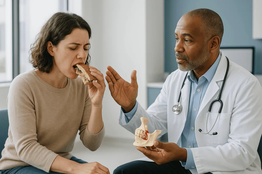
Pain in the TMJ rarely has a single cause. According to the Mayo Clinic, the disc can erode or move out of alignment, the joint cartilage can be damaged by arthritis, or the joint can be injured by a blow or whiplash. Often, though, the pain comes from the surrounding muscles rather than the joint itself: sustained clenching, tooth grinding at night (bruxism) and habits such as holding a phone with the shoulder keep the masseter and temporalis muscles in a low-grade cramp. The NHS adds stress and uneven bite as contributing factors and notes that in many cases no clear cause is found.
This distinction between joint pain and muscle pain is the single most useful thing to understand before any scan. MRI and CT image the joint. They are far less helpful for muscle-driven pain, which is diagnosed by examination. The National Institutes of Health notes that temporomandibular disorders (TMDs) are more common in women than in men and that symptoms often ease without surgery. A scan is therefore a tool for specific questions, not a routine first step for a sore jaw.
What happens during a TMJ MRI with open and closed mouth
Most people arrive expecting a scan of the whole head. In practice the technologist positions two small surface coils, flat plastic pads containing antennae, one over each jaw joint, because the structures of interest are only a couple of centimetres across and need a close-range signal. You lie on your back, the table slides into the magnet, and the machine knocks and hums as it acquires images.
The first series is taken with the teeth lightly together. Then comes the part that makes a TMJ MRI with open and closed mouth different from almost any other scan: you are asked to open as wide as is comfortable and hold still, often with a small plastic spacer or bite block between the front teeth so the opening stays constant for several minutes. Some centres add a short series at intermediate openings, sometimes called a pseudo-dynamic or cine study, to show the disc’s journey rather than just its start and finish.
Both joints are usually imaged in the same appointment, even when only one side hurts, because the opposite joint provides a comparison and because disc problems are frequently bilateral. According to MedlinePlus, MRI examinations can last anywhere from about 15 minutes to more than an hour depending on the number of sequences; a two-position TMJ study generally sits in the middle of that range.
There are no needles for a standard study. Contrast is rarely needed and reserved for specific situations covered later. Hearing protection is provided, and anyone who struggles with enclosed spaces should say so when booking, since positioning, music or a mirror prism can make the experience more tolerable. The most useful preparation is simply practising holding your mouth open for a few minutes beforehand.
What type of MRI is used for TMJ?
There is no special machine for the jaw joint. The difference lies in the protocol, the recipe of image sequences and viewing angles that the radiologist specifies. A TMJ protocol has three defining features.

First, the planes are oblique. Because each condyle sits at an angle to the skull, images are tilted to run along and across the long axis of the condyle itself, giving a true side view (oblique sagittal) and a true front view (oblique coronal) of the disc rather than a slanted slice through it.
Second, the sequences are chosen to separate tissues. Proton-density and T1-weighted images show anatomy crisply and make the dark, low-water disc stand out against brighter fat behind it; these are the pictures used to judge disc position. T2-weighted images make free fluid glow white, so they reveal joint effusion, swelling within the bone marrow and the inflamed, thickened lining seen in some arthritis. Fat-suppressed variants sharpen that contrast further.
Third, the fields of view are small and the slices thin, typically a few millimetres, to resolve a disc that is only a few millimetres thick itself. Higher-field scanners and dedicated coils help here, though image quality depends as much on the patient keeping still as on the hardware.
Gadolinium-based contrast agents, dyes injected into a vein that highlight areas of increased blood flow, are not part of a routine disc study. A clinician may request them when inflammatory arthritis is suspected, especially in children with juvenile idiopathic arthritis, because contrast makes active synovitis, inflammation of the joint lining, visible before bone damage appears. Whether contrast is appropriate is a decision for the referring clinician and radiologist, weighing kidney function and the specific question being asked.
How to tell if a TMJ disc is displaced: reading a TMJ disc displacement MRI
In a healthy joint with the mouth closed, the thick back band of the disc sits on top of the condyle, roughly at the twelve o’clock position, like a beret resting squarely on a head. As the mouth opens, condyle and disc glide forward together and the disc stays interposed between the two bones.
Displacement nearly always means the disc has slipped forward, so that with the mouth closed it lies in front of the condyle and the condyle rests on the stretched tissue behind it instead. What happens next on the open-mouth images defines the two patterns a TMJ disc displacement MRI is designed to distinguish:
- Displacement with reduction. On opening, the condyle slides forward, catches up with the disc and pops back on top of it. That recapture is the click many people hear and feel. On the open images, the disc looks normally positioned.
- Displacement without reduction. The disc stays ahead of the condyle throughout opening, acting as a doorstop. The condyle cannot translate fully, and the jaw opening is limited. Clinically this is often called a closed lock, and the once-familiar click may have disappeared.
Radiologists also describe the disc’s shape. A normal disc looks like a bow tie or dumbbell in side view; one that has been displaced for a long time may fold, thicken or shrink into a rounded lump. Disc perforation and adhesions, where the disc sticks to the socket, are suspected on MRI but confirmed only at arthroscopy.
One honest caution: disc displacement is also found in people with no jaw symptoms at all, a fact repeatedly noted in the radiology literature indexed by the National Library of Medicine. A displaced disc on a scan explains pain only when the examination findings match it.
When is a CT scan for TMJ the better choice?
Computed tomography, or CT, rotates an X-ray source around you and reconstructs cross-sectional slices. It images bone with a sharpness MRI cannot match, because the dense mineral of bone gives a clean, bright signal while cartilage and disc fade into shadow. That trade-off dictates when a CT scan for TMJ earns its place.
Clinicians typically reach for CT when the question is about hard tissue. After a fall or a sporting collision, CT is the standard way to look for a fracture of the condyle or its neck, which MRI can miss. In long-standing arthritis, CT maps the surface of the condyle and socket, showing flattening, bone spurs (osteophytes, small ridges of extra bone at the joint margin), erosions, hardening beneath the surface (sclerosis) and tiny cysts. It also helps when the bite has changed, which can signal shortening of the condyle. Before joint surgery, CT provides a three-dimensional model for planning.
Many dental and oral surgery settings now use cone-beam CT, which captures the whole volume in a single sweep with a cone-shaped beam. According to MedlinePlus, CT exposes you to ionizing radiation, which is why it is not used to follow soft-tissue disc problems that MRI can monitor without any radiation. Cone-beam machines are designed to deliver less radiation than conventional medical CT for a comparable field, though the exact figure depends on settings, and the referring clinician should be able to explain why the exposure is justified in your case.
A CT appointment is brief, usually a matter of minutes in the scanner, and poses no problem for people with pacemakers or metal implants that would exclude them from MRI. What it cannot tell you is whether your disc has slipped. For that, the two scans are complementary, not competing.
MRI, CT or X-ray for the jaw joint: a side-by-side table
Choosing a scan is a matter of matching the question to the tool. The panoramic dental X-ray, the wide, curved image many people have had at a routine check-up, deserves a mention because it is often the first picture a dentist already holds. It gives a rough view of both condyles and can flag an obvious fracture or large cyst, but it overlaps structures and misses early arthritis, and it shows nothing of the disc.
| Question | MRI | CT / cone-beam CT | Panoramic X-ray |
|---|---|---|---|
| Is the disc displaced? Does it reduce? | Yes, the reference method | No | No |
| Fluid or inflammation in the joint? | Yes (T2, sometimes with contrast) | No | No |
| Early bone erosion, spurs, flattening? | Moderate; may understate | Yes, the reference method | Limited |
| Fracture after trauma? | Possible but not first choice | Yes | Screening only |
| Bone marrow swelling? | Yes | No | No |
| Ionizing radiation? | None | Yes | Yes, low dose |
| Typical time in scanner | Roughly 30–60 minutes | Minutes | Under a minute |
| Usual barriers | Pacemakers, some implants, claustrophobia | Pregnancy caution | Few |
Two patterns emerge. For a clicking or locking jaw where the exam points to the disc, MRI alone usually suffices. For a jaw that has changed shape, hurts after an injury or shows signs of arthritis, CT answers the structural question, and MRI is added if inflammation or disc damage also needs mapping. Timing ranges in the table reflect MedlinePlus descriptions of each modality and vary by centre.
What arthritis looks like inside the jaw joint
Arthritis is a broad word for inflammation or wear of a joint, and the jaw is not exempt. The Cleveland Clinic lists osteoarthritis and rheumatoid arthritis among the recognised causes of temporomandibular disorders, and each leaves a distinct signature on imaging.
Osteoarthritis, the degenerative or wear-related form, usually shows first on CT as a loss of the condyle’s smooth, rounded contour. The surface flattens, the bone beneath it hardens and brightens, and small spurs appear at the front edge. Later come erosions, pits in the surface, and shortening of the condyle. On MRI, the same joint may show a displaced or deformed disc, a thin rim of fluid, and patchy brightness in the bone marrow on T2 images that radiologists interpret as a stress response. Osteoarthritis of the TMJ often coexists with long-standing disc displacement, and which came first is frequently impossible to say from pictures alone.
Rheumatoid arthritis and related inflammatory conditions attack the joint lining. Early on, CT may look normal, which is exactly when contrast-enhanced MRI is most informative: the enhancing, thickened synovium and joint effusion announce active inflammation before the bone is harmed. If untreated, inflammation erodes the condyle, sometimes aggressively, and the bite can open at the front as the jaw rotates backwards.
In children, juvenile idiopathic arthritis matters particularly because the condyle contains a growth centre; inflammation there can alter the growth of the lower jaw and the symmetry of the face. The NIH identifies TMJ involvement as a recognised feature of this condition, and MRI with contrast is the usual tool for detecting it, since young joints may be inflamed with few symptoms.
Across all forms, the imaging grade of arthritis is a poor predictor of pain. Some heavily remodelled joints are quiet; some near-normal ones hurt daily.
Who is usually referred for imaging, and who is usually asked to wait
The NHS and the Mayo Clinic both describe temporomandibular disorder as a condition that is usually diagnosed by listening to the history and examining the jaw, and that in most cases improves with time and simple self-care. Imaging is therefore selective. A scan is generally considered when the picture inside the joint would change what the team does next.
Referral is common when:
- The jaw locks, either stuck open or unable to open fully, and the examination suggests a disc that no longer reduces.
- There has been a blow to the face or a whiplash injury and a fracture must be ruled out.
- The bite has shifted, the chin has drifted to one side, or the face looks newly asymmetric.
- A systemic inflammatory condition such as rheumatoid or juvenile idiopathic arthritis is known or suspected.
- Symptoms have persisted despite a reasonable period of conservative care and the team is weighing injections or surgery.
- There is swelling, numbness or a mass that could indicate something other than TMD.
People are usually asked to wait when the jaw clicks painlessly, when the pain is clearly muscular and reproduced by pressing on the chewing muscles rather than the joint, or when symptoms are recent and still settling. A painless click, on its own, is common and does not require a scan; the NHS notes that clicking without pain is not a cause for concern.
Waiting is not the same as doing nothing. Soft diet, warmth, gentle stretching, awareness of clenching and stress management are the first-line measures described by the NHS and Mayo Clinic, and their response over a few weeks is itself diagnostic information. The decision to image, and when, sits with the dentist or clinician who has examined you.
What the days and weeks after the scan usually look like
Walking out of the MRI suite, most people feel exactly as they did walking in. There is no recovery from the scan itself, and holding the mouth open for a few minutes rarely aggravates a sore joint for more than an hour or two. If contrast was given, the technologist will usually ask you to drink normally over the following day.
The images then go to a radiologist, a doctor who specialises in interpreting scans, who writes a structured report describing disc position in both positions, the shape of the disc, any fluid, the state of the bone and marrow, and a comparison between the two sides. That report is sent to the clinician who referred you, not usually to you directly, although many services now release results through a patient portal. Reporting times vary between services, and it is reasonable to ask at the appointment how and when you will hear.
The follow-up conversation is where the scan earns its value. Expect your clinician to lay the findings alongside what they felt and heard during the examination. A displaced disc that reduces, in a jaw that clicks but opens fully, usually confirms a reassuring diagnosis and a continued conservative plan. A disc that does not reduce, with limited opening, may prompt a discussion about jaw exercises, a splint, or in persistent cases a procedure such as arthrocentesis, washing out the joint with sterile fluid, or arthroscopy. Arthritis findings may lead to a referral to a rheumatologist and blood tests.
Over the following weeks, the most useful thing you can do is keep a simple diary: opening width in finger-breadths, pain on waking, clicks, foods avoided. Those notes, more than any repeat scan, show whether the plan is working. Repeat imaging is generally reserved for a change in symptoms or for planning a procedure.
What doctor treats jaw arthritis and disc problems?
The jaw joint sits in a professional borderland, which is why people often bounce between appointments before anyone takes ownership. The Mayo Clinic suggests starting with your dentist or primary care clinician, and that is sensible: both can examine the joint, rule out dental causes such as a cracked tooth or an abscess, and decide whether imaging is warranted.
From there, the pathway depends on what is found. An oral and maxillofacial surgeon, a specialist trained in both dentistry and surgery of the face and jaws, manages locked joints, fractures, severe degenerative change and the small number of cases that need arthrocentesis, arthroscopy or open joint surgery. Many also oversee splint therapy and injections. An orofacial pain specialist, a dentist with advanced training in chronic pain of the head and jaw, focuses on muscle-driven and persistent pain, where behavioural strategies and medicines that act on nerve and muscle pathways matter more than pictures.
Jaw arthritis that is part of a body-wide inflammatory disease belongs with a rheumatologist, a physician who treats conditions such as rheumatoid and juvenile idiopathic arthritis. Medicines in this field work by damping the immune signals that drive synovitis, and they are chosen and adjusted by the rheumatologist based on the whole disease, not the jaw alone. Children are seen by pediatric rheumatologists, often alongside orthodontists who monitor facial growth.
Physical therapists with jaw expertise, ear, nose and throat surgeons who exclude ear disease, and neurologists for headache overlap all have roles. If this sounds like a committee, that is sometimes what persistent TMD needs. What matters is that one clinician coordinates, and that every decision about treatment, including whether to start, pause or change any medicine, rests with the team that has examined you.
What people often get wrong about TMJ imaging MRI
Myth one: a displaced disc on the scan means the joint is damaged and will get worse. The evidence does not support that. Disc displacement is found in many people who have never had jaw pain, and the Mayo Clinic describes most TMJ-related pain as temporary. A displaced disc is a finding, not a prognosis.
Myth two: a normal MRI means the pain is imaginary. Muscle pain, the most common driver of TMD, is invisible on MRI. A clean joint scan in a sore jaw is useful information: it points the team towards the muscles, clenching habits and stress, and away from the joint.
Myth three: CT is the better scan because it is sharper. CT is sharper for bone. It cannot see the disc at all. Asking for a CT scan for TMJ to check disc position is like asking for a weather satellite to read a street sign.
Myth four: the click needs to be fixed before it causes arthritis. A painless click is a reducing disc doing what reducing discs do; the NHS specifically reassures that clicking without pain does not need treatment. Surgery to silence a click is not supported by guideline-level evidence.
Myth five: you must have a scan before any treatment. Conservative care is routinely started on clinical grounds alone, and its response often makes a scan unnecessary.
Myth six: the scan is uncomfortable or involves radiation. A TMJ MRI with open and closed mouth is noisy and requires a few minutes of jaw opening, but it involves no radiation and usually no injection. CT does involve radiation, but a jaw study is brief and targeted.
The thread running through all six is the same: the picture serves the examination, never the other way round.
Questions to ask your care team
A scan appointment is short; the conversation before and after it is where understanding happens. Taking a written list helps, and so does bringing a companion to hear the answers. These are the questions experienced clinicians say they wish more people asked.
- What specific question are you hoping this scan will answer, and how would the result change my plan?
- Is MRI, CT or both the right choice for that question, and why?
- Will both joints be imaged, and will images be taken with the mouth open and closed?
- Will I need contrast, and if so, what are you looking for that a standard study would not show?
- Do I have any implant, device or medical condition that affects whether MRI is safe for me?
- How and when will I receive the report, and who will explain it to me?
- If the disc is displaced, what does that mean for me specifically, given my symptoms and jaw opening?
- If the scan shows arthritis, who will manage it, and do I need blood tests or a rheumatology referral?
- If the scan is normal, what is the most likely cause of my pain and what is the next step?
- What conservative measures should I continue or start while waiting for results?
- At what point would you consider injections, arthrocentesis or surgery, and what would need to be true on the scan and in the clinic?
- Would a repeat scan ever be useful, or will you judge progress by examination?
Notice that none of these ask for a prediction. Clinicians cannot promise a particular outcome from a scan or a treatment, and anyone who does is overstepping the evidence. What they can offer is a clear reason for each step, a named person responsible for the next one, and an honest account of what the images did and did not show. If you leave the follow-up appointment able to explain your own scan in two sentences, the appointment did its job.
When to call your doctor
Most jaw pain is uncomfortable rather than dangerous, and the NHS advises seeing a dentist or general practitioner when symptoms persist or interfere with eating and sleeping. Some situations, though, should not wait for the next routine slot. Contact your doctor or dentist promptly, or seek urgent care, if any of the following occur:
- Your jaw locks open or shut and you cannot free it, especially if this is new or has lasted more than a few hours.
- Severe pain follows a blow to the face, a fall or a road accident, or your teeth suddenly no longer meet the way they did.
- The joint area is swollen, hot or red, or you have a fever alongside jaw pain, which can indicate infection rather than TMD.
- You notice numbness or tingling in the lip, chin or cheek, weakness of the facial muscles, or a change in vision.
- A lump appears in front of the ear or in the jaw, or there is unexplained weight loss or persistent night-time pain.
- New hearing loss, dizziness or discharge from the ear accompanies the jaw symptoms.
- Jaw or facial pain arrives with chest pressure, breathlessness, sweating or pain spreading to the arm, which can be a sign of a heart problem rather than a jaw problem; the American Heart Association lists jaw pain among possible heart attack symptoms, and this is an emergency.
- You are a child or caring for a child whose jaw growth, facial symmetry or ability to open the mouth seems to be changing.
Call sooner rather than later if pain is escalating despite conservative care, if you are unable to eat enough to maintain your weight, or if sleep is consistently broken. None of these signs means a scan is automatically needed; they mean a clinician should examine you and decide. If a scan has already been done and new symptoms appear that the report did not explain, say so clearly, because the picture may need to be revisited in light of what has changed.
Frequently asked questions
What type of MRI is used for TMJ?
A standard MRI scanner with a dedicated TMJ protocol is used, not a special machine. Small surface coils are placed over each jaw joint, images are angled along the condyle in oblique sagittal and coronal planes, and proton-density or T1 sequences show disc position while T2 sequences show fluid and inflammation. Images are taken with the mouth closed and open. Contrast is added only when inflammatory arthritis is suspected.
How can you tell if a TMJ disc is displaced?
Only MRI can confirm disc displacement, by showing the disc sitting in front of the condyle with the mouth closed. Clinically, a reciprocal click on opening and closing suggests a disc that displaces and reduces, while a jaw that suddenly cannot open fully, with the click gone, suggests a disc that no longer reduces. Because displacement also occurs in people without symptoms, the examination and scan are always interpreted together.
Is an MRI for jaw pain always needed?
No. The NHS and Mayo Clinic describe temporomandibular disorder as a diagnosis made mainly by history and examination, and most people improve with simple measures over time. An MRI for jaw pain is usually reserved for locking, suspected arthritis, planned procedures, or symptoms that persist despite conservative care. Muscle-driven pain, the most common type, is not visible on MRI, so a scan would not change the plan.
Can a TMJ MRI show arthritis?
Partly. MRI shows joint fluid, swelling in the bone marrow, a deformed disc and, with contrast, inflamed synovial lining, all of which point to arthritis. It is less sharp than CT for the bone surface, so early flattening, spurs and erosions can be understated. When arthritis is the main question, clinicians often pair CT or cone-beam CT for bone detail with MRI for inflammation.
Is a cone-beam CT scan for TMJ safe?
Cone-beam CT uses ionizing radiation, as MedlinePlus notes for all CT scans, but a focused jaw study is brief and the exposure is small compared with many medical CT examinations. The referring clinician should explain why the information justifies the exposure, and it is reasonable to ask. It is generally avoided in pregnancy unless essential and is not used to follow soft-tissue disc problems that MRI can track without radiation.
What if my MRI is normal but my jaw still hurts?
A normal scan is useful information, not a dead end. It makes a structural joint problem unlikely and points the team towards the chewing muscles, clenching or grinding, stress and posture, which are the most common drivers of jaw pain and are invisible on MRI. The next step is usually a focused muscle examination and conservative measures, with referral to an orofacial pain specialist if symptoms persist.
Can a displaced TMJ disc go back into place?
In displacement with reduction, the disc returns to position every time the mouth opens, which is what produces the click. A disc that no longer reduces rarely returns permanently to its original position on its own, but many people regain comfortable opening over time as the tissue behind the disc adapts. Treatment decisions about exercises, splints or procedures depend on symptoms and function, not on the picture alone.
Why are both jaw joints scanned when only one side hurts?
The two temporomandibular joints move as a pair, and disc problems are often present on both sides even when only one hurts. Imaging both allows the radiologist to compare the painful joint with the quieter one and to detect silent changes that may affect how the whole jaw moves. It adds only a few minutes to the scan and is standard practice in a TMJ protocol.
How long does a TMJ MRI with open and closed mouth take?
MedlinePlus notes that MRI examinations range from about 15 minutes to more than an hour depending on the sequences requested. A two-position TMJ study of both joints typically falls in the middle of that range, with several minutes of holding the mouth open on a bite block. Adding contrast or extra intermediate-opening series lengthens the appointment. Your imaging centre can give an estimate for its own protocol.
Who should not have a TMJ MRI?
People with certain pacemakers, cochlear implants, some aneurysm clips or metal fragments in the eye may not be able to have MRI safely, as Johns Hopkins Medicine outlines. Severe claustrophobia and inability to lie still are practical barriers. Dental fillings and most orthodontic braces are usually acceptable, though they can distort images. Anyone with these concerns should discuss alternatives such as CT with the referring clinician and the radiology team.
References
- NHS: Temporomandibular disorder (TMD)
- NIH National Institute of Dental and Craniofacial Research: TMD (Temporomandibular Disorders)
- Cleveland Clinic: Temporomandibular Disorders (TMD)
- MedlinePlus: MRI Scans
This article is for general information only and is not a substitute for professional medical advice. Please consult a qualified doctor about your individual situation.

More from the Blog

What Porcelain Veneers Can Change and What Still Depends on Your Gums: Realistic Results
Porcelain veneers are thin ceramic shells bonded to the front of teeth. They can reliably change color, small chips, minor spacing and the shape…
How a Dentist Bonds Dental Crystals (Tooth Gems) Without Drilling the Enamel
A dentist bonds a tooth gem the same way tooth-colored fillings are bonded: the enamel is cleaned, briefly etched with a mild acid gel…

Can Gum Disease Kill You? The Links to Heart and Whole-Body Health
Gum disease itself rarely kills, but advanced periodontitis is associated with higher rates of heart attack, stroke, and poorly controlled diabetes, and in rare…

After Halitosis Treatment: Hydration, Tongue Cleaning and the Daily Routine Dentists Recommend
After halitosis treatment, dentists usually recommend a simple halitosis daily routine: brush twice a day for two minutes with fluoride toothpaste, clean between the…

Tongue Scraping: What It Actually Does (and Doesn’t) for Your Mouth
Tongue scraping removes the bacteria-rich coating on the tongue's surface and, in small clinical studies, modestly reduces the sulfur compounds behind bad breath for…

When Bruxism Needs Treatment: Worn Enamel, Morning Jaw Pain and Headaches
Bruxism needs treatment when grinding or clenching is causing measurable harm or persistent symptoms: enamel that is visibly flattening or chipping, jaw muscles that…