

What Protons Change: How Proton Therapy Stops at the Tumor Instead of Passing Through

Key Takeaways
- Photon beams lose energy gradually and exit the body; proton beams release most of their energy at a set depth called the Bragg peak and then stop, which is the single physical difference behind every proton therapy claim.
- Protons do not eliminate dose to skin or to any tissue in front of the tumor, so fatigue, skin reactions and entry-path side effects occur with both beam types.
- The dose prescribed to the tumor itself is designed to be the same regardless of beam, so protons change what happens around the cancer, not tumor control.
- The same stopping power that spares tissue makes proton plans sensitive to weight loss, tumor shrinkage and organ filling, which is why mid-course re-scanning and re-planning are routine.
- Guideline-supported proton indications cluster around pediatric tumors, skull-base and spinal tumors, eye tumors and re-treatment of irradiated tissue, where a photon plan cannot keep a critical organ within safe limits.
- For left-sided breast cancer, photon techniques such as deep-inspiration breath hold already reduce heart dose substantially, and a randomized proton-versus-photon trial with cardiac outcomes was still underway at the time of writing.
Proton therapy and photon radiation both treat cancer with high-energy beams, but they behave differently inside the body. Photon (X-ray) beams deposit dose along their whole path and exit the far side of the patient; proton beams release most of their energy at a set depth, then stop. That can lower dose to healthy tissue beyond the tumor. Whether that physical difference improves outcomes depends on the tumor's location, and the treating team weighs it case by case.
Two folders sit on the kitchen table. One holds the radiation oncologist’s plan for six weeks of daily treatment at the center twenty minutes away. The other holds a printout a cousin sent, headlined with the words proton therapy, and a question scribbled in the margin: is this the better kind? The person reading them has already absorbed the diagnosis. Now they are being asked to absorb physics.
The proton therapy vs photon radiation question sounds like a brand comparison, and the internet often treats it that way. It is not. It is a question about what a beam does after it reaches the tumor, and whether that matters for the particular tissue sitting on the far side.
Most people never learn what their radiation actually is. This is a good moment to find out, because the answer is concrete, testable and far less mysterious than the marketing around it suggests.
Why a beam that stops at the tumor changes the conversation
Start with the two words themselves. A photon is a packet of light energy with no mass and no electrical charge; the photons used in radiation therapy are high-energy X-rays. A proton is a charged particle pulled from the nucleus of a hydrogen atom; it has mass, and that mass is the whole point.
When a photon beam enters the body, it is absorbed gradually. Dose peaks a short distance beneath the skin, then tapers slowly as the beam travels onward, through the tumor and out the other side. Think of a flashlight shone through fog: it dims with distance but never simply switches off. Tissue behind the tumor receives what physicists call exit dose.
A proton behaves more like a ball rolled across thick carpet. It travels fast at first, depositing relatively little energy. As it slows, it interacts more with the atoms around it, and in the last few millimeters of its path it releases a sharp burst of energy, then comes to rest. That burst is the Bragg peak, named for the physicist who described it. Beyond the peak, essentially nothing is deposited.
Treatment planners can set the depth of that peak by adjusting the beam’s energy. To cover a tumor several centimeters thick, they layer many peaks of slightly different energies, one behind the other, building what is called a spread-out Bragg peak that blankets the target and stops at its back edge. The National Cancer Institute and Mayo Clinic both describe this as the core physical distinction in the proton therapy vs photon radiation comparison: protons can spare tissue beyond the target in a way photons physically cannot.
That is the change protons make. Everything else in this article is about when that change actually helps.
What photon radiation still does very well
It would be easy to read the physics above and conclude that photons are the outdated option. That reading is wrong, and the people who deliver photon treatment every day would be the first to say so.
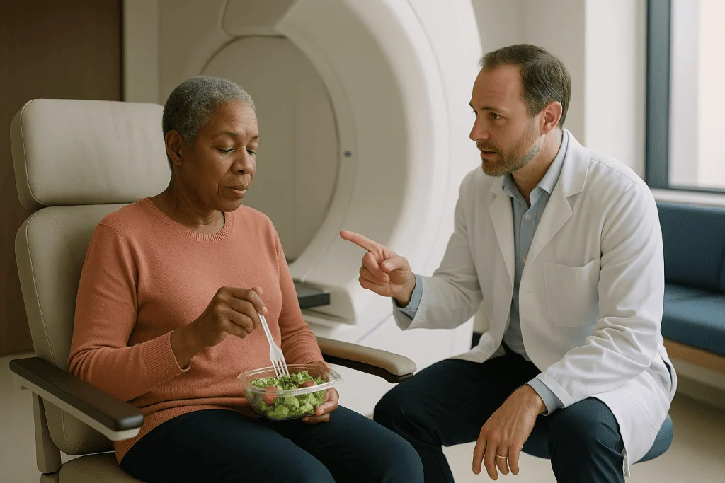
Modern photon radiation is delivered by a linear accelerator, a machine that generates X-rays and rotates around the patient. Techniques with acronyms you may see on a treatment plan, including IMRT (intensity-modulated radiation therapy) and VMAT (volumetric modulated arc therapy), shape the beam from many angles so that the high-dose region wraps tightly around the tumor. The exit dose does not disappear, but it is spread thinly across a wider volume of tissue rather than concentrated behind the target.
Photon machines also carry built-in imaging that checks the patient’s position before every session, sometimes during it. Decades of follow-up data exist for almost every tumor type. Treatment schedules, expected side effects and long-term risks are well characterized because millions of people have been treated this way. Machines are widely available, so treatment can usually start promptly, which matters for cancers where delay carries risk.
For many tumors, the tissue beyond the target is either unimportant or already receiving a low dose that modern planning keeps within safe limits. In those situations, the extra precision of protons has no organ left to protect. The Cleveland Clinic makes this point plainly: proton therapy is not superior in every case, and photon radiation remains standard care for the majority of people who need radiation.
So when a care team recommends photons, it is rarely because protons were unavailable. It is usually because, for this tumor and this anatomy, the physics offered no meaningful gain.
Proton therapy vs photon radiation: how the delivery differs day to day
From the treatment couch, the two experiences look remarkably alike, which surprises many patients who expected something dramatically different.
Both begin with a planning session. A CT scan is taken in the exact position that will be used for treatment, and a custom immobilization device, often a mesh mask for head and neck treatment or a molded cushion for the body, is made so the position can be repeated precisely each day. Small skin marks or tiny tattoo dots may be placed as alignment references. The planning team then spends days designing the beam arrangement, whether photon or proton.
The machines themselves differ behind the walls. Photons come from a linear accelerator that fits in a standard shielded room. Protons must be accelerated to a substantial fraction of the speed of light in a cyclotron or synchrotron, a circular accelerator the size of a room, then steered through magnets to the treatment head. That infrastructure is why proton centers are fewer and larger.
Most modern proton systems use pencil beam scanning, in which a narrow beam is swept across the tumor spot by spot and layer by layer, like painting with a fine brush. Older systems shaped a broad beam with custom-machined blocks and compensators.
Once in the room, the routine is familiar: positioning, imaging to confirm alignment, then the beam. Neither type of beam can be felt. Mayo Clinic notes that a proton session commonly takes around 15 to 30 minutes, with most of that time spent on positioning and only a few minutes of actual beam delivery. Photon sessions are of similar length. Both are typically given once a day, on weekdays, over a course of weeks, according to Mayo Clinic and the National Cancer Institute.
You walk in, you lie still, you walk out. The difference lives in the dose map, not the waiting room.
Proton therapy vs photon radiation at a glance
A table helps here because the two approaches diverge on a handful of specific points and overlap on almost everything else. Read the right-hand columns as descriptions of typical practice, not as promises about your own plan.
| Feature | Photon (X-ray) radiation | Proton therapy |
|---|---|---|
| What the beam is | High-energy X-rays, no mass or charge | Charged particles with mass |
| Dose beyond the tumor | Present; beam exits the body at reduced intensity | Essentially none past the Bragg peak |
| Dose in front of the tumor | Present, including skin | Present, including skin; can be similar or higher at the entry surface |
| Machine | Linear accelerator | Cyclotron or synchrotron with beam line |
| Availability | Widespread | Limited to specialized centers |
| Typical session | Minutes of beam, longer for setup | Similar; around 15–30 minutes per Mayo Clinic |
| Sensitivity to anatomy changes | Moderate | Higher; weight loss or organ filling can shift the stopping point |
| Length of outcome data | Decades, across nearly all cancers | Shorter and concentrated in specific tumor types |
| Common side-effect categories | Fatigue, skin change, site-specific effects | Same categories; some may be reduced when the spared tissue matters |
Two rows deserve emphasis. The dose in front of the tumor is not eliminated by protons; the beam must still enter through skin and whatever lies in its path. And sensitivity to anatomy changes is a genuine trade-off, not a footnote: the same stopping power that spares tissue also means the beam stops in the wrong place if the tissue in front of it changes.
Notice what the table does not contain: a row saying which is better. That row would need to be filled in differently for a child’s brain tumor, a prostate cancer and an early breast cancer, which is exactly why the decision belongs to a team looking at one person’s scans.
Who proton therapy is usually for, and who is usually asked to wait
Radiation oncologists tend to think about protons in terms of neighborhoods. The question is not what kind of cancer, but what lives next door.
Situations where the treating team most often considers protons, according to Mayo Clinic, the Cleveland Clinic and the National Cancer Institute, include:
- Cancers in children, whose growing tissues are more vulnerable to radiation and who have decades of life in which late effects could appear.
- Tumors at the base of the skull or along the spine, such as chordoma and chondrosarcoma, where the target sits against the brainstem or spinal cord.
- Tumors near the eye, including some eye melanomas, where the optic nerve and retina are millimeters away.
- Certain brain tumors where sparing memory or hormone-producing structures is a priority.
- Some head and neck, lung, esophageal and liver cancers, particularly when a critical organ lies directly behind the target.
- Re-treatment of an area that has already received radiation, where the surrounding tissue has little tolerance left.
Then there are the people usually steered toward photons, or asked to hold off on protons. Someone whose tumor has no sensitive organ behind it gains nothing from the missing exit dose. Someone whose treatment must begin urgently may not be able to wait for a slot at a distant center. Someone whose tumor moves substantially with breathing, or whose anatomy is expected to change quickly, may be a poor fit for a beam that depends on a fixed stopping depth. And for many common cancers, the honest position is that protons are being studied in trials because the evidence of benefit is not yet settled; a team may recommend enrolling in one of those trials rather than choosing protons outside it.
None of this is a ranking of patients. It is a matching exercise between a specific dose map and a specific body.
Is proton therapy better than radiation with photons? What the evidence actually shows
The phrase sounds like a yes-or-no question. The evidence answers it in three layers, and the layers do not always agree.
The first layer is dosimetry, the measurement of where dose lands. Here the case for protons is straightforward and consistently demonstrated: for most anatomical sites, a proton plan delivers less radiation to healthy tissue than an equivalent photon plan, because the exit dose is gone. Planning studies comparing the two on the same patients show this again and again. Dosimetry, however, measures radiation, not people.
The second layer is side effects. Lower dose to a healthy organ should mean less harm to it, and in several settings comparative studies have reported fewer severe acute side effects with protons, particularly when chemotherapy is given at the same time. Other studies, including some measuring what patients themselves report about symptoms and quality of life, have found little difference. The National Cancer Institute characterizes this evidence as promising in specific situations but still incomplete, with relatively few randomized trials completed.
The third layer is tumor control and survival. For most cancers, no high-quality evidence shows that protons control cancer better than photons. This is expected: the dose delivered to the tumor itself is designed to be the same. Protons change what happens around the tumor, not what happens inside it.
So is proton therapy better than radiation of the photon kind? Physically, it spares more tissue. Clinically, that translates into a measurable benefit only when the spared tissue was at real risk. For a tumor pressed against a child’s brainstem, the answer leans one way. For many adult cancers, the honest answer is that large randomized trials are still underway, and your team should be able to tell you which category your situation falls into.
Proton therapy for breast cancer: which radiation is best?
Breast cancer generates more proton questions than almost any other diagnosis, partly because it is common and partly because the organ behind a left-sided breast is the heart.
Photon radiation is the established standard after breast-conserving surgery and in many cases after mastectomy. Its long-term outcomes are among the best documented in all of oncology. The concern that drives interest in protons is heart dose: radiation reaching the heart’s arteries and muscle has been linked, in long-term follow-up studies, to a small increase in later cardiac problems, with the risk rising as heart dose rises.
Here the important nuance is that photon techniques have already been reshaped around this concern. Deep-inspiration breath hold, in which the patient takes and holds a deep breath so the heart drops away from the chest wall during the beam, can substantially reduce heart dose. Prone positioning, lying face down so the breast falls away from the chest, does something similar for some patients. Modern planning constrains heart and lung dose to strict limits. For many people, these methods bring heart dose so low that protons have little further room to improve on it.
Where protons are studied most seriously is in situations where photon plans struggle: left-sided cancers involving lymph nodes behind the breastbone, unusual chest anatomy, or existing heart disease. A large randomized trial in the United States is directly comparing protons and photons for breast cancer with cardiac events as its primary outcome; results were not yet mature at the time of writing, and this article will not guess at them.
Skin is the other side of the ledger. Because protons deliver dose without an exit path, the entry surface can receive a relatively higher share, and some series report more skin reaction with protons in breast treatment. Which radiation is best for breast cancer is therefore not a general question. It depends on which side, which nodes, which heart, and whether a photon plan can already meet the safety targets. The treating team can show you those numbers for your own plan.
Proton therapy side effects: what changes and what doesn't
A persistent belief is that proton therapy is somehow gentle radiation. It is not. It is radiation, delivered at a dose designed to destroy cancer cells, and the tissue it passes through on the way in is affected exactly as it would be by photons.
The side effects that occur are therefore the same in kind. According to the NHS and Mayo Clinic, people receiving either form of external beam radiation commonly experience:
- Fatigue that builds over the course of treatment and often lingers for weeks afterward.
- Skin changes in the treated area, from mild pinkness to soreness, peeling or darkening.
- Effects specific to the region treated: a sore mouth or altered taste for head and neck treatment, cough or swallowing discomfort for chest treatment, bowel or bladder irritation for pelvic treatment, hair loss only where the beam enters the scalp.
What protons can change is the frequency and severity of effects in tissue beyond the target. If a photon plan would have sent exit dose through the rectum, the small bowel, the heart or the healthy brain, a proton plan that stops short of those structures may reduce the symptoms arising from them. This is the mechanism behind the reduced-toxicity findings in some comparative studies, and it is a real effect where it applies.
What protons do not change is skin dose at the entry point, fatigue, or effects in any organ the beam must cross to reach the tumor. Nor do they eliminate late effects, the changes that can appear months or years after treatment, though sparing more normal tissue is expected to reduce the volume at risk.
The NHS notes that most acute side effects ease within a few weeks of finishing treatment, whichever beam was used. Proton therapy side effects are, in short, ordinary radiation side effects with a smaller footprint behind the tumor, not a different category of experience.
The downsides of proton therapy that rarely make the brochure
Any honest account of what protons change has to include what they make harder. Several of these downsides flow directly from the same physics that creates the advantage.
Range uncertainty comes first. A proton stops at a depth determined by the density of everything in front of it. Planners calculate that density from the CT scan, but the conversion carries a small margin of error, and the body does not hold still over six weeks. Weight loss, tumor shrinkage, a sinus that fills with fluid, a bowel loop that fills with gas, a bladder fuller or emptier than on planning day: any of these can shift the stopping point by millimeters. With photons, a millimeter shift changes dose gradually. With protons, it can move the sharp edge of the Bragg peak off the back of the tumor or into an organ. Teams manage this with extra margins, frequent imaging and re-planning when anatomy changes, but it is a genuine vulnerability.
Biological uncertainty is the second issue. Protons are slightly more biologically effective per unit of dose than photons, and this effect increases right at the end of the proton’s path. Planners use an average correction factor, but the true value at the very edge of the beam is not precisely known, which is one reason teams are cautious about placing that edge against critical structures.
Access and timing are practical realities. Proton centers are few, waits can be longer, and many people would need to relocate for weeks. For cancers where delay carries risk, that alone can tip the decision toward photons.
Evidence depth is the last downside. Long-term outcome data exist for far fewer people treated with protons than with photons, so rare late effects are less well characterized. Mayo Clinic and the National Cancer Institute both state that for many cancers the comparative benefit remains unproven. Knowing the downsides of proton therapy is not a reason to dismiss it; it is the context that makes the decision meaningful.
What is the safest form of radiation?
People type this question hoping for a single answer, and there is a temptation to hand them the word protons. Resist it, because the question contains a hidden assumption: that safety is a property of the beam rather than of the whole plan.
Radiation safety in cancer treatment means delivering enough dose to the tumor while keeping every nearby organ below the dose at which it is likely to be harmed. Those organ limits are published, guideline-level thresholds, and every plan, photon or proton, is checked against them before a single session. A photon plan that meets every limit with comfortable margin is not less safe than a proton plan that also meets them. The organs simply do not experience the difference.
Where protons become the safer choice is where a photon plan cannot meet a limit, or can meet it only barely. This is the situation for tumors wrapped around the brainstem, for children whose entire body is at heightened long-term risk, and for re-treatment of previously irradiated tissue. In those cases the missing exit dose is not a refinement; it is the difference between a plan that works and one that cannot be delivered.
Where photons may be the safer choice is less intuitive but equally real. A tumor that moves with breathing, an anatomy expected to change, or a target where the end-of-range biological uncertainty would land on a critical nerve can all make the sharp proton edge a liability rather than an asset. In those cases the gradual fall-off of photons is more forgiving.
There are also other forms of radiation entirely: brachytherapy places a sealed radioactive source inside or beside the tumor, and stereotactic techniques deliver very high doses in a few sessions with either photons or protons. Each has a niche where it is the safest tool.
The safest form of radiation, then, is the one whose dose map best fits your anatomy, delivered by a team that checks that map daily. Ask to see yours.
What the following days and weeks usually look like
Whichever beam is chosen, treatment is a routine before it is anything else, and knowing the shape of that routine helps.
The first week is mostly logistics. After planning, there is typically a gap of a week or more while the plan is designed and checked; the NHS describes this planning interval as a normal part of radiotherapy rather than a delay. The first session includes extra imaging and takes longer than the rest. Many people feel nothing at all during the first several days and wonder whether anything is happening. It is. Radiation damage to cells accumulates, and symptoms follow the accumulation.
By the second and third weeks, fatigue usually begins. Mayo Clinic and the NHS describe it as the most common effect of any external radiation, building gradually and often peaking near the end of treatment. Skin in the treated area may pink or feel warm. Site-specific effects appear on a similar timeline: a dry mouth in head and neck treatment, mild urinary urgency in pelvic treatment.
Through the middle weeks, the team watches for anatomy changes. This matters more with protons, where a scan showing weight loss or tumor shrinkage may prompt a new plan mid-course. That re-planning is a safety step, not a sign that something has gone wrong.
The final week often feels the hardest, because effects have accumulated while the finish line is visible. Then a counterintuitive thing happens: side effects frequently continue to build for one to two weeks after the last session before they begin to ease, because the tissue reaction lags behind the dose. The NHS notes that most acute effects settle over the weeks following treatment, though fatigue can take longer.
Follow-up begins some weeks after completion, with imaging timed to let post-treatment inflammation subside so that scans reflect the tumor rather than the reaction. Your team will set that schedule; it varies by cancer type and is not something to compare against a neighbor’s.
What people often get wrong about proton therapy vs photon radiation
The myths here are not foolish. Most of them are reasonable inferences from true facts, pushed one step too far.
“Protons are radiation without the radiation.” The exit dose is gone; the entrance dose and the tumor dose are not. Every cell the beam crosses on the way in is irradiated exactly as photons would irradiate it. Skin reactions, fatigue and effects in organs in front of the tumor still occur.
“Protons treat the cancer more effectively.” The dose prescribed to the tumor is designed to be the same regardless of beam. Protons change what happens to healthy tissue, not the tumor itself. No large body of evidence shows better tumor control from protons for most cancers, as the National Cancer Institute makes clear.
“Newer and more complex must mean better.” Complexity buys precision, and precision is only valuable where there is something to protect. A proton plan for a tumor with nothing important behind it is an elaborate solution to a problem that did not exist.
“Proton therapy means fewer sessions.” The number of sessions is set by the biology of the tumor and the tissue around it, not by the beam type. Shortened courses exist for both photons and protons, and long courses exist for both.
“Proton therapy is experimental.” For established indications such as certain pediatric, skull-base and eye tumors, it is standard care with guideline support. For other cancers, its comparative benefit is still being tested. Both statements are true at once, about different tumors.
“If my team didn’t offer protons, they were cutting corners.” In most cases they were reading a dose map and finding no organ that needed the sparing. Asking them to walk you through that map is a fair request. Assuming the worst is not.
Each correction points the same direction: the beam is a tool, and the choice of tool depends on the job.
Will Medicare pay for proton therapy? Coverage questions without the sales pitch
This question arrives in nearly every proton consultation, and it deserves a plain answer even though this article will not discuss costs or prices.
Coverage for proton therapy in the United States, whether through Medicare or a private insurer, is generally decided on the basis of medical necessity for a specific diagnosis rather than as a blanket yes or no. Payers typically maintain lists of indications they consider supported by evidence; tumors in children, tumors at the base of the skull and along the spine, and eye tumors commonly appear on those lists, reflecting the same evidence discussed above. For cancers where the comparative benefit is still being studied, coverage decisions are more variable and may depend on whether treatment is delivered within a clinical trial, whether a photon plan was shown to exceed an organ dose limit, or on regional policy.
Prior authorization, a process in which the treating team submits the plan and supporting evidence before treatment begins, is the norm. Radiation oncology departments are accustomed to this and will often prepare a side-by-side comparison of the proton and photon plans as part of the request, because demonstrating that the photon plan could not protect a specific organ is frequently what a payer needs to see.
What this means practically: the question is not really whether Medicare pays for proton therapy, but whether your specific diagnosis and your specific plan meet your specific payer’s criteria. Only the treating team and the payer can answer that, and they should be asked early, before travel or scheduling decisions are made.
A caution belongs here. Coverage is not a verdict on medical value in either direction. A covered treatment is not automatically the right one, and a treatment that is hard to get covered is not automatically better. Keep the clinical question and the coverage question in separate folders, and let the care team address the first before anyone opens the second.
Questions to ask your care team
A good consultation about beam choice is a conversation about your anatomy, not a lecture about machines. These questions tend to move it in that direction.
- Which healthy organs sit closest to my tumor, and which of them is your plan most worried about protecting?
- With a photon plan, would every one of those organs stay within its safe dose limit? By how much?
- If a proton plan were made for me, which organ would receive meaningfully less dose, and what symptom or late effect would that be expected to prevent?
- Is there a randomized trial comparing protons and photons for my cancer, and would I be eligible to join it?
- How would my anatomy be monitored during treatment, and how likely is a mid-course re-plan?
- Does my tumor move with breathing, and how does that affect the choice?
- How quickly do I need to start, and would waiting for a proton slot introduce any risk?
- What are the expected side effects of my specific plan, and which of them would protons change or not change?
- Are there other options, such as brachytherapy or a shortened stereotactic course, that would suit my situation?
- Has the plan been reviewed at a multidisciplinary tumor board?
Notice that none of these asks the team to declare which beam is best. They ask the team to show its reasoning, which is the more useful request. Most radiation oncologists welcome the chance to put the dose map on the screen and point.
Bring someone to take notes. Ask for a copy of the dose-volume summary if one exists; it is a table of how much of each organ receives how much dose, and it is the real answer to the proton therapy vs photon radiation question for your body. If the team cannot make a proton plan themselves, ask whether a referral for a second opinion at a center that can would be reasonable. Asking is not disloyalty. It is how the decision gets made well, and it still ends with the team you trust.
When to call your doctor
Radiation side effects usually build slowly and predictably, and your treatment team will see you at least weekly during the course. Certain changes should not wait for that visit. Contact the radiation oncology team, or seek urgent care if you cannot reach them, if you notice any of the following, whichever type of beam you are receiving:
- A fever, chills or shaking, especially if you are also receiving chemotherapy, which can lower your infection defenses.
- Skin in the treated area that is blistering, weeping, bleeding or shows spreading redness, warmth or pus, which may signal infection.
- New or worsening difficulty breathing, chest pain or a racing heartbeat, particularly during chest or breast treatment.
- Inability to swallow liquids, or signs of dehydration such as very dark urine, dizziness on standing or not passing urine for many hours.
- Severe or persistent vomiting or diarrhea that does not respond to the measures your team has advised.
- Sudden new weakness, numbness, severe headache, confusion or a seizure, especially during treatment of the brain or spine.
- Pain that is escalating quickly or is no longer controlled by what your team has prescribed.
- Any bleeding that does not stop with gentle pressure.
Sudden chest pain, severe breathing difficulty, signs of stroke or a seizure warrant emergency services rather than a clinic call.
Less dramatic changes also deserve a mention at your next review or a call between sessions: noticeable weight loss, a mask or mold that no longer fits the same way, new swelling in the treated region, or any change in how your body feels on the treatment couch. With protons especially, these small shifts can affect where the beam stops, and the team would rather re-scan than guess.
The Cleveland Clinic and the NHS both emphasize that teams expect these calls and would rather hear from you unnecessarily than not at all. Every treatment decision, including whether to pause, adjust or continue, rests with the clinicians who can see your scans and examine you. This article can explain what protons change. It cannot see your dose map, and it was never meant to replace the people who can.
Frequently asked questions
Is proton therapy better than radiation with photons?
Not as a general rule. Protons spare more tissue beyond the tumor, and that can reduce side effects when a critical organ sits in the exit path. For most cancers, no high-quality evidence shows better tumor control with protons, because the tumor dose is designed to be the same. Whether the sparing translates into a real benefit depends on your anatomy, and your treating team can show you the comparison for your own plan.
What are the downsides of proton therapy?
The main downsides are range uncertainty, biological uncertainty at the end of the beam, limited availability and shorter long-term outcome data. Because protons stop at a depth set by the tissue in front of them, changes such as weight loss or a full bladder can shift the stopping point, requiring extra margins and re-planning. Centers are few, waits can be longer, and for many cancers the comparative benefit over photons is still being tested in trials.
What is the safest form of radiation?
There is no single safest form; safety depends on whether a plan keeps every nearby organ below its published dose limit. A photon plan that meets all limits is not less safe than a proton plan that also meets them. Protons become the safer option when photons cannot protect an organ, such as the brainstem or a child’s developing tissues. Photons may be safer when a tumor moves or anatomy is expected to change.
Which radiation is best for breast cancer?
Photon radiation is the established standard for breast cancer and has the longest outcome record. Interest in protons centers on lowering heart dose for left-sided tumors, especially those involving nodes behind the breastbone. Photon techniques such as deep-inspiration breath hold already reduce heart dose for many patients. A large randomized trial is directly comparing the two; until results mature, the choice rests on your individual dose map and your team’s judgment.
Will Medicare pay for proton therapy for cancer?
Coverage is decided on medical necessity for a specific diagnosis rather than as a blanket rule. Indications with stronger evidence, such as pediatric, skull-base and eye tumors, are commonly covered; others vary by policy, trial enrollment and whether a photon plan was shown to exceed an organ dose limit. Prior authorization is standard. Only your treating team and your payer can confirm coverage for your plan, and they should be asked early.
What are proton therapy side effects like compared with photons?
They fall into the same categories: fatigue, skin changes in the treated area and effects specific to the region treated. Protons can reduce the frequency or severity of effects arising from tissue beyond the tumor, which some comparative studies have reported, particularly with concurrent chemotherapy. They do not reduce entry-path effects or fatigue. The NHS notes that most acute effects from either beam ease within weeks of finishing treatment.
Does proton therapy mean fewer treatment sessions?
No. The number of sessions is determined by the tumor type, the surrounding tissue and the treatment strategy, not by the beam. Shortened courses exist for both photons and protons, and longer courses exist for both. Mayo Clinic describes both forms as typically given on weekdays over a course of weeks, with each session lasting roughly 15 to 30 minutes, most of that spent on positioning.
Why did my doctor recommend photons instead of protons?
Most often because the dose map showed no critical organ behind your tumor that needed sparing, so protons would offer no meaningful gain. Other reasons include a tumor that moves with breathing, anatomy expected to change, the need to start treatment promptly, or a cancer where the comparative evidence is unsettled and trial enrollment is the recommended route. Asking to see the dose-volume summary is a reasonable way to understand the reasoning.
Can I feel the difference between a proton and a photon beam?
No. Neither beam can be felt during delivery, and the experience on the treatment couch is nearly identical: positioning in a custom mold or mask, imaging to confirm alignment, then a few minutes of beam time. The machines differ behind the walls, with protons requiring a large circular accelerator, but from the patient’s side the routine, the sounds and the sensations are essentially the same.
Is proton therapy still considered experimental?
For established indications such as certain pediatric tumors, skull-base and spinal tumors and eye tumors, proton therapy is standard care with guideline support. For many other cancers, including most adult prostate, breast and lung cancers, its comparative benefit over photons is still being evaluated in randomized trials. Both statements are true at once about different tumor types, which is why the answer depends on your specific diagnosis.
References
This article is for general information only and is not a substitute for professional medical advice. Please consult a qualified doctor about your individual situation.

More from the Blog
Follow-Up After Testicular Cancer: Why Marker Checks and Scans Continue for Years
After testicular cancer, follow-up typically continues for five years or longer because most relapses appear within the first two years but a small number…

Before Bladder Tumor Resection (TURBT): Urine Tests, Cystoscopy and Blood Thinner Review
Preparing for TURBT usually involves three checks: a urine test to rule out infection, a cystoscopy (camera look inside the bladder) to map the…

Why Does Cancer Treatment Sometimes Include a Neck Dissection? Lymph Node Spread Explained
Cancer treatment sometimes includes a neck dissection because many head and neck cancers spread first to the lymph nodes of the neck, and those…

Preparing for Therapeutic Apheresis: Hydration, a Light Meal and Your Vascular Access Plan
To prepare for plasmapheresis, drink normally the day before and the morning of the session, eat a light meal a few hours ahead rather…
Surgery or Radiotherapy First for Neck Cancer? How the Multidisciplinary Team Decides
There is no single right order. For neck and throat cancers the multidisciplinary team weighs where the tumor sits, how far it has spread,…

Infection Risk After a Stem Cell Transplant: Protective Isolation, Masks and When to Call
Infection risk after a stem cell transplant is highest in the first few weeks, when conditioning treatment has wiped out white blood cells and…