Placenta Accreta
Placenta accreta occurs when the placenta grows too deeply into the uterus. Learn about symptoms, causes, how it is diagnosed, and treatment options.
Quick answer
Placenta accreta is a pregnancy complication in which the placenta grows too deeply into the wall of the uterus and does not separate normally after birth, which can cause severe bleeding. It is most common after previous cesarean deliveries, is usually detected by ultrasound, and is typically managed with a planned cesarean delivery, often with hysterectomy.
What is placenta accreta?
Placenta accreta is a pregnancy complication in which the placenta, the organ that supplies oxygen and nutrients to the baby, grows too deeply into the wall of the uterus (womb). In a typical pregnancy, the placenta attaches to the inner lining of the uterus and separates on its own shortly after the baby is born. With placenta accreta, part or all of the placenta stays firmly attached because it has grown into, or through, the muscle layer of the uterus. This can cause heavy bleeding during or after delivery.
Doctors often use the broader term placenta accreta spectrum to describe three related conditions that differ by how deeply the placenta has grown:
- Placenta accreta: the placenta attaches directly to the uterine muscle rather than to the lining.
- Placenta increta: the placenta grows into the uterine muscle.
- Placenta percreta: the placenta grows through the full thickness of the uterine wall and may reach nearby organs such as the bladder.
Placenta accreta affects pregnant women and is most often seen in those who have had one or more previous cesarean deliveries (surgical births through an incision in the abdomen and uterus). It has become more common in recent decades, which many experts link to the rising number of cesarean births worldwide. It is considered a serious, high-risk pregnancy condition, and care is usually coordinated by specialists in maternal-fetal medicine, also called perinatology.
Placenta accreta symptoms
Many women with placenta accreta have no noticeable symptoms during pregnancy. The condition is frequently found on a routine ultrasound rather than because of a complaint. When placenta accreta symptoms do occur, they may include:
- Vaginal bleeding during the third trimester (the last three months of pregnancy), especially without pain
- Vaginal bleeding earlier in pregnancy in some cases
- Pelvic or lower abdominal pain, which is less common
- Blood in the urine, which can occur if the placenta has grown toward the bladder (placenta percreta)
- Heavy or prolonged bleeding after delivery when the placenta does not separate
- Signs of significant blood loss after birth, such as dizziness, a fast heartbeat, or feeling faint
Bleeding in the third trimester is a recognized warning sign, in part because placenta accreta often occurs together with placenta previa, a condition in which the placenta lies low in the uterus and covers the cervix (the opening of the uterus). It is placenta previa, rather than the accreta itself, that most often causes painless bleeding before labor.
Symptoms may vary by type. The milder form, placenta accreta, may cause no symptoms until delivery, when the placenta fails to come away and bleeding follows. Deeper forms, such as increta and percreta, carry a higher chance of complications and, when the bladder is involved, may cause blood in the urine or urinary discomfort. Because symptoms are often absent or mild, screening with ultrasound in women who have risk factors is a key part of detecting the condition early.
Causes and risk factors
The exact placenta accreta causes are not fully understood, but the leading explanation involves damage or scarring of the uterine lining. Normally, a layer of tissue called the decidua sits between the placenta and the muscle wall and helps the placenta separate after birth. When this layer is thin, damaged, or missing, usually because of a previous scar, the placenta may attach directly to the muscle and grow into it.
Recognized risk factors include:
- Previous cesarean delivery: this is the strongest risk factor, and the risk rises with each additional cesarean birth.
- Placenta previa: particularly when combined with a prior cesarean scar.
- Other uterine surgery: such as removal of fibroids (myomectomy), dilation and curettage (D&C, a procedure to remove tissue from inside the uterus), or repair of the uterine wall.
- Maternal age: the condition is more common in women over 35.
- Multiple previous pregnancies: having given birth several times is associated with higher risk.
- Uterine conditions: such as fibroids, scarring from infection, or certain congenital (present from birth) differences in the shape of the uterus.
- Assisted reproductive technology: pregnancies conceived through in vitro fertilization (IVF) appear to carry a somewhat higher risk.
- Previous placenta accreta: having had the condition before increases the chance in a later pregnancy.
Placenta accreta can also occur in women with none of these risk factors, although this is less common. Having a risk factor does not mean the condition will develop; it means your doctor may recommend closer monitoring.
Placenta accreta diagnosis
Placenta accreta diagnosis usually begins with a review of your medical history, particularly any previous cesarean deliveries or uterine surgery, and the location of the placenta on routine scans. If risk factors are present, your doctor may arrange targeted imaging.
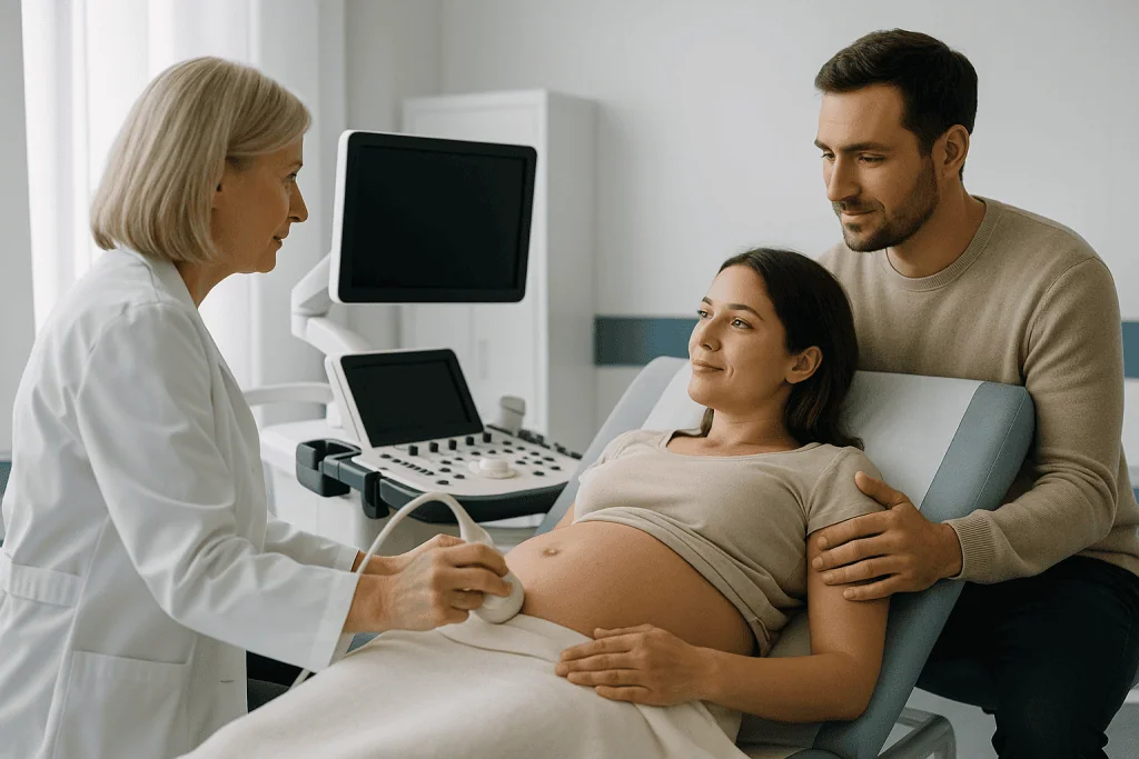
- Ultrasound: this is the main tool for diagnosis. A detailed ultrasound, sometimes performed through the vagina (transvaginal ultrasound) as well as through the abdomen, looks for signs such as irregular spaces within the placenta, thinning or loss of the normal boundary between the placenta and the uterine wall, and abnormal blood flow patterns seen with color Doppler (a setting that shows blood movement). Screening is typically performed in the second trimester and repeated later in pregnancy.
- Magnetic resonance imaging (MRI): MRI uses magnets and radio waves to create detailed pictures without radiation. It may be used when ultrasound findings are unclear, when the placenta is located on the back wall of the uterus where ultrasound is less reliable, or when doctors need to assess how deeply the placenta has grown and whether nearby organs are involved.
- Blood tests: certain proteins measured in routine prenatal screening, such as alpha-fetoprotein, may be higher than expected in some cases, but no blood test can confirm or rule out the condition on its own.
- Findings at delivery: sometimes placenta accreta is first recognized during a cesarean birth or when the placenta fails to separate after a vaginal birth. In these situations, the surgical team assesses the uterus directly.
- Pathology examination: after delivery, laboratory examination of the placenta and, if removed, the uterus can confirm the diagnosis and the depth of invasion.
No imaging test is perfect. Ultrasound and MRI can suggest the diagnosis with reasonable confidence, but they may miss some cases or suggest the condition when it is not present. For this reason, doctors typically plan delivery for suspected placenta accreta as though it were confirmed, so that the right team and resources are available.
Placenta accreta treatment options
Placenta accreta treatment focuses on delivering the baby safely, controlling bleeding, and protecting the mother’s health. There is no medication that can loosen a placenta that has grown into the uterine wall, and the condition cannot be corrected during pregnancy. Instead, management centers on careful planning and a coordinated delivery.
Monitoring during pregnancy
Once placenta accreta is suspected, you will usually be followed closely with repeat ultrasounds and regular visits. Your doctor may advise avoiding sexual intercourse and vigorous activity, particularly if placenta previa is also present, and will discuss what to do if bleeding occurs. In some cases, especially after an episode of bleeding, admission to the hospital for observation may be recommended in the later weeks of pregnancy.
Planned delivery
Delivery is almost always by cesarean section and is often scheduled before the due date, commonly in the late preterm period, to reduce the chance of going into labor or bleeding unexpectedly. Your doctor may recommend corticosteroid injections before an early delivery to help the baby’s lungs mature. Delivery is ideally carried out at a hospital that has a multidisciplinary team, including obstetricians experienced in the condition, anesthesiologists, blood bank services, and often urologists or other surgeons, with blood products prepared in advance. In many hospital groups, this care is organized through a dedicated Perinatology (High-Risk Pregnancy) Department; at Acibadem, for example, perinatology specialists coordinate the planning of such deliveries.
Cesarean hysterectomy
The most widely accepted treatment for confirmed placenta accreta is a cesarean hysterectomy. In this operation, the baby is delivered through an incision that avoids the placenta, and then the uterus is removed with the placenta still inside it. Leaving the placenta undisturbed avoids the severe bleeding that can happen when doctors try to separate it. This approach means that future pregnancy is not possible, which can be a difficult reality for many women, and counseling before delivery is an important part of care.
Conservative (uterus-preserving) management
In selected situations, doctors may consider approaches that aim to keep the uterus. These may include leaving the placenta in place after delivery and allowing the body to absorb it over weeks to months, or removing only the affected portion of the uterine wall. These options carry risks of delayed heavy bleeding, infection, and the need for later surgery, and they require very close follow-up. They are generally reserved for women who strongly wish to preserve fertility and whose situation makes it reasonably safe to try, and they are not suitable for everyone. Your doctor can explain whether this is an option in your case.
Managing bleeding
Because significant blood loss is the main danger, the team prepares for transfusion of blood and blood products. Additional measures may include medications that help the uterus contract, techniques to temporarily reduce blood flow to the pelvis, and procedures performed by interventional radiologists (doctors who use imaging to guide treatments through blood vessels) to block bleeding vessels. Which measures are used depends on the individual situation and hospital resources.
Recovery
Recovery after a cesarean hysterectomy is generally longer than after a routine cesarean. Some women need a stay in an intensive care unit, and iron supplements or further transfusion may be needed to treat anemia (low red blood cell count). Physical recovery usually takes several weeks, and support for feeding and caring for the newborn during this time is helpful. Emotional support and, where wanted, counseling can help with the experience of a complicated delivery and, for many women, the loss of future fertility.
Living with placenta accreta and outlook
The outlook for placenta accreta has improved considerably as awareness and prenatal detection have increased. When the condition is identified before delivery and the birth is planned with an experienced team, most women recover well, although the delivery itself still carries real risks, including major blood loss, injury to the bladder or other organs, blood clots, infection, and, rarely, death. Outcomes tend to be less favorable when the condition is unexpected at delivery, which is why prenatal diagnosis matters.
For the baby, the main concern is early delivery. Babies born in the late preterm period may need extra monitoring or a short stay in a neonatal unit, but many do well. The condition itself does not usually harm the baby’s development directly.
After a hysterectomy, menstrual periods stop and pregnancy is no longer possible. The ovaries are usually left in place, so hormone levels are typically unaffected and early menopause is not expected. Women who have had uterus-preserving treatment remain at increased risk of placenta accreta in any later pregnancy and will need early and careful monitoring if they conceive again.
Emotional recovery is an important and sometimes overlooked part of living with placenta accreta. Feelings of grief, anxiety, or trauma after a complicated birth are common and understandable. Talking with your care team, a mental health professional, or a support group of women who have been through similar experiences may help. Partners and family members may also benefit from support.
Frequently asked questions
What are the first signs of placenta accreta?
Often there are no early signs at all, and the condition is found on ultrasound. When symptoms occur, the most common is painless vaginal bleeding in the third trimester, usually related to an accompanying placenta previa. Any bleeding in pregnancy should be assessed promptly, but bleeding does not by itself mean placenta accreta is present.
What causes placenta accreta?
The most accepted explanation is scarring or damage to the uterine lining, most commonly from a previous cesarean delivery, which allows the placenta to attach directly to the muscle wall. Other uterine surgery, placenta previa, older maternal age, and IVF pregnancies are also associated with higher risk, though the condition can occur without any known cause.
How is placenta accreta diagnosed during pregnancy?
Diagnosis relies mainly on detailed ultrasound, including Doppler assessment of blood flow, usually performed in women with risk factors. MRI may be added when the ultrasound is unclear or when doctors need to judge how deep the placenta has grown. A definite diagnosis is often only confirmed at delivery or by laboratory examination afterward.
Can placenta accreta be treated without a hysterectomy?
In selected cases, doctors may attempt uterus-preserving management, such as leaving the placenta in place to be absorbed over time. These approaches carry risks of delayed bleeding and infection and require close follow-up, so they are offered only when the situation makes them reasonably safe and the woman strongly wishes to keep her uterus.
Is placenta accreta dangerous for the baby?
The condition usually does not affect the baby’s growth directly. The main risk to the baby is planned early delivery, which may lead to a stay in a neonatal unit. Your doctor weighs the risks of prematurity against the risks of unexpected bleeding when choosing the timing of delivery.
Can I have another baby after placenta accreta?
If the uterus was removed, pregnancy is not possible. If the uterus was preserved, pregnancy may be possible, but the chance of placenta accreta happening again is higher, and any future pregnancy would be considered high risk and would need close monitoring from early on.
Can placenta accreta be prevented?
There is no reliable way to prevent placenta accreta once a pregnancy has begun. Because prior cesarean delivery is the strongest risk factor, avoiding cesarean births that are not medically needed may lower the risk in future pregnancies. Early ultrasound screening in women with risk factors helps ensure the condition is detected and planned for.
When to see a doctor
If you are pregnant and have risk factors such as a previous cesarean delivery or placenta previa, discuss placenta accreta with your prenatal care provider so that appropriate screening can be arranged. Seek urgent medical care, without waiting for your next appointment, if you experience any of the following red-flag warning signs:
- Any vaginal bleeding during pregnancy, especially bright red bleeding in the second or third trimester
- Heavy bleeding that soaks through a pad in an hour or less
- Severe or persistent abdominal or pelvic pain
- Blood in the urine
- Dizziness, fainting, a racing heartbeat, or feeling very weak, which can indicate serious blood loss
- Regular contractions or signs of labor before your planned delivery date
- A noticeable decrease in your baby’s movements
- After delivery: heavy or increasing vaginal bleeding, fever, foul-smelling discharge, or worsening pain, particularly if the placenta was left in place
Bleeding in pregnancy always needs prompt assessment, even if it stops on its own. Acting quickly gives your care team the best chance to protect both you and your baby.
Medically reviewed by the Acıbadem International Medical Board September 13, 2026
See our medical review board →
Update history
- PublishedSeptember 13, 2026
- Medical review approvedSeptember 13, 2026
- Last content updateSeptember 13, 2026