Pterygium
Pterygium is a non-cancerous eye growth linked to UV exposure. Learn symptoms, causes, diagnosis, treatment and when to see a doctor.
Quick answer
Pterygium is a non-cancerous growth of tissue on the eye’s surface that can cause redness, irritation, and blurred vision if it extends over the cornea. At Acibadem in Turkey, evaluation includes a detailed eye examination, and treatment ranges from lubricating drops and protection from irritation to surgery when the growth affects vision, comfort, or appearance.
What is pterygium?
A pterygium (pronounced tuh-RIJ-ee-um) is a raised, wedge-shaped growth of fleshy tissue on the surface of the eye. It develops on the conjunctiva, which is the thin, clear membrane that covers the white part of the eye, and it can gradually extend onto the cornea, the transparent front window of the eye that light passes through. A pterygium is benign, which means it is not cancer, and it does not spread to other parts of the body. Even so, it can cause irritation, redness, and cosmetic concerns, and in more advanced cases it can affect vision.
People sometimes call this condition “surfer’s eye” because it is closely linked to long-term exposure to sunlight, wind, and dust, conditions common in outdoor and water sports. Understanding what is pterygium and how it behaves helps patients decide, together with an eye doctor, whether observation, medication, or surgery is the right approach.
Pterygium can affect anyone, but it is more common in adults, particularly those between roughly 20 and 50 years of age, and in people who live or work in sunny, dry, dusty, or windy environments. It occurs more frequently in regions close to the equator, where ultraviolet (UV) light exposure is higher. Men who work outdoors are affected somewhat more often in many populations, likely because of greater cumulative sun exposure rather than any biological difference. A pterygium usually appears on the inner corner of the eye, nearest the nose, although it can also form on the outer side. It may affect one eye or both eyes.
A pterygium is different from a pinguecula, which is a small, yellowish bump on the conjunctiva that does not grow onto the cornea. A pinguecula can sometimes develop into a pterygium over time, but many never do.
Symptoms of pterygium
Pterygium symptoms vary widely. Some people have a small growth for years with no discomfort at all, while others experience ongoing irritation or, in advanced cases, blurred vision. Common pterygium symptoms include:
- A visible growtha pink, fleshy, triangular or wedge-shaped patch of tissue on the white of the eye, most often on the side closest to the nose
- Rednessthe growth and surrounding conjunctiva may look inflamed, especially after sun, wind, or dust exposure
- Foreign body sensationa feeling of grittiness or sand in the eye
- Dryness, burning, or itching in the affected eye
- Tearingthe eye may water more than usual as it tries to relieve irritation
- Blurred or distorted visionusually only when the growth is large enough to change the shape of the cornea or cover part of the pupil
- Difficulty wearing contact lensesthe raised tissue can interfere with lens fit and comfort
Symptoms often depend on the stage of the growth. An early, small pterygium confined to the conjunctiva may cause little more than occasional redness or mild irritation. As the tissue advances onto the cornea, it can pull on the corneal surface and create astigmatisman irregular curvature of the cornea that blurs or distorts vision. In advanced cases, the growth may extend far enough to partially block the visual axis, the central zone of the cornea that light must pass through to reach the retina, which can noticeably reduce sight in that eye.
Doctors also distinguish between a quiet, or “atrophic,” pterygium, which appears pale, thin, and stable, and an active, or “fleshy,” pterygium, which looks thicker, redder, and more vascular (containing many visible blood vessels). An active-looking pterygium is generally more likely to grow and to cause symptoms, although the appearance alone cannot predict its behavior with certainty.
Causes and risk factors
The exact cause of pterygium is not fully understood, but the strongest and most consistent factor is long-term exposure to ultraviolet (UV) light from the sun. UV radiation is thought to damage cells at the edge of the cornea and trigger abnormal growth and blood-vessel formation in the conjunctival tissue. Chronic irritation from wind, dust, sand, smoke, and dry conditions appears to contribute as well. Recognized pterygium causes and risk factors include:
- Ultraviolet light exposurethe leading risk factor; people who spend many hours outdoors without eye protection are at higher risk
- Living near the equatorsunnier climates carry higher cumulative UV exposure
- Outdoor occupations and hobbiesfarming, construction, fishing, sailing, surfing, and skiing (snow reflects UV light strongly)
- Chronic eye surface irritationfrom wind, dust, sand, pollen, or smoke
- Dry eyea poorly lubricated eye surface may be more vulnerable to irritation and abnormal tissue growth
- Age and sexpterygium is more common in adults, and in many studies more common in men, likely reflecting exposure patterns
- Family historya genetic tendency may play a role in some families, although environment appears to be more important
Pterygium is not contagious. It does not result from an infection, and it cannot be passed from one person to another. It is also not caused by reading, screen use, or eye strain, although these can worsen dryness and make an existing pterygium feel more irritated.
Diagnosis
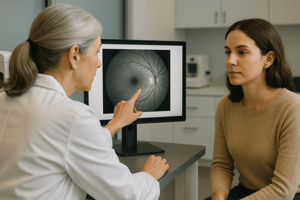
Pterygium diagnosis is usually straightforward and is made during a standard eye examination. In most cases, an eye doctor can recognize the growth by its typical appearance and location. The evaluation commonly includes:
- Slit-lamp examinationthe key test. A slit lamp is a microscope with a bright, focused light that lets the doctor examine the surface of the eye in magnified detail. It shows the size, thickness, and vascularity of the growth and how far it extends onto the cornea.
- Visual acuity testinga standard eye chart to check whether the pterygium is affecting sight.
- Corneal topography or keratometrypainless imaging or measurement of the shape of the cornea. These tests can detect astigmatism caused by the pterygium pulling on the corneal surface and can help track changes over time.
- Photographic documentationphotographs of the eye surface may be taken at intervals so the doctor can compare the size of the growth from visit to visit and determine whether it is progressing.
Part of the diagnostic process is ruling out other conditions that can look similar. A pinguecula, certain inflammatory lesions, and, rarely, ocular surface tumors such as conjunctival intraepithelial neoplasia (an abnormal, potentially precancerous growth of surface cells) can resemble a pterygium. If a growth has an unusual appearance, grows rapidly, or looks suspicious in any way, the doctor may recommend removing it and sending the tissue for histopathology, laboratory examination under a microscope, to confirm the diagnosis. This is a standard precaution, and the great majority of pterygia are confirmed to be benign.
Treatment options for pterygium
Pterygium treatment depends on the size of the growth, whether it is growing, how much discomfort it causes, and whether it threatens or affects vision. Many small, stable pterygia never need surgery. Care for this condition is typically provided by an eye specialist within an ophthalmology department; at hospital groups such as Acibadem, pterygium is managed by ophthalmologists as part of routine eye care.
Watchful waiting
If a pterygium is small, not growing, and causing few or no symptoms, doctors often recommend simple observation with periodic eye examinations. During this time, protecting the eyes from UV light and irritants is important, because reducing exposure may help slow further growth. Measures usually advised include wearing sunglasses that block UVA and UVB rays (wraparound styles offer better side protection), wearing a wide-brimmed hat outdoors, and avoiding or protecting against dusty and windy conditions when possible.
Medications for symptom relief
Medications do not remove a pterygium or reverse its growth, but they can relieve irritation and inflammation. Options your doctor may suggest include:
- Lubricating eye drops (artificial tears) and lubricating gels or ointments, to relieve dryness and the gritty, foreign-body feeling
- Short courses of anti-inflammatory eye drops, such as mild steroid drops, when the pterygium becomes red and inflamed; steroid drops are used cautiously and under medical supervision because long-term use can raise eye pressure or cause other side effects
- Decongestant (“redness-relief”) drops, which some patients use occasionally for appearance; doctors generally advise against regular use, as these drops can cause rebound redness
Surgery
Surgical removal is considered when a pterygium is growing steadily toward the center of the cornea, is causing significant astigmatism or reduced vision, causes persistent irritation that medication does not control, or is a major cosmetic concern for the patient. Pterygium surgery is usually performed as an outpatient procedure under local anesthesia, meaning the eye is numbed and the patient goes home the same day.
Simply cutting the growth away and leaving the bare white of the eye exposed (the “bare sclera” technique) is associated with a relatively high chance of the pterygium growing back, so modern surgery usually combines removal with a graft:
- Conjunctival autograftafter the pterygium is removed, a thin piece of the patient’s own healthy conjunctiva, usually taken from under the upper eyelid, is placed over the area where the growth was removed. This technique substantially lowers the chance of recurrence and is widely regarded as a standard approach.
- Amniotic membrane graftin some cases, especially for larger or recurrent growths, a graft of processed amniotic membrane (a donor tissue that supports healing) may be used to cover the surgical area.
- Adjunctive medicationfor pterygia that have already recurred or appear aggressive, the surgeon may apply an anti-scarring medication such as mitomycin C during surgery to reduce the risk of regrowth. This is used selectively because it carries its own risks.
- Tissue glue or suturesgrafts may be secured with fine stitches or with a special tissue adhesive; the choice depends on the surgeon’s judgment and the individual eye.
After surgery, the eye is typically red, scratchy, and mildly sore for days to a few weeks while the surface heals. Patients usually use antibiotic and anti-inflammatory eye drops for several weeks and attend follow-up visits so the doctor can monitor healing. It is important to understand that a pterygium can grow back after surgery. Recurrence is more likely in younger patients, in people with continued heavy UV exposure, and after certain surgical techniques, which is why graft-based methods and strict sun protection after surgery are emphasized. A recurrent pterygium can sometimes behave more aggressively than the original, so the decision to operate is weighed carefully against the option of continued observation.
Living with pterygium and outlook
For most people, the outlook with a pterygium is good. Many growths remain small and stable for years, and permanent vision loss is uncommon, particularly when the condition is monitored and treated appropriately. A pterygium is not cancer and does not turn into cancer, although any unusual eye-surface growth should be evaluated to confirm the diagnosis.
Day-to-day management focuses on comfort and prevention of progression:
- Wear UV-blocking sunglasses whenever you are outdoors, including on cloudy days and in winter
- Use a wide-brimmed hat for additional shade
- Use lubricating eye drops regularly if your eyes feel dry or gritty
- Protect your eyes from dust, sand, and wind with appropriate eyewear when working or playing outdoors
- Attend the follow-up eye examinations your doctor recommends so the size of the growth can be tracked over time
If a pterygium does progress and cause astigmatism or encroach on the line of sight, timely surgery can usually remove the growth and, in many cases, improve or stabilize vision. However, if a pterygium has grown far across the cornea before removal, some corneal scarring or irregularity can persist, and vision may not return fully to what it was. No treatment can guarantee that a pterygium will never return, but with modern graft techniques and good sun protection, many patients remain free of recurrence long term. Your ophthalmologist can discuss the likely outcome in your particular situation.
Frequently asked questions
What is pterygium in simple terms?
A pterygium is a benign, fleshy growth of tissue on the white of the eye that can slowly extend onto the cornea, the clear front surface of the eye. It is strongly linked to long-term sun, wind, and dust exposure, which is why it is sometimes called “surfer’s eye.” It is not cancer and is not contagious, but it can cause redness, irritation, and, when large, blurred vision.
Can a pterygium go away on its own?
A pterygium does not usually disappear on its own. The redness and irritation can come and go: an inflamed pterygium may calm down with lubricating drops, sun protection, or a short course of anti-inflammatory drops, but the tissue itself generally remains. The only way to remove the growth is surgery, which doctors typically reserve for pterygia that are growing, affecting vision, or causing persistent symptoms.
How serious is a pterygium?
In most cases, a pterygium is a nuisance rather than a serious threat to eye health. Many stay small and cause only occasional irritation. The main concern is that a growing pterygium can distort the cornea and cause astigmatism, and a very advanced one can cover part of the pupil and reduce vision. Regular eye examinations allow doctors to detect progression early, before vision is significantly affected.
What does pterygium surgery recovery involve?
After surgery, the eye is usually red, watery, and scratchy for days to a few weeks while the surface heals; some discomfort in the first few days is common. Patients typically use prescribed eye drops for several weeks, may wear an eye patch or shield briefly, and are advised to avoid rubbing the eye, swimming, and dusty environments during early healing. Most people return to everyday activities within days, but full healing of the eye surface and stabilization of vision can take longer. Your surgeon will give you specific instructions and follow-up appointments.
Can a pterygium grow back after it is removed?
Yes, recurrence is a well-known possibility after pterygium surgery. The risk is higher with simple excision alone and lower when the surgeon uses a conjunctival autograft or other modern techniques, and it also depends on factors such as age and ongoing UV exposure. Consistent use of UV-blocking sunglasses and a hat after surgery is generally recommended to help reduce the chance of regrowth, although no measure can eliminate the risk entirely.
Is a pterygium the same as a cataract?
No. A pterygium is a growth on the surface of the eye, while a cataract is a clouding of the lens inside the eye. They are separate conditions with different causes and treatments, although both become more common with age and both can be influenced by long-term UV exposure. A person can have either condition alone or both at the same time, and an eye examination can easily tell them apart.
How can I prevent a pterygium from getting worse?
The most useful step is consistent protection from ultraviolet light: wear sunglasses that block UVA and UVB rays whenever you are outdoors, ideally a wraparound style, along with a wide-brimmed hat. Protecting the eyes from wind, dust, and smoke, treating dry eye with lubricating drops, and attending regular eye check-ups also help. These measures do not guarantee the growth will stop, but they address the main factors thought to drive progression.
When to see a doctor
Any new or changing growth on the surface of the eye should be examined by an eye doctor, even if it is painless, so the diagnosis can be confirmed and the growth monitored. In addition, seek medical attention promptly if you notice any of the following warning signs:
- A noticeable change in visionnew blurring, distortion, or difficulty seeing from the affected eye
- Rapid growtha pterygium that appears to be enlarging quickly or advancing toward the center of the eye
- Severe or worsening eye paina pterygium usually causes irritation, not significant pain; marked pain suggests another problem that needs evaluation
- Intense redness, discharge, or swellingpossible signs of infection or significant inflammation
- Sudden sensitivity to light that is new or severe
- An unusual appearancea growth that looks irregular, pigmented, ulcerated, or otherwise different from a typical pterygium
- Symptoms after surgeryincreasing pain, worsening redness, discharge, or decreasing vision during recovery from pterygium removal
An ophthalmologist can confirm whether the growth is a pterygium, rule out less common but more serious conditions, and recommend the monitoring or treatment plan that fits your situation. Early evaluation gives you the widest range of options and the best chance of protecting your vision.
Medically reviewed by the Acıbadem International Medical Board September 2, 2026
See our medical review board →
Update history
- PublishedJune 8, 2026
- Medical review approvedSeptember 2, 2026
- Last content updateSeptember 2, 2026