

1St Trimester Screening: How It Works, Results and What to Expect

1st trimester screening is usually performed between 11 weeks and 13 weeks plus 6 days of pregnancy. It commonly includes a nuchal translucency ultrasound and blood tests measuring pregnancy-related markers.
Key Takeaways
- 1st trimester screening is usually performed between 11 weeks and 13 weeks plus 6 days of pregnancy.
- It commonly includes a nuchal translucency ultrasound and blood tests measuring pregnancy-related markers.
- A higher-chance result is not a diagnosis and should be discussed with a qualified prenatal care professional.
- Cell-free DNA screening and diagnostic tests such as chorionic villus sampling or amniocentesis may be offered after an increased-chance result.
- The ultrasound portion also helps assess early fetal development, dating and the number of fetuses.
1st trimester screening is an optional prenatal assessment that uses an ultrasound and maternal blood test to estimate the chance of certain chromosome conditions and, in some settings, early pregnancy complications. It does not diagnose a condition, but its results can help a pregnant person and their care team decide whether further testing may be useful.
Overview: What Is 1st Trimester Screening?
1st trimester screening is an optional prenatal screening process performed early in pregnancy. It estimates the likelihood that a fetus has certain chromosome conditions, especially trisomy 21 (Down syndrome), trisomy 18 and trisomy 13. Depending on local practice and the individual pregnancy, it may also contribute information about the chance of pre-eclampsia or other pregnancy-related concerns.
The screening usually combines two parts: a detailed ultrasound measurement called nuchal translucency and a maternal blood test. The results are considered alongside factors such as gestational age, maternal age and whether there is one fetus or more. Screening estimates probability; it cannot confirm or rule out a chromosome condition with certainty.
Many people choose this assessment because it provides information early enough to allow time for counselling, further testing and considered decision-making. Others may choose a different screening approach or may decide not to have screening. A prenatal care professional can explain the available options in a way that respects each person’s values and circumstances.
Timing and Who May Consider Screening
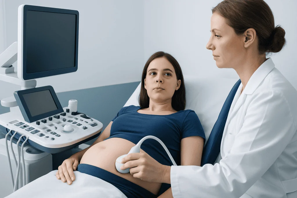
The usual 1st trimester screening weeks are from 11 weeks to 13 weeks and 6 days of pregnancy. The scan is timed carefully because the nuchal translucency measurement is validated only within a particular fetal size range. People wondering, “first trimester screening how many weeks?” should contact their maternity team early, as appointment availability and local screening pathways vary.
1st trimester screening in pregnancy may be offered to people of all ages and risk levels. It can be especially helpful for those who would like early information about chromosome conditions, have a family or personal history that affects pregnancy planning, have had a prior pregnancy with a chromosome condition, or have an ultrasound finding that needs closer evaluation.
Screening choices differ between healthcare systems. Some programs offer combined first-trimester screening as standard care, while others commonly use cell-free DNA screening, also known as non-invasive prenatal testing (NIPT), as a primary or follow-up option. A clinician can discuss which approach is appropriate based on gestational age, medical history, ultrasound findings and personal preferences.
How the Procedure Works Step by Step
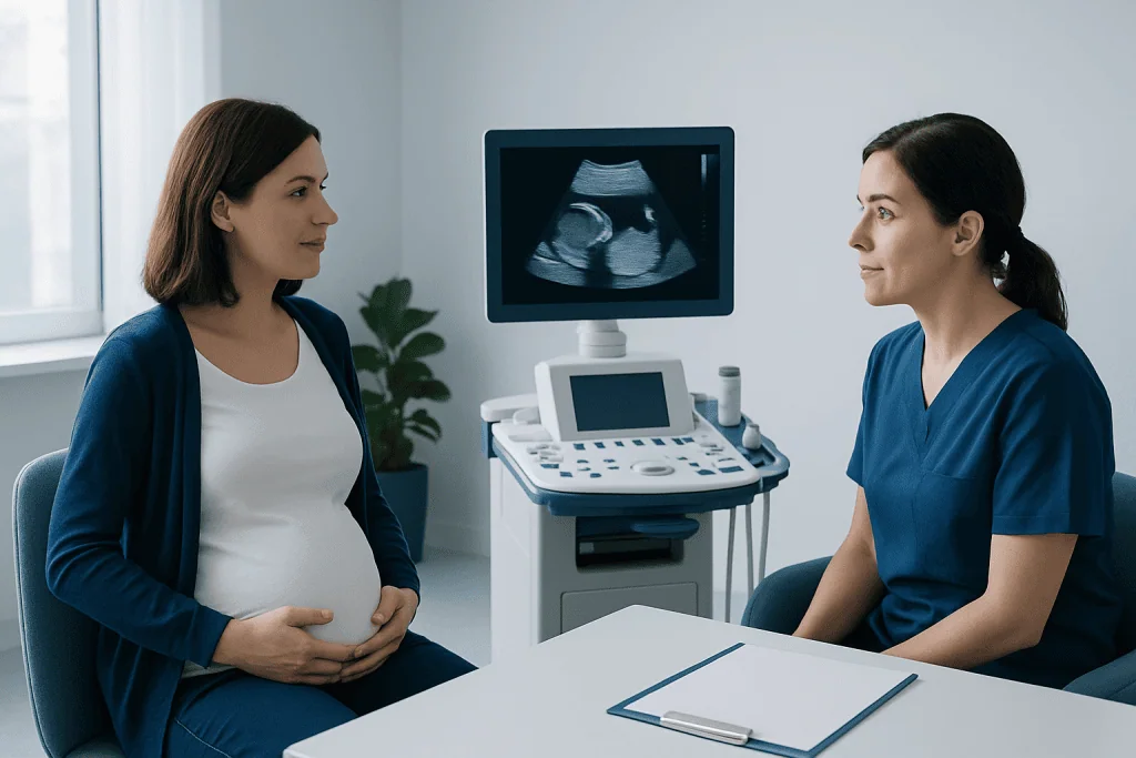
Before the appointment, the care team confirms the estimated gestational age and reviews relevant health and pregnancy history. No special fasting or medication changes are usually needed for the blood test or ultrasound, unless the healthcare team provides individual instructions. It may be useful to bring previous scan reports or information about prior pregnancies if available.
During the ultrasound, a trained sonographer or fetal medicine specialist uses an abdominal probe to obtain images of the uterus and fetus. In some cases, a transvaginal ultrasound may be recommended if clearer images are needed. The clinician checks fetal heartbeat, measures the fetus to confirm dating, identifies the number of fetuses and measures the fluid-filled space at the back of the fetal neck, known as nuchal translucency.
A blood sample is then collected from a vein in the arm. The laboratory measures markers that can include pregnancy-associated plasma protein A (PAPP-A) and free beta-human chorionic gonadotropin (free beta-hCG). A laboratory algorithm combines these results with the ultrasound measurement and other relevant details to calculate an individualized chance estimate.
Ultrasound is also an important part of early prenatal imaging. If the scan identifies a structural concern or needs more specialist review, the care team may recommend follow-up imaging and consultation through pregnancy follow-up and monitoring.
Understanding Results and Next Steps
Results are often reported as a lower-chance or higher-chance estimate, sometimes using a ratio such as 1 in a certain number. A lower-chance result means the calculated likelihood is below the threshold used by that healthcare program. It reduces, but does not eliminate, the possibility of the screened conditions. It also does not assess every genetic, developmental or health condition.
A higher-chance result does not mean that the fetus definitely has a chromosome condition. Most importantly, it is a screening result rather than a diagnosis. The care team may offer genetic counselling to explain what the result means, answer questions and discuss the possible next steps at an appropriate pace.
Further options can include cell-free DNA screening, which is another screening test based on placental DNA fragments in maternal blood, or diagnostic testing. Diagnostic tests may include chorionic villus sampling (CVS), generally performed in the first trimester, or amniocentesis later in pregnancy. These tests can provide a more definite answer but involve different considerations, including a small procedure-related risk that should be reviewed with a fetal medicine specialist.
Not all 1st trimester screenings produce a clear result. The test may be inconclusive because of timing, laboratory limitations, multiple pregnancy or other factors. Repeating an assessment, using another screening method or arranging specialist review may then be advised.
Benefits, Limitations and Possible Risks
A main benefit of 1st trimester screening is that it offers information early in pregnancy. The ultrasound can also confirm fetal viability, refine pregnancy dating, identify multiple pregnancy and sometimes detect early signs of structural differences. For many families, receiving clear explanations of normal findings or timely support after an unexpected result can be valuable.
However, screening has limitations. It does not diagnose chromosome conditions, and it may produce false-positive or false-negative results. A result can also cause uncertainty or worry, particularly while waiting for confirmatory testing. Genetic counselling can help people understand both the test’s accuracy and the meaning of a result for their own pregnancy.
The ultrasound examination does not use radiation and is considered safe when performed for medical purposes by appropriately trained professionals. A routine blood draw may cause brief discomfort, bruising, light-headedness or rarely infection at the needle site. The screening itself does not increase the risk of miscarriage.
If diagnostic testing is considered, its benefits and risks are different from screening. The specialist team should explain the procedure, what information it can provide, what results may be possible and any potential complications before a decision is made.
Preparing for the Appointment and Aftercare
Most people can return to their usual activities immediately after 1st trimester screening. There is no recovery period after the ultrasound or blood test. If a transvaginal scan is used, mild temporary discomfort can occur, but it should not be painful. Any persistent pain, bleeding or other concerning symptom during pregnancy should be discussed promptly with a maternity care professional.
It can help to prepare a few questions in advance, such as what conditions are included in the local screening panel, when results will be available, what a higher-chance result would mean and whether a genetic counsellor is available. Some people prefer to bring a partner, friend or family member for support, although this depends on the clinic’s policies.
After results are available, the care team explains whether routine antenatal follow-up is appropriate or whether further testing is worth considering. Continuing regular prenatal appointments remains important even after a lower-chance result, because routine care monitors the health of both the pregnant person and the developing fetus throughout pregnancy.
Acibadem International’s multidisciplinary obstetric, fetal medicine and genetic specialists support international patients with pregnancy assessment and follow-up in JCI-accredited hospitals, with care plans based on individual clinical needs.
When to Seek Medical Care
Contact a prenatal care professional soon if an early-pregnancy scan is needed, if there is uncertainty about pregnancy dates, or if a screening result is difficult to understand. Prompt discussion is particularly useful when a report describes a higher-chance result, an increased nuchal translucency measurement, an unclear result or an ultrasound finding that requires review.
Urgent medical advice is appropriate for heavy vaginal bleeding, severe or worsening abdominal pain, fainting, shoulder pain, fever, or symptoms that make the pregnant person feel significantly unwell. These symptoms are not caused by screening itself in most cases, but they need timely assessment in pregnancy.
Emotional support is also part of prenatal care. Waiting for results or deciding about further testing can feel stressful. A maternity clinician, fetal medicine specialist or genetic counsellor can provide balanced information and help families make decisions that fit their circumstances.
Frequently asked questions
What is included in 1st trimester screening?
It commonly includes a nuchal translucency ultrasound and a maternal blood test. The ultrasound measures fluid at the back of the fetal neck and checks early development, while the blood test measures pregnancy-related markers. Together, these findings provide an estimate of the chance of certain chromosome conditions.
When is 1st trimester screening done?
It is generally done between 11 weeks and 13 weeks plus 6 days of pregnancy. The exact timing matters because the ultrasound measurement is assessed within a specific developmental window. Booking early helps ensure the test can be performed at the appropriate time.
Is 1st trimester screening the same as NIPT?
No. Combined first-trimester screening uses an ultrasound and maternal blood markers, whereas NIPT analyzes cell-free DNA in the pregnant person’s blood. Both are screening tests, not diagnostic tests, although NIPT can be more accurate for some common chromosome conditions.
Does a high-risk 1st trimester screening result mean the baby has a condition?
No. A higher-chance result means the calculated likelihood is above a program’s threshold for further assessment; it is not a diagnosis. A clinician may discuss additional screening, genetic counselling or diagnostic testing to obtain clearer information.
Are 1st trimester screenings safe?
The standard ultrasound and blood test are considered safe when used appropriately in prenatal care. Ultrasound does not use radiation, and blood sampling usually causes only minor, temporary effects such as brief discomfort or bruising. The screening process itself does not increase miscarriage risk.
What happens if the result is low chance?
A lower-chance result usually means routine prenatal care can continue, unless there are other clinical concerns. It does not guarantee that every health or developmental condition has been excluded. Regular antenatal appointments and the recommended mid-pregnancy anatomy scan remain important.
References
- American College of Obstetricians and Gynecologists
- Society for Maternal-Fetal Medicine
- National Health Service
- International Society of Ultrasound in Obstetrics and Gynecology
- World Health Organization
This article is for general information only and is not a substitute for professional medical advice. Please consult a qualified doctor about your individual situation.
Explore treatments in Turkey — costs, top hospitals & a free quote
JCI-accredited · board-certified surgeons · reply within 24h
Add us as a Preferred Source to see more of our trusted health content across Google Search, AI Overviews and Discover.

More from the Health Library


Related Specialists

Prof. Dr. Sabahattin Kaymakoğlu
Gastroenterology
Dr. Hakan Kutlu
Infectious Diseases & Clinical Microbiology
Dr. Ali Uçar
Emergency Service
Dr. Nuri Soysal
Aesthetic Plastic & Reconstructive Surgery