AI in Medical Imaging: How Algorithms Support Diagnosis

AI can help identify patterns in medical images, highlight possible abnormalities, and support faster triage of urgent findings. Radiologists and physicians remain responsible for interpreting results in the context of a patient’s symptoms, history, and other tests.
Key Takeaways
- AI can help identify patterns in medical images, highlight possible abnormalities, and support faster triage of urgent findings.
- Radiologists and physicians remain responsible for interpreting results in the context of a patient’s symptoms, history, and other tests.
- AI tools are trained on large datasets, but their performance depends on data quality, validation, and appropriate clinical use.
- Benefits may include workflow support, measurement consistency, and improved detection of subtle findings in selected applications.
- Patients should understand that AI is one part of a diagnostic process and should ask their doctor how imaging results will guide care.
AI in medical imaging uses computer algorithms to help healthcare professionals detect, measure, and prioritize findings on scans such as X-rays, CT, MRI, ultrasound, and mammography. These tools support, rather than replace, radiologists and other specialists by making image review more efficient and consistent.
Overview
AI in medical imaging refers to the use of computer algorithms that analyze medical scans and assist healthcare professionals in detecting, measuring, or prioritizing findings. These systems are most often used in radiology, pathology, cardiology, oncology, emergency medicine, and other fields where images play an important role in diagnosis and treatment planning.
In practice, artificial intelligence does not “read” a scan in the same way a doctor does. Instead, it searches for patterns in image data that it has learned from large numbers of previously labeled images. For example, an algorithm may be trained to flag a possible lung nodule on a chest CT, estimate the size of a brain bleed, or identify mammograms that need closer review.
The goal is to support clinical decision-making, not to replace medical expertise. A radiologist or another qualified physician reviews the images, considers the patient’s symptoms and medical history, compares prior studies when available, and communicates the final interpretation. AI can be useful as a second set of digital “eyes,” especially when imaging volumes are high or when subtle findings require careful attention.
How AI Algorithms Work in Imaging
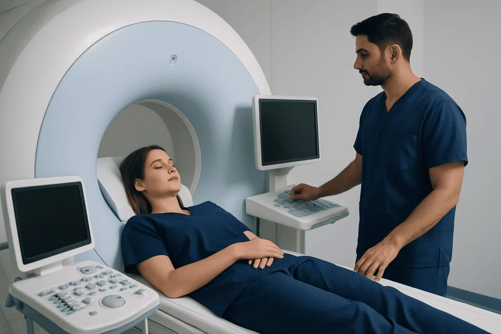
Most AI imaging tools use machine learning, a method in which computers learn patterns from examples rather than following only fixed instructions. A common approach is deep learning, which uses layered mathematical models called neural networks. These models can be trained to recognize visual features such as edges, shapes, textures, density differences, or patterns that may not be obvious at first glance.
Training begins with datasets of medical images that have been labeled by experts. For example, a dataset might include CT scans marked to show where a stroke, lung nodule, fracture, or tumor is located. The algorithm adjusts itself repeatedly until it can identify similar patterns in new images. Before clinical use, reliable systems should be tested on separate datasets and evaluated in real-world healthcare settings.
AI tools can perform several imaging-related tasks, including:
- Detection: highlighting a possible abnormality, such as a fracture, hemorrhage, lesion, or polyp.
- Classification: suggesting whether a finding has features that are more likely benign or suspicious.
- Segmentation: outlining an organ, tumor, vessel, or other structure to support measurement or treatment planning.
- Quantification: measuring volume, density, calcification, blood flow, or changes over time.
- Triage: prioritizing scans that may need urgent review, such as suspected intracranial bleeding.
Common Uses of AI in Diagnostic Imaging
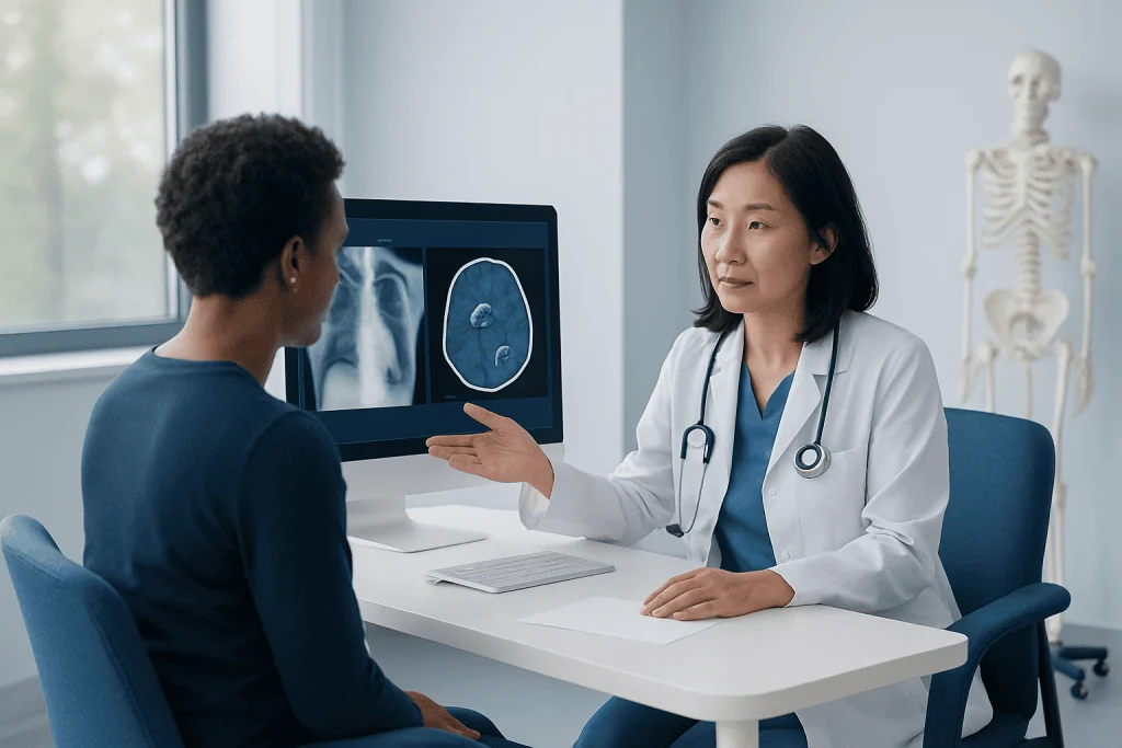
AI-assisted tools are being used or studied across many types of imaging. In X-ray imaging, algorithms may help detect fractures, pneumonia patterns, collapsed lung, or changes related to bone and joint disease. In CT, AI may assist with identifying bleeding in the brain, lung nodules, blood clots, coronary artery calcification, or injuries after trauma.
In MRI, AI may support brain, spine, prostate, breast, liver, and musculoskeletal imaging by improving image reconstruction, highlighting lesions, or measuring structures. In mammography, algorithms can help radiologists review breast images and prioritize cases that may need additional attention. Ultrasound applications include support for thyroid, liver, obstetric, vascular, and cardiac assessments, although ultrasound can be more challenging because image quality depends strongly on the operator and scanning technique.
AI is also used behind the scenes to improve workflow and image quality. Some systems can help reduce scan time, enhance low-dose images, reconstruct images from raw data, or automatically route studies to the correct specialist. These functions may not be visible to patients, but they can support a more efficient diagnostic process when used appropriately.
Benefits for Patients and Healthcare Teams
One of the main benefits of AI in medical imaging is that it can help manage large numbers of scans more efficiently. In busy hospitals, some algorithms can flag potentially urgent findings so they are reviewed sooner. This may be especially helpful in emergency settings, where timely interpretation can guide rapid treatment decisions.
AI can also improve consistency in measurement. For patients with cancer, heart disease, neurological conditions, or chronic lung disease, small changes over time may matter. Algorithms that measure lesion size, organ volume, or tissue characteristics in a standardized way can help clinicians compare current and previous scans more accurately, although physician oversight remains essential.
Another potential benefit is support for early or subtle detection in selected situations. Some findings are small, complex, or easily missed when many images must be reviewed. AI may highlight an area for closer inspection, prompting the radiologist to evaluate it carefully. However, a highlighted area is not automatically a diagnosis, and an unflagged image is not automatically normal.
For healthcare teams, AI may reduce repetitive tasks and allow more time for complex interpretation, patient communication, and multidisciplinary planning. The best use of AI is often as part of a broader system that includes skilled imaging technologists, experienced radiologists, referring physicians, and appropriate follow-up care.
Limitations and Safety Considerations
AI tools are powerful, but they are not perfect. An algorithm can produce a false positive, meaning it highlights something that is not clinically significant, or a false negative, meaning it misses an important finding. The chance of error depends on the type of scan, the condition being evaluated, image quality, the patient population, and how the tool was trained and validated.
Bias is another important concern. If an AI system is trained mainly on images from one population, scanner type, age group, or clinical setting, it may perform less well in other groups. For safe use, tools should be tested across diverse patients and imaging equipment. Hospitals and imaging centers also need clear quality control processes to monitor performance after implementation.
AI cannot understand the full clinical picture by itself. A scan finding may have different meanings depending on a patient’s symptoms, lab results, previous imaging, medications, and medical history. For example, a small lung nodule may be assessed differently in a young nonsmoker than in an older person with a history of smoking or cancer. This is why the final diagnosis and care plan must remain in the hands of qualified clinicians.
Privacy and data protection are also essential. Medical images are sensitive health information. Institutions using AI should follow applicable laws and ethical standards for data storage, security, consent, and access. Patients can ask how their imaging data is protected and whether AI tools are used in their diagnostic pathway.
What Patients Can Expect During AI-Assisted Imaging
For most patients, the imaging appointment feels the same whether AI is used or not. The scan is performed by a trained radiology technologist or sonographer according to standard protocols. Depending on the examination, the patient may be asked to remove metal objects, change position, hold their breath briefly, or receive a contrast agent if clinically indicated.
After the scan, the images are processed and sent to the radiology system. If an AI tool is part of the workflow, it may analyze the images before or during the radiologist’s review. The algorithm might mark areas of interest, provide measurements, or assign a priority level. The radiologist then evaluates the complete study and prepares a report for the referring doctor.
Patients usually receive results from the doctor who ordered the test, although procedures vary by hospital and country. It is helpful to ask what the result means, whether follow-up imaging is needed, and how the finding fits with symptoms or other tests. If the report mentions AI-generated measurements or computer-assisted analysis, the patient can ask the physician to explain how that information was used.
Questions to Ask Before or After Imaging
Patients do not need to be technology experts to benefit from AI-assisted imaging. A few practical questions can help them understand the process and feel informed. The most important point is that AI should be used as a support tool within a medically supervised diagnostic pathway.
Useful questions may include:
- What type of imaging test is recommended, and why?
- Will AI or computer-assisted analysis be used in reviewing the images?
- Who will provide the final interpretation of the scan?
- What are the possible next steps depending on the result?
- Is follow-up imaging needed, and if so, when?
- How will my imaging data and personal information be protected?
Patients should also tell their care team about pregnancy, kidney disease, allergies to contrast agents, implanted devices, previous reactions during imaging, and any prior scans that may be available for comparison. Sharing complete information helps clinicians choose the safest and most useful imaging approach.
When to Consult a Doctor
A doctor should be consulted whenever imaging is recommended to investigate symptoms, monitor a known condition, plan treatment, or follow up on a previous finding. AI may support the analysis, but it does not replace a clinical consultation. The decision to order imaging should be based on medical history, physical examination, risk factors, and the expected benefit of the test.
Patients should seek prompt medical advice for symptoms such as sudden weakness, severe chest pain, shortness of breath, new neurological symptoms, significant injury, unexplained weight loss, persistent pain, or a new lump or swelling. These symptoms do not always mean a serious condition is present, but they deserve timely assessment by a qualified healthcare professional.
For international patients, Acibadem International’s multidisciplinary specialists and JCI-accredited hospitals provide diagnostic imaging services and clinical evaluation for many conditions. Patients should discuss their individual situation with a physician, including whether AI-supported imaging tools are appropriate for their case.
Frequently asked questions
Does AI replace the radiologist?
No. AI is designed to support radiologists and other physicians, not replace them. The final interpretation of an imaging study should be made by a qualified medical professional who considers the patient’s symptoms, history, and other test results.
Is AI in medical imaging safe?
AI can be safe and useful when it is properly validated, monitored, and used by trained healthcare teams. Like any medical technology, it has limitations and can make errors. This is why physician oversight, quality control, and appropriate clinical judgment are essential.
Will patients know if AI was used to review their scan?
Practices vary between hospitals, health systems, and countries. Some reports may mention computer-assisted detection or AI-based measurements, while others may not. Patients can ask their doctor or imaging center whether AI tools are used and how they contribute to the review process.
Can AI find cancer earlier?
In some imaging applications, AI may help highlight subtle findings that need closer review, including findings that could be suspicious for cancer. However, AI cannot diagnose cancer by itself. Diagnosis may require expert image interpretation, comparison with previous scans, laboratory tests, biopsy, or other clinical evaluation.
Can AI make imaging results faster?
AI may help prioritize urgent scans or automate measurements, which can support faster workflow in certain settings. The time needed for results still depends on the type of examination, clinical urgency, availability of specialists, and whether additional review is required.
What should patients do if an imaging report mentions an AI finding?
Patients should discuss the report with the doctor who ordered the test or with a relevant specialist. The physician can explain whether the finding is important, whether it matches symptoms, and what follow-up is recommended. Patients should avoid drawing conclusions from technical report language without medical guidance.
References
- World Health Organization
- U.S. Food and Drug Administration
- American College of Radiology
- European Society of Radiology
- Radiological Society of North America
This article is for general information only and is not a substitute for professional medical advice. Please consult a qualified doctor about your individual situation.
Explore treatments in Turkey — costs, top hospitals & a free quote
JCI-accredited · board-certified surgeons · reply within 24h
Add us as a Preferred Source to see more of our trusted health content across Google Search, AI Overviews and Discover.