AI in Medical Imaging: What Patients Should Know About Bias and Accuracy
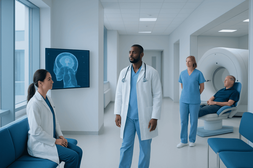
AI in medical imaging helps detect patterns on scans, but it does not replace a radiologist or treating doctor. Accuracy can vary depending on the quality of the images, the patient population used to train the system, and how the tool is used in practice.
Key Takeaways
- AI in medical imaging helps detect patterns on scans, but it does not replace a radiologist or treating doctor.
- Accuracy can vary depending on the quality of the images, the patient population used to train the system, and how the tool is used in practice.
- Bias can occur if an AI system performs better for some groups than others because of uneven training data or design limitations.
- Hospitals reduce risks through validation, human oversight, and regular monitoring of AI performance.
- Patients can ask how AI supported their scan interpretation and how final decisions are confirmed by clinicians.
AI in medical imaging can help doctors analyze scans more quickly and consistently, but it is not perfect. Patients benefit most when AI is used as a support tool alongside expert medical judgment, quality controls, and clear communication.
Overview
AI in medical imaging refers to computer systems designed to help analyze images such as X-rays, CT scans, MRI scans, ultrasound, and mammograms. These tools are trained to recognize patterns that may suggest normal findings or possible disease. In many settings, AI is used to support radiologists by highlighting areas of concern, prioritizing urgent cases, or measuring features that would otherwise take more time to assess manually.
For patients, the most important point is that AI is usually an aid, not a replacement for medical expertise. A radiologist still interprets the scan in the context of symptoms, medical history, physical examination, and other test results. AI can make workflows faster and may improve consistency in some tasks, but the final diagnosis and treatment plan should still involve qualified clinicians.
Interest in AI has grown because imaging produces large amounts of information, and some findings can be subtle. AI may help identify small abnormalities, compare current and prior scans, or reduce delays in reviewing urgent images. However, the value of any AI tool depends on how well it has been developed, tested, and integrated into real-world care.
How AI Is Used in Imaging
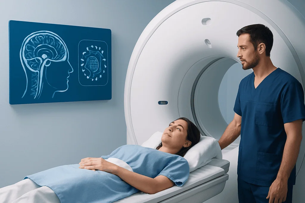
Different AI systems are designed for different jobs. Some act as a second reader, flagging suspicious areas for the radiologist to review more closely. Others help classify findings, calculate organ volumes, measure tumors over time, or sort scans by urgency so that potentially critical cases can be reviewed sooner.
AI may be used in screening, diagnosis, treatment planning, and follow-up. For example, it can assist with reading mammograms, identifying possible stroke-related changes on brain scans, or assessing lung nodules on chest imaging. In clinical practice, these tools are often combined with established imaging methods such as MRI scans, CT scans, or mammography.
Even when AI performs well in studies, it is only one part of the care process. Image quality, patient movement, unusual anatomy, prior surgery, implants, and overlapping medical conditions can all affect interpretation. That is why imaging results still need careful review by a trained professional who understands the full clinical picture.
What Accuracy Means for Patients
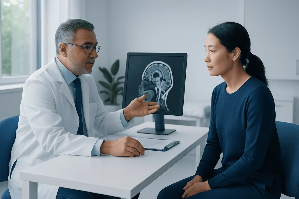
When people hear that an AI tool is “accurate,” it can sound as if it always gives the right answer. In reality, accuracy is more nuanced. A tool may be good at identifying many true abnormalities, but it can still miss some problems or flag normal findings as suspicious. Performance is often described using measures such as sensitivity and specificity, which reflect how well a test detects disease and how well it avoids false alarms.
For patients, false negatives and false positives both matter. A false negative means a condition may be overlooked, while a false positive can lead to anxiety, repeat imaging, or additional procedures. Whether an AI system is helpful depends on the clinical setting, the condition being evaluated, and how the tool’s strengths and weaknesses are balanced by human review.
Accuracy can also change outside the environment where the software was first studied. A tool trained on high-quality images from one type of scanner or one hospital may not perform exactly the same way in another setting. Differences in equipment, imaging protocols, disease prevalence, and patient demographics can all influence results. For this reason, hospitals should validate AI systems locally before relying on them in routine care.
Understanding Bias in AI
Bias in medical AI does not necessarily mean intentional unfairness. Often, it means that a system performs better for some groups than for others. This can happen if the data used to train the tool did not include enough diversity in age, sex, ethnicity, body size, underlying health conditions, or imaging from different machines and healthcare settings.
For example, an AI model developed mostly from one population may be less reliable when used in another. Bias can also arise if certain diseases look different at different stages, or if historical healthcare patterns led to uneven labeling or diagnosis in the training data. In imaging, subtle technical factors such as image resolution, contrast use, and positioning can also influence how a system learns.
Bias matters because it can contribute to unequal care. If an AI system is less accurate for one patient group, it could increase the risk of missed findings or unnecessary follow-up for that group. This is why responsible AI development includes diverse datasets, transparent reporting, subgroup testing, and continued monitoring after a tool is introduced into practice.
Patients may not be able to judge algorithm design directly, but they can feel reassured when a healthcare team explains that AI tools are reviewed for fairness and are not used without physician oversight. Good clinical practice treats AI results as one source of information rather than an unquestioned answer.
How Hospitals and Doctors Improve Safety
Safe use of AI in imaging depends on multiple layers of quality control. Before a tool is adopted, it should be assessed for its intended purpose, tested on relevant patient groups, and checked to make sure it works with the hospital’s scanners and workflows. Ongoing audits are also important because performance may shift over time as patient populations, equipment, or clinical practices change.
Human oversight remains central. Radiologists review AI suggestions, confirm or reject flagged findings, and interpret results in context. If an AI tool highlights a possible abnormality, the radiologist decides whether it is clinically meaningful. If the AI misses something, an experienced clinician may still identify it. This partnership is one of the main safeguards for patient safety.
Hospitals may also use standardized reporting, peer review, and multidisciplinary discussion to improve quality. In complex cases, imaging findings may be reviewed alongside pathology, laboratory tests, and specialist input. Depending on the clinical question, a patient may need further evaluation with studies such as PET-CT scanning or targeted procedures after the initial image review.
Near the end of the diagnostic pathway, communication is just as important as technology. Patients should receive results in a way that explains what was found, how certain the interpretation is, and what the next steps may be. At centers such as Acibadem International, multidisciplinary specialists in JCI-accredited hospitals use advanced imaging and clinical review together when caring for international patients.
Questions Patients Can Ask
Patients do not need technical expertise to take an active role in their care. It is reasonable to ask whether AI was used in reading a scan and what role it played. In most cases, the answer will be that AI served as a support tool and that a radiologist or treating physician made the final interpretation.
It can also help to ask how uncertain findings are handled. If a scan result is unclear, the next step may be comparison with older images, repeat imaging after a period of time, or evaluation with another modality such as ultrasound. This kind of follow-up is common in medicine and does not necessarily mean something serious has been found.
Useful questions may include:
- Was AI used to assist with my scan interpretation?
- Who reviewed the final images and report?
- How confident is the team in the result?
- Do I need additional tests or follow-up imaging?
- Are there any limitations in my scan that could affect interpretation?
Clear answers can help patients understand both the benefits and limits of AI-supported imaging. Shared decision-making is especially valuable when follow-up options include observation, repeat testing, or referral to another specialist.
What This Means for the Future of Care
AI in medical imaging is likely to become more common, not less. As systems improve, they may help reduce repetitive tasks, support earlier detection of some conditions, and make imaging services more efficient. This could be especially useful in busy departments where rapid review of urgent scans can improve care coordination.
At the same time, trust in AI will depend on careful regulation, transparency, and evidence from real clinical use. Patients benefit when healthcare organizations choose tools based on proven performance, monitor them continuously, and remain open about what AI can and cannot do. Responsible use means recognizing that technology should support good medicine, not replace thoughtful clinical judgment.
The future is likely to involve collaboration rather than substitution. Radiologists, technologists, physicists, software developers, ethicists, and treating physicians all have a role in making AI safe and effective. For patients, the most reassuring message is that the best imaging care still combines advanced tools with experienced human interpretation and individualized medical decision-making.
Frequently asked questions
Does AI replace the radiologist?
No. In most healthcare settings, AI is used to support the radiologist, not replace them. A trained physician still reviews the images, considers the patient's history, and makes the final interpretation.
Can AI make mistakes on scans?
Yes. Like any medical tool, AI can miss abnormalities or incorrectly flag normal findings. That is why human oversight and follow-up testing, when needed, remain important parts of safe care.
Why can AI be biased?
Bias can happen when the data used to train an AI system does not represent all patient groups or clinical situations well enough. As a result, the tool may perform better in some populations than others. Careful testing and monitoring are needed to reduce this risk.
Should patients be worried if AI was used in their imaging?
Not necessarily. When used responsibly, AI can be a helpful support tool that improves workflow and assists image review. The key issue is that it should be combined with expert clinical judgment rather than used alone.
How can patients know if an AI-supported result is reliable?
Patients can ask who reviewed the scan, whether the findings are clear or uncertain, and whether additional tests are needed. Reliability is strongest when AI is part of a system that includes quality checks, experienced radiologists, and clinical follow-up.
What happens if AI and the doctor do not agree?
The clinician's judgment guides the final decision. If there is uncertainty, the team may review the images again, compare prior scans, discuss the case with other specialists, or recommend further testing. This is a normal part of careful medical practice.
References
- World Health Organization
- U.S. Food and Drug Administration
- Radiological Society of North America
- American College of Radiology
- European Society of Radiology
This article is for general information only and is not a substitute for professional medical advice. Please consult a qualified doctor about your individual situation.
Explore treatments in Turkey — costs, top hospitals & a free quote
JCI-accredited · board-certified surgeons · reply within 24h
Add us as a Preferred Source to see more of our trusted health content across Google Search, AI Overviews and Discover.