Anisocoria: An Evidence-Based Guide for Patients

Anisocoria is a difference in pupil size, and a small, stable difference can be normal in some people. New or worsening anisocoria should be assessed, especially if it comes with pain, vision changes, eyelid drooping, or neurological symptoms.
Key Takeaways
- Anisocoria is a difference in pupil size, and a small, stable difference can be normal in some people.
- New or worsening anisocoria should be assessed, especially if it comes with pain, vision changes, eyelid drooping, or neurological symptoms.
- Causes range from harmless physiologic anisocoria to medication effects, eye inflammation, nerve problems, or head and neck conditions.
- Diagnosis focuses on history, eye examination, and sometimes imaging or specialist testing.
- Treatment depends on the underlying cause rather than the pupil difference itself.
Anisocoria means the pupils are different sizes. In many people it is harmless, but when it appears suddenly or comes with eye pain, drooping eyelid, double vision, or headache, it may point to an eye or nervous system problem that needs urgent medical care.
Overview: what anisocoria means
Anisocoria is the medical term for pupils that are not the same size. The pupil is the dark opening in the center of the eye that changes size to control how much light enters. A small difference between the pupils can be completely normal, but anisocoria can also be a sign of an eye condition, a medication effect, or a problem involving the nerves that control the pupil.
The key question is not simply whether the pupils look different, but whether the change is new, whether it varies in bright or dim light, and whether other symptoms are present. Doctors use these details to decide whether anisocoria is likely to be benign or whether it may require urgent evaluation.
Many patients first notice anisocoria in a mirror or in a photograph. Others are told about it during a routine eye exam or emergency assessment. Because the causes vary widely, a calm but timely medical review is the safest approach when the difference is new or unexplained.
How the pupils normally work
Pupil size is controlled by a balance between two muscles in the iris and the nerves that supply them. One muscle makes the pupil smaller in bright light, while the other makes it larger in dim light. This automatic response helps protect the eye and optimize vision.
Doctors often assess anisocoria by comparing the pupils in both bright and dark settings. If the larger pupil does not constrict well in light, the problem may involve the pathway that normally makes the pupil smaller. If the smaller pupil does not enlarge well in darkness, the issue may involve the pathway that normally makes the pupil bigger.
This pattern matters because it can point toward specific causes. For example, some conditions affect the iris or the nerves inside the eye, while others involve the sympathetic or parasympathetic nervous system. A careful examination helps identify which pupil is abnormal and what tests may be needed next.
Symptoms and warning signs to notice

Anisocoria itself may cause no symptoms at all. Some people have a mild, lifelong difference in pupil size and normal vision. Others may notice blurred vision, sensitivity to light, difficulty focusing at near distance, or one pupil that seems slow to react.
Symptoms that deserve closer attention include eye pain, redness, drooping of one eyelid, double vision, reduced vision, a severe or unusual headache, neck pain, confusion, weakness, numbness, or a recent head or eye injury. These features can suggest that anisocoria is part of a more serious problem rather than an isolated finding.
It can also help to notice the circumstances. Did the difference begin after using eye drops, a skin patch, an inhaler, or contact with a plant or chemical? Did it follow recent surgery, trauma, or a migraine episode? Small details from daily life can be important clues when a doctor is trying to find the cause.
Common causes and risk factors
One of the most common explanations is physiologic anisocoria, which means a naturally occurring, harmless difference in pupil size. In this situation, the pupils still respond normally to light, the difference is usually small, and there are no troubling symptoms. Many people with physiologic anisocoria have had it for years without realizing it.
Other causes involve the eye itself. These include inflammation inside the eye, prior eye surgery or trauma, iris abnormalities, and certain forms of glaucoma. Some medications can also change pupil size, including prescription eye drops and accidental exposure to substances that affect the pupil muscles. Depending on the cause, the pupil may be larger or smaller than usual.
Anisocoria may also reflect a nerve or brain-related problem. Examples include third cranial nerve palsy, Horner syndrome, migraine-related changes, and less commonly conditions affecting the brain, neck, or chest. Because these causes can overlap with broader neurological disorders, a doctor may consider evaluation for related problems such as stroke when symptoms suggest a sudden nervous system event.
Risk factors depend on the underlying cause and may include recent eye procedures, facial or head trauma, use of certain medications, neurological disease, diabetes affecting nerves, infections, or vascular conditions. Not every patient has a clear risk factor, which is why a structured medical assessment is important.
How doctors diagnose anisocoria
Diagnosis begins with a detailed history. A clinician will ask when the unequal pupils were first noticed, whether the difference is constant or intermittent, and whether there are associated symptoms such as pain, ptosis, double vision, headache, or recent trauma. Medication history is especially important, including eye drops, patches, inhalers, and over-the-counter products.
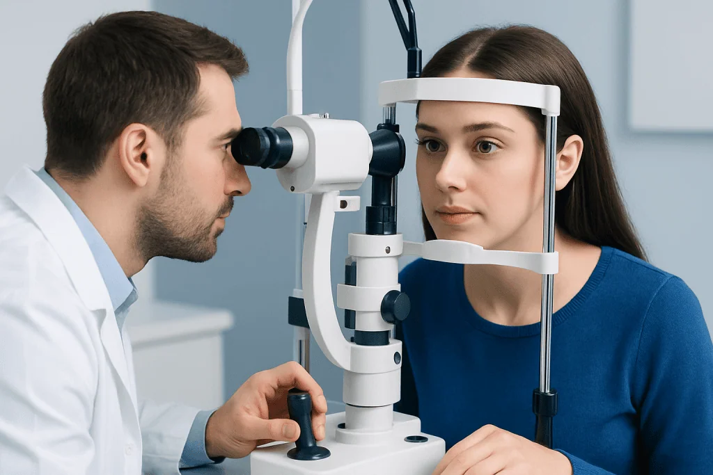
The physical examination usually includes measuring the pupils in bright and dim light, checking how they react, and looking at eye movements, eyelid position, visual acuity, and the front structures of the eye. A slit-lamp eye exam may be used to look for inflammation, injury, or structural changes. If a nerve problem is suspected, a neurological exam is often performed as well.
Additional tests depend on the findings. Some patients need imaging such as MRI scanning or CT imaging to assess the brain, eye socket, or blood vessels. Others may need specialist evaluation in ophthalmology or neurology if the pattern suggests a nerve-related cause. In selected cases, specific eye-drop tests may help distinguish between certain conditions, but these are interpreted in the context of the overall exam.
Treatment options and what recovery depends on
There is no single treatment for anisocoria because the pupil difference is a sign, not a disease by itself. Management focuses on the underlying cause. If the difference is due to physiologic anisocoria and no other problem is found, treatment may not be needed. Reassurance and routine follow-up can be enough.
When anisocoria results from an eye condition, treatment may involve managing inflammation, addressing elevated eye pressure, or treating injury or infection. If a medication or chemical exposure is responsible, symptoms often improve after the effect wears off or the triggering substance is stopped under medical guidance.
If the cause is neurological, treatment may involve urgent care and targeted management of the underlying disorder. For example, third nerve palsy, Horner syndrome, or a vascular problem may require imaging, monitoring, and specialist treatment. In some patients, recovery is quick once the cause is treated; in others, the pupil difference may improve more gradually or persist even after the main condition is stabilized.
For international patients who need coordinated assessment, Acibadem International’s multidisciplinary specialists and JCI-accredited hospitals diagnose and treat eye and neurological conditions related to anisocoria. The exact plan is always based on the patient’s symptoms, examination findings, and test results.
Self-care, monitoring, and prevention
Because anisocoria has many causes, self-care is mainly about observation and safety rather than home treatment. It can be helpful to note when the pupil difference started, whether it changes in light or darkness, and whether symptoms such as headache, eye pain, or drooping eyelid occur at the same time. Photographs taken at different times may also help a clinician understand the pattern.
People should avoid using leftover eye drops or someone else’s medication to try to correct the pupil size. If there has been contact with a new medication, inhaler, patch, or chemical, that information should be shared with a doctor. Protective eyewear during sports, work, or activities with risk of eye injury may help prevent trauma-related causes.
There is no guaranteed way to prevent all forms of anisocoria, but routine eye care, prompt attention to eye injuries, and good management of underlying medical conditions can reduce risk. Patients with known migraines, nerve disorders, or prior eye surgery should mention these factors if a new pupil difference appears.
When to seek medical care
Medical care should be sought promptly if anisocoria is new, clearly increasing, or associated with concerning symptoms. Urgent assessment is especially important when unequal pupils appear with severe headache, eye pain, loss of vision, double vision, drooping eyelid, weakness, numbness, confusion, trouble speaking, or after a head or eye injury.
Even when there are no dramatic symptoms, a new or unexplained difference in pupil size should still be evaluated by a qualified doctor or eye specialist. This is because some causes are harmless while others need timely treatment, and it is not always possible to tell the difference at home.
Immediate emergency care is appropriate if anisocoria develops suddenly with stroke-like symptoms or a severe neurological change. A prompt diagnosis can help guide the right treatment and protect both vision and overall health.
Frequently asked questions
Is anisocoria always serious?
No. A small, stable difference in pupil size can be normal and is called physiologic anisocoria. However, sudden or new anisocoria should be assessed, especially if it comes with pain, vision changes, headache, eyelid drooping, or neurological symptoms.
How much difference in pupil size is normal?
Some healthy people naturally have a slight difference in pupil size. What matters most is whether the difference is longstanding and stable, and whether the pupils still react normally to light. A doctor can determine whether the finding looks normal or needs further investigation.
Can medications cause anisocoria?
Yes. Certain eye drops, patches, inhalers, and accidental exposure to medications or chemicals can change pupil size in one eye. This is one reason doctors ask carefully about all prescription, over-the-counter, and topical products.
What is the difference between anisocoria and Horner syndrome?
Anisocoria is a description of unequal pupil size, not a diagnosis by itself. Horner syndrome is one possible cause of anisocoria and usually involves a smaller pupil, mild drooping of the eyelid, and sometimes reduced sweating on one side of the face.
Can a migraine cause anisocoria?
Sometimes, yes. Some people develop temporary pupil changes during or around migraine episodes. Even so, new anisocoria should not automatically be assumed to be migraine-related until a clinician has considered other possible causes.
Will anisocoria go away on its own?
It depends on the cause. If it is physiologic anisocoria, it may simply remain as a harmless normal variation. If it is caused by a medication effect or a treatable condition, it may improve once the underlying trigger is removed or managed.
This article is for general information only and is not a substitute for professional medical advice. Please consult a qualified doctor about your individual situation.
Explore treatments in Turkey — costs, top hospitals & a free quote
JCI-accredited · board-certified surgeons · reply within 24h
Add us as a Preferred Source to see more of our trusted health content across Google Search, AI Overviews and Discover.

More from the Health Library

Pentoxifylline: A Complete Medical Overview

Anal Fissure: What Patients Need to Know

Juice Boxes — Explained by Medical Evidence, Not Myths
Blisters in Pennis — Explained by Medical Evidence, Not Myths

Ghirardelli Dark Chocolate — Explained by Medical Evidence, Not Myths