Basal Cell Carcinoma: Slow-Growing Skin Cancer and Removal Options

Basal cell carcinoma often appears as a pearly bump, pink patch, non-healing sore, scar-like area, or a spot that bleeds and heals repeatedly. Long-term ultraviolet exposure from sunlight or tanning beds is the main risk factor, but skin type, age, immune status, and previous skin cancers also matter.
Key Takeaways
- Basal cell carcinoma often appears as a pearly bump, pink patch, non-healing sore, scar-like area, or a spot that bleeds and heals repeatedly.
- Long-term ultraviolet exposure from sunlight or tanning beds is the main risk factor, but skin type, age, immune status, and previous skin cancers also matter.
- Diagnosis is made by a dermatologist through skin examination and usually confirmed with a biopsy.
- Treatment may include surgical excision, Mohs surgery, curettage and electrodesiccation, topical medicines, radiation therapy, or other targeted approaches depending on the tumor.
- Regular skin checks, daily sun protection, and prompt evaluation of changing skin spots help reduce complications and detect new lesions early.
Basal cell carcinoma is the most common type of skin cancer and usually grows slowly, most often on sun-exposed skin. With early diagnosis and appropriate treatment, it is highly manageable and many removal options are available.
Overview
Basal cell carcinoma, often shortened to BCC, is a type of nonmelanoma skin cancer that begins in the basal cells of the epidermis, the outer layer of the skin. It is usually slow-growing and is most often found on areas that receive frequent sun exposure, such as the face, scalp, ears, neck, shoulders, and arms. Although it is a cancer, it rarely spreads to distant parts of the body when detected and treated appropriately.
BCC can look different from person to person. Some lesions are raised and shiny, while others are flat, pink, scaly, or similar to a pale scar. Because it can resemble eczema, a pimple, a small wound, or an age-related skin change, people may not recognize it right away. A spot that does not heal, keeps returning, or slowly enlarges should be assessed by a dermatologist.
The main goal of care is to remove or destroy the cancerous cells while preserving as much healthy skin and function as possible. Treatment choice depends on the size, depth, subtype, location, previous treatments, and the patient’s overall health. Many basal cell carcinomas can be treated in an outpatient setting, and follow-up care is important because having one BCC increases the chance of developing another skin cancer in the future.
Symptoms and What Basal Cell Carcinoma Can Look Like
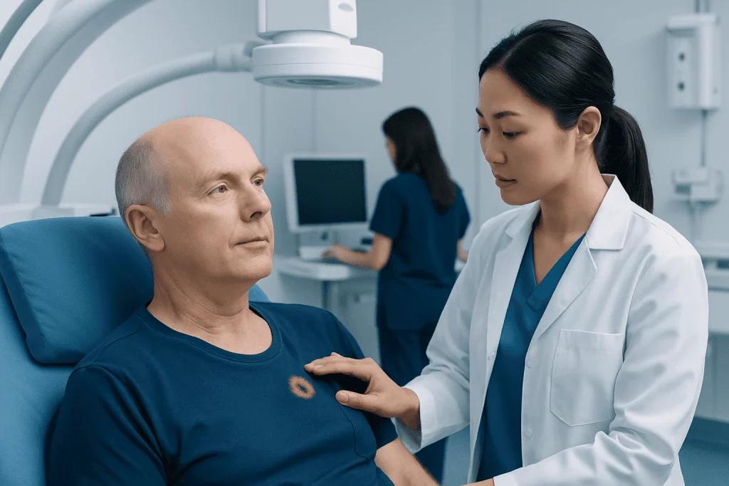
Basal cell carcinoma commonly develops on sun-exposed areas, especially the nose, cheeks, eyelids, forehead, ears, scalp, and neck. However, it can also occur on the trunk, legs, or other areas. It usually grows gradually over months or years, which is why changes may be subtle at first.
Possible signs of BCC include a shiny or pearly bump, a pink or red patch, a flat scaly area, a sore that does not heal, or a lesion that bleeds easily after minor irritation. Some BCCs have small visible blood vessels on the surface. Others may appear waxy, white, yellowish, brown, blue-black, or similar to a scar, particularly in more fibrous subtypes.
Common features that should prompt medical evaluation include:
- A new skin growth that persists for more than a few weeks.
- A wound that heals and then opens again.
- A spot that bleeds, crusts, oozes, or becomes tender without clear reason.
- A slowly enlarging patch or bump with a rolled, raised, or shiny border.
- A scar-like area where there was no previous injury.
Not every suspicious spot is cancer, and many benign skin conditions can look similar. Still, a professional skin examination is the safest way to tell the difference. Early evaluation often allows simpler treatment and a better cosmetic result, especially on the face and other delicate areas.
Causes and Risk Factors
The most important cause of basal cell carcinoma is damage to skin-cell DNA from ultraviolet radiation. This damage can build up over time from sunlight, tanning beds, or repeated sunburns. BCC is especially linked with cumulative sun exposure, but intermittent intense exposure and childhood sunburns may also contribute.
Anyone can develop BCC, but certain factors increase risk. These include fair skin that burns easily, light-colored eyes, blond or red hair, older age, a personal or family history of skin cancer, and living or working in sunny or high-altitude environments. People who spend significant time outdoors for work or recreation may have higher lifetime ultraviolet exposure.
Other risk factors include a weakened immune system, such as after organ transplantation or during certain immune-suppressing treatments, previous radiation therapy to the skin, chronic wounds or scars, and rare inherited syndromes that predispose a person to multiple BCCs. A history of one basal cell carcinoma is also an important risk factor for future BCCs or other skin cancers.
Risk is not limited to people with lighter skin. Basal cell carcinoma is less common in darker skin tones, but it can occur and may be diagnosed later if changes are overlooked. Any persistent, changing, bleeding, or non-healing skin lesion deserves attention regardless of skin color.
Diagnosis
Diagnosis begins with a careful skin examination by a dermatologist or qualified clinician. The doctor reviews the patient’s medical history, sun exposure, previous skin cancers, medications, and immune status. A dermatoscope, a handheld tool that magnifies and illuminates the skin, may be used to examine structures that are not visible to the naked eye.
If basal cell carcinoma is suspected, the usual next step is a skin biopsy. During a biopsy, a small sample or the entire visible lesion is removed under local anesthesia and sent to a pathology laboratory. The pathologist examines the tissue under a microscope to confirm the diagnosis and identify the subtype, such as nodular, superficial, infiltrative, micronodular, or morpheaform BCC.
The pathology result helps guide treatment. For example, superficial BCCs on low-risk areas may be suitable for less invasive options, while aggressive subtypes, recurrent tumors, or lesions on the nose, eyelids, lips, ears, hands, or genitals may require more precise surgical planning. In most cases, imaging tests are not needed because BCC is usually localized, but additional assessment may be considered for very large, deeply invasive, or unusual tumors.
Patients may also receive a full-body skin check because people with one suspicious lesion may have other sun-related skin changes. This examination can identify additional basal cell carcinomas, squamous cell carcinomas, precancerous lesions, or melanomas that need separate attention.
Treatment and Removal Options
Treatment is individualized. The best option depends on the tumor’s size, location, depth, subtype, borders, whether it is new or recurrent, and the patient’s preferences and medical condition. The aim is to clear the cancer while maintaining appearance and function, especially on cosmetically sensitive areas such as the face.
Surgical excision is a common treatment. The doctor removes the visible tumor along with a margin of normal-looking skin, and the tissue is examined to check whether the edges are clear of cancer cells. This approach is widely used for many low- and moderate-risk BCCs and often provides both diagnosis confirmation and treatment in one procedure.
Mohs micrographic surgery is a specialized technique used when tissue preservation and complete margin assessment are especially important. During Mohs surgery, thin layers of tissue are removed and examined step by step until no cancer cells remain at the edges. It is often considered for BCCs on the nose, eyelids, lips, ears, scalp, hands, feet, genitals, recurrent tumors, tumors with poorly defined borders, or aggressive histologic subtypes.
Other treatment options may include curettage and electrodesiccation, in which the tumor is scraped and treated with heat; cryotherapy in selected superficial cases; topical prescription medicines for certain superficial BCCs; photodynamic therapy for selected thin lesions; and radiation therapy when surgery is not suitable or would be difficult. For rare advanced cases, targeted medicines or immunotherapy may be considered by oncology and dermatology specialists. Patients should discuss benefits, limitations, healing time, scarring, and follow-up needs before choosing a treatment.
Recovery, Follow-Up, and Possible Complications
Recovery depends on the treatment method and the location of the lesion. After surgery, patients usually receive instructions on wound care, dressing changes, activity limits, and signs of infection. Mild soreness, swelling, bruising, or tightness can occur and typically improves as healing progresses. Scars often mature over months, and sun protection helps reduce discoloration of healing skin.
Most basal cell carcinomas are successfully treated with appropriate local therapy, but follow-up is important. A BCC can return if cancer cells remain, and a person who has had one BCC has a higher chance of developing another skin cancer later. Follow-up intervals vary based on risk level, number of lesions, immune status, and the dermatologist’s assessment.
Potential complications are usually related to local growth rather than distant spread. If left untreated for a long time, BCC can enlarge and affect nearby skin, cartilage, nerves, or bone, particularly on the face. This is why timely diagnosis is recommended even though the cancer is generally slow-growing.
Patients should keep a personal record of previous biopsy and treatment sites, especially if they receive care in different countries or health systems. Photographs, pathology reports, and operative notes can help future doctors understand the history of the lesion and plan follow-up appropriately.
Prevention and Self-Care
Sun protection is the most practical way to reduce the risk of basal cell carcinoma and other sun-related skin damage. Protection should be consistent, not only during holidays or beach days. Ultraviolet radiation can reach the skin during daily activities, cloudy weather, and through reflection from water, sand, snow, or pavement.
Helpful prevention habits include using a broad-spectrum sunscreen, wearing a wide-brimmed hat and UV-protective sunglasses, choosing long sleeves or tightly woven clothing when outdoors, and seeking shade during peak sunlight hours. Tanning beds should be avoided because they expose the skin to ultraviolet radiation that can damage DNA.
Self-skin checks are also valuable. People can examine their skin from head to toe once a month, using mirrors or help from a partner for the scalp, back, and behind the ears. The goal is not to make a diagnosis at home, but to notice new, changing, bleeding, or non-healing spots and arrange medical review.
People with a history of BCC may benefit from personalized prevention advice. A dermatologist can recommend follow-up frequency, review sun-protection habits, and evaluate precancerous or suspicious lesions early. For international patients seeking assessment or treatment, Acibadem International’s multidisciplinary specialists and JCI-accredited hospitals can provide diagnosis and treatment planning for skin cancers, including basal cell carcinoma.
When to See a Doctor
A doctor should evaluate any skin spot that does not heal, repeatedly bleeds or crusts, grows slowly, changes color or texture, or looks different from surrounding skin. It is also wise to seek care for a new lesion on the face, eyelids, ears, lips, scalp, hands, or any area where delayed treatment could affect appearance or function.
People with previous skin cancer, many sun-damaged spots, a weakened immune system, or a strong family history of skin cancer should follow a dermatologist’s recommended screening schedule. Earlier and more regular checks may be needed after a transplant, during immune-suppressing therapy, or after multiple BCCs.
Medical review is usually not an emergency, but it should not be postponed for months if a lesion is persistent or changing. A simple biopsy can provide clarity, and treatment is often more straightforward when BCC is diagnosed at an early stage. Anyone unsure about a skin change should consult a qualified dermatologist rather than trying to remove or treat it at home.
Frequently asked questions
Is basal cell carcinoma dangerous?
Basal cell carcinoma is usually slow-growing and rarely spreads to distant organs. However, it can continue to enlarge and damage nearby tissue if not treated. Early diagnosis and appropriate removal or treatment are important for the best medical and cosmetic outcome.
How is basal cell carcinoma different from melanoma?
Basal cell carcinoma begins in basal cells of the outer skin layer and usually grows locally and slowly. Melanoma begins in pigment-producing cells and has a higher risk of spreading if not detected early. Both conditions require medical evaluation, but their behavior and treatment plans are different.
Does every basal cell carcinoma need surgery?
Not always. Many BCCs are treated surgically, but selected superficial or low-risk lesions may be treated with topical medicines, photodynamic therapy, curettage and electrodesiccation, cryotherapy, or radiation therapy. The best choice depends on the lesion and should be decided with a dermatologist.
What is Mohs surgery and when is it used?
Mohs surgery is a precise technique that removes the cancer layer by layer while checking the tissue edges under a microscope during the procedure. It is often used for BCCs in high-risk or cosmetically sensitive areas, recurrent tumors, aggressive subtypes, or lesions with unclear borders. It helps preserve healthy tissue while confirming complete removal.
Can basal cell carcinoma come back after removal?
Yes, recurrence is possible, especially if the tumor was large, aggressive, recurrent, or located in a high-risk area. Regular follow-up helps detect recurrence or new skin cancers early. Patients should also monitor the treated area and report any new bump, bleeding, crusting, or non-healing change.
Can sunscreen prevent basal cell carcinoma completely?
Sunscreen reduces ultraviolet exposure and is an important prevention tool, but it cannot remove all risk. Best protection comes from combining sunscreen with shade, protective clothing, hats, sunglasses, and avoiding tanning beds. People with a history of BCC still need regular skin checks.
Will removal of basal cell carcinoma leave a scar?
Any procedure that removes skin can leave some degree of scarring, but doctors choose techniques that aim to clear the cancer and preserve appearance and function. Scar size and visibility depend on the lesion, location, treatment method, wound care, and individual healing. Patients can discuss reconstruction and scar-care options before treatment.
References
- American Academy of Dermatology
- National Cancer Institute
- World Health Organization
- British Association of Dermatologists
- European Academy of Dermatology and Venereology
This article is for general information only and is not a substitute for professional medical advice. Please consult a qualified doctor about your individual situation.
Basal Cell Carcinoma in Turkey — costs, top hospitals & a free quote
JCI-accredited · board-certified surgeons · reply within 24h
Add us as a Preferred Source to see more of our trusted health content across Google Search, AI Overviews and Discover.

Medical Oncology Department
Medical treatment of cancer with chemotherapy, immunotherapy and targeted therapies under a multidisciplinary tumor board.
60 specialists in this unit