Biplane Angiography: Why Two-Angle Imaging Matters in Vascular Procedures
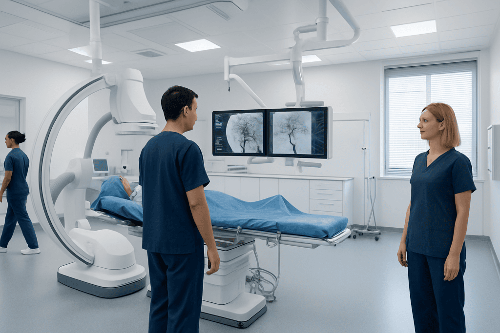
Biplane angiography provides two simultaneous imaging angles during a procedure. It is commonly used for complex vascular, cardiac, and neurovascular interventions.
Key Takeaways
- Biplane angiography provides two simultaneous imaging angles during a procedure.
- It is commonly used for complex vascular, cardiac, and neurovascular interventions.
- The technique can improve anatomical understanding and support accurate device placement.
- It may reduce the need to repeatedly reposition imaging equipment during treatment.
- Patients still need preparation and follow-up similar to other catheter-based angiography procedures.
Biplane angiography is an advanced imaging technique that captures two X-ray views at the same time during vascular procedures. This dual perspective helps doctors see blood vessels, catheters, and treatment devices more clearly and guide interventions with greater precision.
Overview of Biplane Angiography
Biplane angiography is a specialized medical imaging system used during procedures that involve blood vessels. Unlike standard single-plane angiography, which shows one X-ray view at a time, a biplane system captures two views simultaneously from different angles. This gives the medical team a more complete understanding of complex anatomy in real time.
The technique is especially valuable when doctors need to navigate delicate or branching vessels. Seeing the same area from two directions at once can make it easier to follow the path of a catheter, identify vessel narrowing or abnormal connections, and position treatment devices accurately. It is often used in highly specialized procedure rooms for image-guided interventions rather than for routine imaging alone.
Biplane angiography is not a separate disease treatment itself. Instead, it is an imaging technology that supports diagnosis and minimally invasive therapy. It may be used in the brain, heart, and other vascular areas where precision matters, such as during evaluation of aneurysms, arteriovenous malformations, or blocked arteries.
Why Two-Angle Imaging Matters

The main advantage of biplane angiography is its simultaneous two-angle view. Blood vessels are three-dimensional structures, but standard fluoroscopy displays them on a flat image. When only one angle is available, overlapping vessels or devices may make interpretation more difficult. With a second view shown at the same time, doctors can better judge depth, direction, and spatial relationships.
This can be particularly helpful in procedures involving small, tortuous, or highly sensitive vessels. For example, in neurovascular work, accurate visualization is important when treating conditions such as brain aneurysm or complex vascular malformations. In these settings, even small differences in angle can change how clearly anatomy is seen.
Two-angle imaging may also improve workflow. Because the images are acquired simultaneously, the medical team may not need to stop as often to rotate the imaging arm and repeat contrast injections from multiple positions. This can support efficient decision-making during technically demanding procedures while maintaining a clear view of both the target vessel and the devices being used.
Common Uses in Vascular Procedures

Biplane angiography is most often used in interventional neuroradiology, cardiology, and vascular procedures where exact navigation is important. In the brain, it may help doctors diagnose and treat aneurysms, stenosis, fistulas, and arteriovenous malformations. In the heart and major vessels, it can support certain catheter-based evaluations and interventions.
Examples of procedures that may benefit from biplane imaging include angiography, embolization, and image-guided treatment of vascular narrowing or bleeding. In neurovascular care, it may be part of planning or performing aneurysm treatment when detailed vessel mapping is needed. The technology helps the physician see both the lesion and the path of the instruments in real time.
Biplane systems may also be used during treatment of arteriovenous malformation and some stroke-related interventions. Not every patient or every vascular procedure requires this technology. The decision depends on the anatomy involved, the complexity of the case, and the type of treatment being planned.
What Patients Can Expect Before and During the Procedure
From a patient’s perspective, having a procedure performed with biplane angiography is often similar to having other catheter-based angiographic procedures. Before treatment, the care team usually reviews medical history, medications, allergies, kidney function, and whether contrast dye can be used safely. Patients may be asked not to eat or drink for a certain period before the procedure.
During the procedure, the patient lies on a specialized table while the imaging system moves into position around the body. A doctor inserts a thin catheter, usually through an artery in the wrist or groin, and guides it to the area being examined or treated. Contrast dye is injected so blood vessels become visible on X-ray images. Sedation or anesthesia depends on the procedure type, the treatment area, and the patient’s overall condition.
The dual imaging arms of a biplane system may appear more complex than a standard setup, but they are designed to assist the medical team rather than cause added discomfort. The care team continuously monitors the patient’s heart rate, blood pressure, and oxygen levels. After the procedure, the catheter is removed and the insertion site is observed for bleeding or swelling.
Benefits and Possible Limitations
Biplane angiography offers several practical benefits. It can provide a more complete anatomical view, support accurate catheter navigation, and help physicians confirm device position from two perspectives at once. In complex procedures, this may improve confidence in treatment decisions and reduce the need for repeated repositioning of the imaging equipment.
Another advantage is that the technology can be especially useful in intricate brain and vascular procedures, where millimeter-level precision matters. Better visualization may also help when vessels overlap on one projection but become clearer on another. In some cases, the ability to obtain both views simultaneously may streamline the procedure and reduce the need for repeated contrast runs.
However, biplane angiography is not necessary for every patient. It is typically reserved for procedures where added imaging detail is likely to improve guidance. As with other X-ray-based procedures, it involves radiation exposure and the use of contrast dye. The medical team works to keep imaging as efficient and safe as possible, balancing image quality with the lowest practical exposure.
Safety, Risks, and Recovery
The risks associated with biplane angiography are generally related to angiography and catheter-based intervention rather than to the two-angle system itself. Potential concerns can include bleeding or bruising at the catheter insertion site, contrast dye reactions, blood vessel injury, infection, or kidney strain in susceptible patients. The exact level of risk depends on the procedure being performed and the patient’s health status.
Radiation safety is an important part of planning and performing these procedures. Specialized teams use protocols and shielding strategies to minimize exposure while still obtaining the images needed for accurate treatment. Patients can ask their doctor why a biplane system is being recommended and how the team manages radiation and contrast use in their particular case.
Recovery varies depending on whether the procedure was diagnostic only or involved treatment. Some patients go home the same day, while others stay in the hospital for observation, especially after neurovascular or complex vascular interventions. At discharge, patients are usually advised about rest, hydration, medication use, and signs of complications that should prompt medical attention.
When to Ask About Biplane Angiography
Patients do not usually need to request a specific imaging platform on their own, but it can be helpful to understand why one may be recommended. A doctor may prefer biplane angiography when the planned procedure involves small vessels, branching anatomy, or treatments that require exact device placement. This is often the case in neurovascular interventions and some advanced vascular procedures.
Questions patients may wish to ask include whether two-angle imaging is expected to improve accuracy, whether the procedure is diagnostic or therapeutic, and what alternatives exist. It is also reasonable to ask about sedation, recovery time, and whether special precautions are needed because of kidney disease, diabetes, or blood-thinning medication.
For international patients seeking advanced image-guided care, Acibadem International’s multidisciplinary specialists and JCI-accredited hospitals diagnose and treat vascular conditions using modern interventional technologies, including biplane angiography where appropriate. The most suitable approach is always based on the individual patient’s anatomy, diagnosis, and treatment goals.
Frequently asked questions
What is the difference between biplane angiography and standard angiography?
Standard angiography usually shows one imaging angle at a time, while biplane angiography shows two angles simultaneously. This can help doctors better understand complex vessel anatomy during a procedure. The patient experience is often similar, but the imaging system is more advanced.
Is biplane angiography only used for brain procedures?
No, although it is especially useful in neurovascular procedures. It may also be used in selected cardiac and other vascular interventions where a dual view helps guide treatment. The exact use depends on the anatomy and the goals of the procedure.
Does biplane angiography expose patients to more radiation?
Biplane systems use X-rays, so radiation exposure is part of the procedure. However, the total exposure depends on many factors, including the complexity and length of the intervention. Medical teams use established safety practices to keep radiation as low as reasonably achievable while maintaining image quality.
Will the procedure be painful?
Patients may feel brief discomfort from the catheter insertion site and sometimes a warm sensation when contrast dye is injected. Sedation, local anesthesia, or general anesthesia may be used depending on the procedure. The care team monitors comfort closely throughout the intervention.
Why would a doctor choose biplane angiography for one patient but not another?
The choice depends on how complex the vessels are, what treatment is planned, and how much detail is needed for safe guidance. In straightforward cases, single-plane imaging may be enough. In more intricate procedures, two-angle imaging can offer a clearer and more practical view.
How long does recovery take after a procedure using biplane angiography?
Recovery time depends more on the type of procedure than on the imaging system itself. Some patients recover within hours after a diagnostic angiogram, while others need hospital monitoring after an interventional treatment. The doctor will give personalized instructions based on the procedure performed.
References
- World Health Organization
- Radiological Society of North America
- American Heart Association
- Society of Interventional Radiology
- American College of Radiology
This article is for general information only and is not a substitute for professional medical advice. Please consult a qualified doctor about your individual situation.
Explore treatments in Turkey — costs, top hospitals & a free quote
JCI-accredited · board-certified surgeons · reply within 24h
Add us as a Preferred Source to see more of our trusted health content across Google Search, AI Overviews and Discover.