Brain Anatomy Diagram: How to Read Labeled Brain Structures

A brain anatomy diagram usually labels the cerebrum, cerebellum, brainstem, and major lobes. Different diagram views, such as side, top, or cross-sectional images, show different structures more clearly.
Key Takeaways
- A brain anatomy diagram usually labels the cerebrum, cerebellum, brainstem, and major lobes.
- Different diagram views, such as side, top, or cross-sectional images, show different structures more clearly.
- Knowing basic directional terms like frontal, posterior, left, right, superior, and inferior helps with reading labels.
- Brain structures work together to support movement, sensation, language, memory, and automatic body functions.
- Medical scans such as MRI and CT provide more detailed information than a simplified educational diagram.
A brain anatomy diagram helps readers identify the main parts of the brain and understand what each structure does. Learning how labels are arranged can make medical images, health articles, and doctor explanations easier to follow.
Overview: What a Brain Anatomy Diagram Shows
A brain anatomy diagram is a visual guide to the main structures of the brain. It may be a simple educational drawing with a few labels or a detailed medical illustration showing deeper regions, nerves, blood vessels, or cross-sections. Most diagrams are designed to help readers understand where each brain part is located and how those parts relate to important functions such as thinking, movement, balance, speech, and breathing.
The brain is part of the central nervous system, together with the spinal cord. In many labeled images, the largest visible regions are the cerebrum, cerebellum, and brainstem. Within the cerebrum, diagrams often identify the frontal, parietal, temporal, and occipital lobes. Some may also label internal structures such as the thalamus, hypothalamus, hippocampus, pituitary gland, corpus callosum, and ventricles.
Educational brain diagrams are simplified on purpose. They help build a basic understanding, but they do not replace a doctor’s interpretation of imaging tests. If a person is reviewing a scan because of symptoms or a diagnosed condition, a neurologist, neurosurgeon, or radiologist can explain what is normal and what may need further evaluation.
How to Read Labels on a Brain Diagram
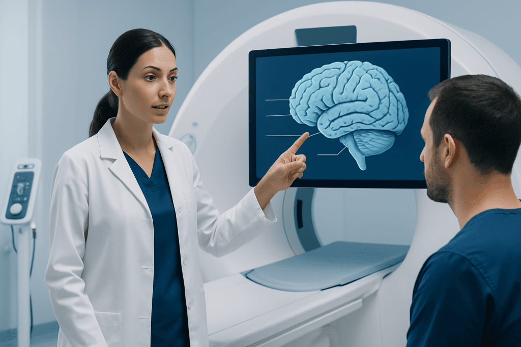
The first step is to identify the view of the image. A side view, also called a lateral view, is common in textbooks because it clearly shows the outer surface of the brain, including the lobes and cerebellum. A top view highlights the two hemispheres, while a front view can show left-right symmetry. Cross-sectional images, such as sagittal, coronal, or axial views, display deeper structures that cannot be seen from the outside.
It also helps to know a few directional terms. Frontal or anterior means toward the front of the head. Posterior means toward the back. Superior means upper, and inferior means lower. Medial refers to structures closer to the middle, while lateral refers to those farther out to the sides. In imaging, right and left may appear reversed depending on the viewing orientation, so the label guide is important.
Many diagrams use color coding to separate regions. For example, one lobe may be shaded differently from another, or sensory and motor areas may be highlighted. A labeled line usually points to the outer edge or center of a structure. When learning from a diagram, it is often easier to begin with the largest parts and then move inward to smaller structures and pathways.
- Start by finding the cerebrum, cerebellum, and brainstem.
- Identify the view: side, top, front, or cross-section.
- Read directional labels before interpreting position.
- Then locate the lobes and deeper structures one by one.
Main Brain Structures Commonly Labeled

The cerebrum is the largest part of the brain and fills most of the skull. It is divided into right and left hemispheres and is responsible for higher functions such as thought, voluntary movement, sensation, language, planning, and many aspects of memory and emotion. The folded outer layer is called the cerebral cortex. The folds increase surface area and allow more nerve cells to fit within the skull.
The cerebellum sits at the back and lower part of the brain. It plays a key role in coordination, balance, posture, and fine-tuning movement. Although it is smaller than the cerebrum, it is highly important for smooth and accurate physical activity. The brainstem connects the brain to the spinal cord and includes the midbrain, pons, and medulla. It helps control vital automatic functions such as breathing, heart rate, swallowing, and wakefulness.
Many diagrams also label internal structures. The thalamus acts as a relay center for many sensory signals. The hypothalamus helps regulate body temperature, hunger, thirst, sleep, and hormone-related functions. The hippocampus is associated with learning and memory, while the amygdala is involved in emotional processing. The corpus callosum is a thick bundle of nerve fibers that connects the two hemispheres. Ventricles are fluid-filled spaces that contain cerebrospinal fluid, which cushions and supports the brain.
Understanding the Brain Lobes and Their Functions
The frontal lobe is located at the front of the brain. It is involved in planning, judgment, problem-solving, personality, emotional regulation, and voluntary movement. Important areas for speech production are also found here in many people. Because the frontal lobe supports executive functions, its role is often highlighted in educational diagrams.
The parietal lobe lies near the upper middle part of the brain. It helps process touch, temperature, pain, body position, and spatial awareness. The temporal lobe is located on the sides of the brain near the ears and contributes to hearing, language understanding, memory, and aspects of emotion. The occipital lobe sits at the back of the brain and is mainly responsible for visual processing.
These functions are not isolated in a simple one-structure, one-task way. Brain regions communicate continuously through networks, so most everyday activities involve several areas working together. For example, reading a word aloud may require the occipital lobe for vision, temporal regions for language processing, frontal regions for speech planning, and motor pathways for speaking.
Some diagrams may also point out the motor cortex and sensory cortex, which lie on opposite sides of a central groove called the central sulcus. These areas are often shown to explain how the brain organizes movement and sensation across the body.
What Cross-Sectional Brain Diagrams Reveal
Cross-sectional diagrams show the brain as if it has been sliced in a specific plane. A sagittal section divides the brain into left and right portions and can clearly display midline structures such as the corpus callosum, pituitary gland, brainstem, and cerebellum. A coronal section divides the brain into front and back parts, while an axial section divides it into upper and lower portions. These views are especially useful for understanding anatomy seen on MRI or CT scans.
In cross-sections, readers often encounter labels for structures that are less visible on the outer surface. These can include the basal ganglia, ventricles, thalamus, hypothalamus, pineal region, and deep white matter tracts. Educational diagrams may simplify these structures, but they still provide a useful bridge between textbook anatomy and real medical imaging.
This is also where readers may first notice how closely different parts are arranged. Even small areas can have important functions, which is why doctors carefully correlate imaging findings with symptoms and neurological examination. If a person is being evaluated for a structural problem such as a brain tumor or another neurological condition, imaging is interpreted in clinical context rather than by labels alone.
Brain Diagrams Versus MRI and CT Scans
A brain anatomy diagram is a teaching tool, while MRI and CT scans are diagnostic tools. Diagrams show idealized anatomy in a clear and simplified way. By contrast, real medical images vary from person to person and may be more difficult for a non-specialist to interpret. Differences in angle, slice thickness, contrast, and normal anatomy can make scans look complex even when they are normal.
MRI is often used when doctors need detailed images of soft tissues in the brain. It can help evaluate the brain’s structure in many neurological conditions. CT scans are commonly used in urgent settings because they are fast and particularly useful for detecting some forms of bleeding, injury, or major structural changes. Depending on the situation, doctors may recommend MRI or CT imaging to investigate symptoms.
People should avoid drawing conclusions from scans without professional guidance. A structure that looks unusual in an online comparison may still be normal, while subtle abnormalities may not be apparent to an untrained eye. If there are symptoms such as severe headache, seizures, weakness, confusion, or changes in speech or vision, prompt medical assessment is more helpful than self-interpretation.
Why Brain Anatomy Knowledge Matters
Understanding labeled brain structures can make health information easier to follow. Patients and families often hear terms such as frontal lobe, cerebellum, brainstem, lesion, or hemisphere during consultations. Knowing where these structures are and their general functions can help people ask clearer questions and better understand explanations about symptoms, tests, treatment, or recovery.
This knowledge is especially useful when discussing stroke, injury, epilepsy, tumors, memory disorders, or movement problems. For example, a symptom affecting balance may relate to the cerebellum or connected pathways, while language changes may involve frontal or temporal networks. A diagram does not diagnose the cause, but it helps place medical information into context.
It is also important to remember that the brain is highly interconnected. Symptoms may arise from networks rather than a single isolated spot. In some cases, further evaluation may include neurological examination, imaging, cognitive testing, or procedures such as EEG when electrical brain activity needs assessment. When surgery is considered, specialized teams may evaluate options such as neurosurgical treatment depending on the diagnosis.
When to Seek Medical Advice
Reading a brain anatomy diagram is educational, but it should not be used to diagnose symptoms at home. A person should seek medical advice if they have persistent headaches, new seizures, fainting, numbness, weakness, balance problems, memory decline, confusion, major personality changes, or difficulties with speech, vision, or coordination. These symptoms can have many possible causes, and a qualified doctor can assess them properly.
Urgent medical help is especially important for sudden symptoms, such as abrupt weakness on one side, a sudden severe headache, facial drooping, difficulty speaking, loss of consciousness, or head injury with neurological changes. Fast evaluation can be crucial in emergencies affecting the brain and nervous system.
For patients who need specialist assessment, Acibadem International’s multidisciplinary teams in JCI-accredited hospitals diagnose and treat brain and nervous system conditions for international patients. Whether someone is learning basic anatomy or reviewing a medical finding, the most reliable next step is professional interpretation tailored to the individual case.
Frequently asked questions
What is the easiest way to read a brain anatomy diagram?
The easiest approach is to start with the largest structures first: the cerebrum, cerebellum, and brainstem. After that, identify the view of the image and then look for the lobes and any deeper structures shown.
What are the four main lobes of the brain?
The four main lobes are the frontal, parietal, temporal, and occipital lobes. Each supports different functions, but they work together through connected brain networks rather than acting alone.
Why do MRI or CT scans look different from textbook brain diagrams?
Textbook diagrams are simplified and designed for learning, while MRI and CT scans show real anatomy in slices and shades of contrast. Real scans may look more complex because they display fine detail, natural variation, and the exact viewing angle used during imaging.
What does the brainstem do?
The brainstem connects the brain to the spinal cord and helps control essential automatic functions. These include breathing, heart rate, swallowing, alertness, and some reflexes.
Can a brain anatomy diagram help explain symptoms?
Yes, it can help a person understand where a symptom may relate anatomically, such as balance with the cerebellum or vision with the occipital lobe. However, symptoms are not specific enough for self-diagnosis, so a doctor’s evaluation is still necessary.
Are left and right brain structures always shown the same way on diagrams?
Not always. Educational diagrams may present a side view or a cross-section, and medical images can use conventions that make left and right appear different from what a person expects. Reading the orientation labels is important before interpreting the image.
References
- National Institute of Neurological Disorders and Stroke
- MedlinePlus
- National Cancer Institute
- Radiological Society of North America
- Merck Manual Consumer Version
This article is for general information only and is not a substitute for professional medical advice. Please consult a qualified doctor about your individual situation.
Explore treatments in Turkey — costs, top hospitals & a free quote
JCI-accredited · board-certified surgeons · reply within 24h
Add us as a Preferred Source to see more of our trusted health content across Google Search, AI Overviews and Discover.
Check your numbers in seconds
BMI, calories, due date, blood pressure and 30+ more clinical calculators — free, instant, doctor-reviewed ranges.