Brain Aneurysm Coiling: How It Works, Who It Helps, and Recovery

Brain aneurysm coiling treats some aneurysms from inside the blood vessel without open brain surgery. It is often used for ruptured aneurysms and may also be an option for selected unruptured aneurysms.
Key Takeaways
- Brain aneurysm coiling treats some aneurysms from inside the blood vessel without open brain surgery.
- It is often used for ruptured aneurysms and may also be an option for selected unruptured aneurysms.
- Recovery time is often shorter than with surgical clipping, but follow-up imaging is important.
- Not every aneurysm is suitable for coiling; treatment is individualized.
- Urgent medical care is needed for sudden severe headache or symptoms suggesting a ruptured aneurysm.
Brain aneurysm coiling is a minimally invasive procedure used to treat certain aneurysms in the brain by filling the weakened area with tiny coils. It can help prevent rupture or manage bleeding after a rupture, with treatment decisions based on the aneurysm’s size, shape, location, and the person’s overall health.
Overview: What brain aneurysm coiling is
Brain aneurysm coiling, also called endovascular coiling, is a procedure used to treat an aneurysm in a blood vessel of the brain. An aneurysm is a weakened, bulging area in an artery wall. The aim of coiling is to reduce blood flow into that bulge so the aneurysm is less likely to bleed, or to stop further bleeding if a rupture has already happened.
Instead of opening the skull, a specialist usually reaches the aneurysm through the blood vessels. A thin tube called a catheter is inserted, often through an artery in the wrist or groin, and carefully guided to the arteries in the brain. Very small soft metal coils are then placed inside the aneurysm. These coils encourage clotting within the aneurysm, helping seal it off from normal circulation.
Coiling is one of the main treatments for cerebral aneurysms, alongside surgical clipping. For many people, it offers a less invasive option with a shorter hospital stay and recovery period. However, the best choice depends on detailed imaging, the aneurysm’s anatomy, whether it has ruptured, and the experience of the treating neurovascular team.
Who it helps and when it may be recommended
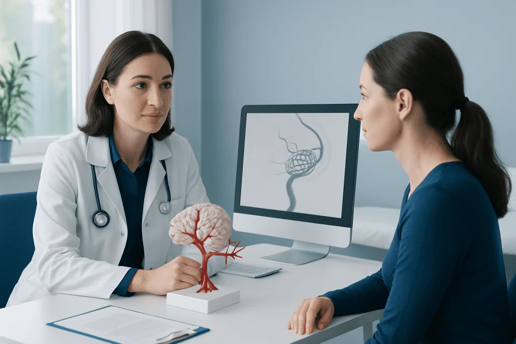
Brain aneurysm coiling may help people with either ruptured or unruptured aneurysms. In an emergency, it is commonly used after a ruptured aneurysm to reduce the chance of rebleeding. In non-emergency situations, it may be offered for an unruptured aneurysm that is judged to have a meaningful risk of future rupture.
Doctors do not recommend treatment based on one factor alone. They consider the aneurysm’s size, neck width, shape, and exact location in the brain, as well as the person’s age, symptoms, general health, family history, and whether the aneurysm has changed over time. Some small unruptured aneurysms may be monitored rather than treated right away.
Coiling is often particularly useful for aneurysms that are difficult to reach with open surgery or for people who may benefit from a less invasive approach. In some cases, additional devices such as stents or balloons are used to help keep the coils in place, especially if the aneurysm has a wide neck. A multidisciplinary discussion between neurosurgery, interventional neuroradiology, and neurology helps guide the safest plan.
How the procedure works
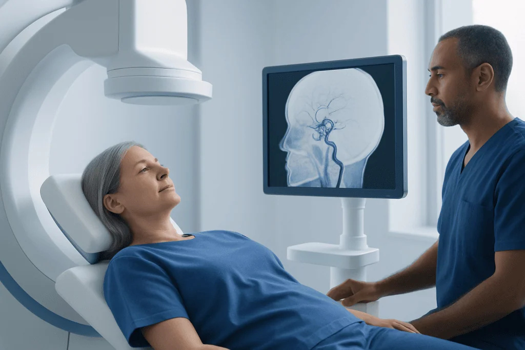
Before the procedure, the medical team reviews brain imaging such as CT angiography, MR angiography, or catheter angiography to map the aneurysm in detail. Coiling is usually done in a specialized angiography suite under general anesthesia, although the exact approach can vary. The skin is cleaned, the artery access site is prepared, and the catheter is guided through the vascular system using live X-ray imaging.
Once the catheter reaches the aneurysm, the specialist advances extremely fine coils into the bulging area. The coils are designed to fit within the aneurysm sac and promote clot formation there. The goal is to pack the aneurysm enough to block blood flow into it while preserving normal flow through the parent artery.
Some aneurysms need supportive techniques. Balloon-assisted coiling temporarily uses a small balloon to help shape the coil placement. Stent-assisted coiling uses a small mesh tube in the artery to support wide-neck aneurysms. In selected cases, other endovascular options may also be discussed as part of brain aneurysm treatment.
After the coils are placed, imaging is repeated to confirm the result. The catheter is removed, and pressure or a closure device is used at the access site. The patient is then monitored closely for neurological changes, blood pressure control, and any signs of complications.
Benefits, limits, and possible risks
The main benefit of brain aneurysm coiling is that it is minimally invasive. Because it avoids a large surgical incision and opening the skull, it often leads to less postoperative discomfort and a faster early recovery than surgical clipping. For many patients, this can mean a shorter stay in hospital and a quicker return to everyday activities.
Coiling can be highly effective, but it is not the best option for every aneurysm. Some aneurysms have shapes or locations that make surgical clipping more durable or safer. Coiled aneurysms may also have a higher chance of reopening over time compared with clipped aneurysms, which is why scheduled follow-up imaging is so important.
As with any procedure involving blood vessels in the brain, there are risks. These may include bleeding, stroke, blood clot formation, vessel injury, infection, contrast-related kidney problems, or a reaction to medications or contrast dye. Rarely, the aneurysm can rupture during the procedure. The treating team discusses these risks in the context of the risks of leaving the aneurysm untreated.
For people whose aneurysm has already bled, the overall situation can be more complex because the hemorrhage itself may cause complications such as vasospasm, hydrocephalus, or brain injury. In those cases, coiling addresses the aneurysm, but recovery also depends on the severity of the initial bleed, such as in subarachnoid hemorrhage.
Diagnosis and planning before coiling
Diagnosis usually begins when an aneurysm is suspected because of symptoms or found incidentally on imaging done for another reason. A ruptured aneurysm often presents as a sudden, severe headache and requires urgent evaluation. Imaging may include a CT scan, CT angiography, MRI, MR angiography, or digital subtraction angiography, which provides very detailed information about the blood vessels.
For an unruptured aneurysm, doctors assess whether treatment or monitoring is more appropriate. They weigh the estimated risk of rupture against the risks of the procedure. This planning is individualized because two aneurysms of similar size may behave differently depending on their shape, location, and the patient’s risk profile.
Before coiling, the team also reviews medical conditions that may affect safety, such as high blood pressure, kidney disease, bleeding disorders, or the use of blood thinners. If a stent is likely to be needed, antiplatelet medication may be required before and after the procedure. Careful preparation helps reduce risk and improves the chance of a good result.
Recovery and follow-up after the procedure
Recovery after brain aneurysm coiling varies depending on whether the aneurysm was ruptured or unruptured. For an unruptured aneurysm treated electively, many people spend a short period in intensive or high-dependency monitoring and then go home within a few days if recovery is smooth. Tiredness, mild headache, and soreness at the access site are common for a short time.
After treatment of a ruptured aneurysm, the hospital stay is often longer because the brain needs close monitoring for complications related to the bleed. The team watches for vasospasm, changes in alertness, hydrocephalus, and blood pressure issues. Rehabilitation may be needed if the hemorrhage caused weakness, speech problems, or changes in thinking.
Once home, patients are usually advised to avoid heavy lifting for a period, take medications exactly as prescribed, and attend follow-up appointments. Follow-up imaging is an essential part of care after coiling because some aneurysms can partially reopen or compact over time. If that happens, additional endovascular treatment may be considered.
People recovering from a ruptured aneurysm may also need support for longer-term symptoms such as fatigue, mood changes, headaches, or cognitive slowing. In selected cases, rehabilitation therapies and specialist follow-up for brain aneurysm recovery can help improve day-to-day function and confidence.
Prevention, self-care, and when to seek medical help
There is no guaranteed way to prevent every brain aneurysm, but managing vascular risk factors may help lower the chance of aneurysm growth or rupture. Important steps include controlling high blood pressure, not smoking, limiting excessive alcohol use, staying active, and following treatment plans for other conditions that affect blood vessels. People with a strong family history should discuss whether screening is appropriate.
Self-care after coiling focuses on protecting overall brain and vascular health. This includes attending all imaging follow-up, taking antiplatelet or other medications as instructed, and asking before stopping any prescribed medicine. A gradual return to activity is often recommended, and patients should follow the advice of their own specialist about driving, work, and exercise.
Urgent medical attention is needed for symptoms that could suggest a ruptured aneurysm or another serious neurological problem. These include a sudden severe headache, loss of consciousness, new weakness or numbness, difficulty speaking, vision changes, seizure, or severe confusion. After a recent procedure, patients should also seek prompt care for worsening headache, new neurological symptoms, or significant swelling or bleeding at the access site.
Specialist evaluation is important because coiling is only one part of comprehensive aneurysm care. Near the end of the treatment journey, some patients may benefit from care in centers with neurointerventional, neurosurgical, and rehabilitation expertise. Acibadem International’s multidisciplinary specialists in JCI-accredited hospitals diagnose and treat this condition for international patients, including advanced interventional neuroradiology and, when needed, neurosurgery.
Frequently asked questions
Is brain aneurysm coiling the same as brain surgery?
Brain aneurysm coiling is a minimally invasive endovascular procedure, not open brain surgery. The doctor usually reaches the aneurysm through a blood vessel in the wrist or groin rather than opening the skull.
How long does recovery after aneurysm coiling take?
Recovery depends greatly on whether the aneurysm was ruptured or unruptured. People treated for an unruptured aneurysm often recover more quickly, while recovery after a rupture may take much longer because the bleeding itself can affect the brain.
Can a coiled aneurysm come back?
A coiled aneurysm can sometimes partially reopen over time. That is why follow-up scans are important, even when the initial treatment result is good. If reopening is seen, the doctor may recommend observation or another procedure.
Who is a good candidate for brain aneurysm coiling?
A good candidate is someone whose aneurysm anatomy and overall health make endovascular treatment a suitable and safe option. The decision depends on factors such as the aneurysm’s size, shape, neck width, location, and whether it has ruptured.
Is aneurysm coiling safer than clipping?
Neither treatment is automatically better for every person. Coiling is less invasive and may allow quicker early recovery, but clipping may be more suitable or more durable for some aneurysms. Specialists compare the risks and benefits for the individual case.
What are the warning signs of a ruptured brain aneurysm?
A ruptured brain aneurysm can cause a sudden severe headache, often described as the worst headache of a person’s life. It may also cause nausea, vomiting, neck stiffness, confusion, fainting, seizure, or weakness. These symptoms need emergency medical attention.
References
- American Heart Association
- National Institute of Neurological Disorders and Stroke
- National Health Service
- Brain Aneurysm Foundation
- Society of NeuroInterventional Surgery
This article is for general information only and is not a substitute for professional medical advice. Please consult a qualified doctor about your individual situation.
Explore treatments in Turkey — costs, top hospitals & a free quote
JCI-accredited · board-certified surgeons · reply within 24h
Add us as a Preferred Source to see more of our trusted health content across Google Search, AI Overviews and Discover.
Check your numbers in seconds
BMI, calories, due date, blood pressure and 30+ more clinical calculators — free, instant, doctor-reviewed ranges.