Brain Aneurysm Coiling: Procedure Steps, Risks, and Recovery

Brain aneurysm coiling treats an aneurysm from inside the blood vessel without open brain surgery. The procedure uses a catheter and tiny coils to reduce blood flow into the aneurysm sac.
Key Takeaways
- Brain aneurysm coiling treats an aneurysm from inside the blood vessel without open brain surgery.
- The procedure uses a catheter and tiny coils to reduce blood flow into the aneurysm sac.
- Recovery is often shorter than with open surgery, but follow-up imaging is important.
- Risks can include bleeding, stroke, aneurysm reopening, or reaction to contrast dye.
- Treatment decisions depend on the aneurysm’s anatomy, symptoms, and whether it has ruptured.
Brain aneurysm coiling is a minimally invasive treatment used to block blood flow into a brain aneurysm. It can help prevent rupture in some unruptured aneurysms or reduce the risk of rebleeding after a rupture, with treatment planning tailored to the aneurysm’s size, shape, and location.
Overview of brain aneurysm coiling
Brain aneurysm coiling, also called endovascular coiling, is a procedure used to treat certain aneurysms in the brain. A brain aneurysm is a weakened area in a blood vessel wall that bulges outward. If the aneurysm ruptures, it can cause bleeding around the brain, which is a medical emergency. Coiling aims to seal off the aneurysm from the inside so blood is less likely to enter it.
Unlike open surgery, coiling is performed through the blood vessels. A specialist guides a thin tube called a catheter, usually from an artery in the wrist or groin, up to the blood vessels of the brain. Tiny soft metal coils are then placed inside the aneurysm. These coils encourage clotting within the aneurysm, helping reduce pressure on its wall and lowering the risk of bleeding.
Coiling is not the right choice for every aneurysm. Some aneurysms have a shape, size, or neck width that may make other treatments more suitable. In some cases, doctors may use additional devices such as stents or balloons to help keep the coils in place. A neuroradiologist, vascular neurologist, or neurosurgeon will choose the most appropriate approach based on imaging findings and the person’s overall health.
When coiling is recommended
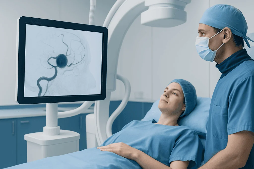
Doctors may recommend brain aneurysm coiling for either a ruptured or unruptured aneurysm. In a ruptured aneurysm, treatment is often urgent because the main goal is to prevent rebleeding. In an unruptured aneurysm, the decision is more individualized. It depends on factors such as the aneurysm’s size, location, shape, growth over time, and the person’s age and medical history.
Some aneurysms are found after symptoms such as a severe sudden headache, double vision, eyelid drooping, or nerve-related symptoms. Others are discovered incidentally during imaging for another reason. Not every unruptured aneurysm needs immediate treatment; some are monitored with repeat scans. If treatment is advised, coiling may be considered when a minimally invasive option is likely to be effective and safe.
Coiling is part of a broader group of interventional neuroradiology treatments used for disorders of the brain’s blood vessels. In some cases, evaluation also includes understanding related conditions such as brain aneurysm and how its features influence the risk of rupture or recurrence.
Procedure steps: what happens during coiling

Before the procedure, the care team reviews brain imaging such as CT angiography, MR angiography, or catheter angiography to map the aneurysm and the surrounding blood vessels. Blood tests are usually performed, and the team will review medications, including blood thinners and antiplatelet drugs. The person is typically asked not to eat or drink for a period before the procedure. Coiling is commonly done under general anesthesia, although the exact approach varies.
During the procedure, the specialist inserts a catheter into an artery, often in the wrist or groin, and carefully advances it to the arteries in the brain using live X-ray guidance. Contrast dye is injected so the blood vessels and aneurysm can be seen clearly. A very small microcatheter is then passed into the aneurysm sac. Through this microcatheter, tiny platinum coils are placed into the aneurysm one by one until it is densely packed.
If the aneurysm has a wide neck, the doctor may use a temporary balloon or a small stent to help support the coils and keep them from protruding into the parent artery. This is sometimes called balloon-assisted or stent-assisted coiling. Once the aneurysm has been treated, the catheters are removed and pressure or a closure device is used at the entry site. Many centers provide this care within stroke and neurovascular unit care because close neurological monitoring after the procedure is important.
Afterward, the patient is observed in a recovery area or intensive care setting, especially if the aneurysm was ruptured. Nurses and doctors monitor blood pressure, neurological status, and the catheter entry site. Additional imaging may be performed to confirm the result and to check for any immediate complications.
Benefits and possible risks
The main benefit of brain aneurysm coiling is that it is minimally invasive. Because it is done through the blood vessels rather than through an opening in the skull, recovery may be faster and there is no surgical incision on the scalp. For many patients, this means a shorter hospital stay, less post-procedure discomfort, and a quicker return to daily activities compared with open surgery.
Coiling can be very effective, but like any procedure it carries risks. Possible complications include bleeding, stroke, injury to the blood vessel, infection, blood clots, reaction to contrast dye, and problems related to anesthesia. There is also a small risk that the aneurysm could rupture during the procedure. These risks vary depending on whether the aneurysm has already ruptured, as well as the aneurysm’s size, location, and shape.
Another important consideration is durability. Some coiled aneurysms can reopen slightly over time, especially if they are large or have a wide neck. For this reason, follow-up imaging is often needed months or years later. If imaging shows significant recurrence, additional treatment may be recommended. The doctor will explain the balance of benefits and risks in the context of the individual case.
Recovery after aneurysm coiling
Recovery after brain aneurysm coiling depends greatly on whether the aneurysm was ruptured. People treated for an unruptured aneurysm may stay in the hospital for a short period and gradually resume normal activities over days to a few weeks. Fatigue is common at first, and mild soreness or bruising at the catheter insertion site may occur. Clear guidance is usually given on activity limits, bathing, driving, and medication use.
Recovery is often longer and more complex after treatment of a ruptured aneurysm because the bleeding itself can affect the brain. In these situations, hospital care may include monitoring for complications such as vasospasm, hydrocephalus, seizures, and changes in sodium balance. Rehabilitation may be needed if the person has weakness, speech changes, balance problems, or cognitive difficulties after the bleed.
Follow-up care is an important part of recovery. The doctor may schedule repeat angiography, CT angiography, or MR angiography to make sure the aneurysm remains sealed. Some patients need medicines for a period after the procedure, particularly if a stent was placed. If symptoms such as worsening headache, new weakness, confusion, fever, or bleeding from the catheter site develop after discharge, prompt medical advice is important.
How coiling compares with other treatments
The main alternative to coiling is surgical clipping. In clipping, a neurosurgeon places a small metal clip across the neck of the aneurysm during open brain surgery. Clipping can be a strong option for some aneurysms, particularly when the anatomy is less favorable for coiling. In other cases, coiling may be preferred because it avoids open surgery. The choice is based on a careful review of anatomy, urgency, age, health conditions, and the expertise of the treating team.
Newer endovascular techniques may also be considered in selected patients. These can include stent-assisted coiling, balloon-assisted coiling, or flow-diverting devices, which redirect blood away from the aneurysm. The best method depends on the aneurysm’s features and treatment goals. Imaging specialists and neurosurgical teams often discuss each case together before making a recommendation.
When complex decision-making is needed, patients may benefit from assessment by teams experienced in neurosurgery and image-guided vascular procedures. Near the end of the care pathway, some international patients seek treatment in centers such as Acibadem International, where multidisciplinary specialists and JCI-accredited hospitals diagnose and treat brain aneurysms using medical, endovascular, and surgical approaches.
Prevention, self-care, and when to seek medical help
It is not always possible to prevent a brain aneurysm, but some risk factors can be managed. Controlling high blood pressure, avoiding smoking, limiting excessive alcohol use, and following treatment plans for vascular risk factors may help reduce the chance of aneurysm growth or rupture. People with a strong family history of aneurysm or certain inherited disorders may be advised to discuss screening with a specialist.
After coiling, self-care focuses on healing and follow-up. It is usually helpful to take medicines exactly as prescribed, keep follow-up imaging appointments, avoid strenuous activity until the doctor says it is safe, and gradually return to normal routines. Good hydration, rest, blood pressure control, and avoiding tobacco are practical recovery steps.
Urgent medical attention is needed for symptoms that may suggest a ruptured aneurysm or another neurological emergency. These include a sudden severe headache unlike usual headaches, loss of consciousness, seizures, new weakness or numbness, trouble speaking, sudden vision changes, or severe confusion. After a recent procedure, immediate care is also important for severe groin or wrist swelling, persistent bleeding, chest pain, shortness of breath, or signs of stroke.
Frequently asked questions
Is brain aneurysm coiling a surgery?
Brain aneurysm coiling is a procedure, but it is not open brain surgery. It is performed from inside the blood vessels using catheters, usually inserted through the wrist or groin. Because it is minimally invasive, recovery may be shorter than with surgical clipping.
How long does aneurysm coiling take?
The length of the procedure varies depending on the aneurysm’s location and complexity. In general, it may take a few hours, especially if additional devices such as a balloon or stent are needed. Preparation and recovery time in the hospital add to the overall time spent in care.
What are the risks of brain aneurysm coiling?
Possible risks include stroke, bleeding, blood vessel injury, reaction to contrast dye, infection, and anesthesia-related complications. In some cases, the aneurysm may reopen over time and need further treatment. The exact level of risk depends on the aneurysm’s anatomy and whether it has already ruptured.
How long is recovery after brain aneurysm coiling?
Recovery after treatment of an unruptured aneurysm is often relatively quick, with many people returning to usual activities within days to weeks. Recovery may be much longer after a ruptured aneurysm because the brain bleed itself can cause lasting symptoms. The doctor will provide a personalized recovery plan and follow-up schedule.
Can a coiled aneurysm come back?
A coiled aneurysm can sometimes reopen or show a small recurrence over time. This is why follow-up imaging is an important part of care after the procedure. If significant reopening is found, doctors may recommend additional treatment.
Will a patient need medicines after coiling?
Some patients need medication after coiling, particularly if a stent was used during the procedure. These medicines may help reduce the risk of clot formation in the treated vessel. The exact medication plan depends on the procedure details and should be followed closely under medical supervision.
References
- American Heart Association
- National Institute of Neurological Disorders and Stroke
- National Health Service
- Society of NeuroInterventional Surgery
- Radiological Society of North America
This article is for general information only and is not a substitute for professional medical advice. Please consult a qualified doctor about your individual situation.
Explore treatments in Turkey — costs, top hospitals & a free quote
JCI-accredited · board-certified surgeons · reply within 24h
Add us as a Preferred Source to see more of our trusted health content across Google Search, AI Overviews and Discover.
Check your numbers in seconds
BMI, calories, due date, blood pressure and 30+ more clinical calculators — free, instant, doctor-reviewed ranges.