Brain AVM vs Aneurysm: Key Differences in Symptoms, Risk, and Treatment
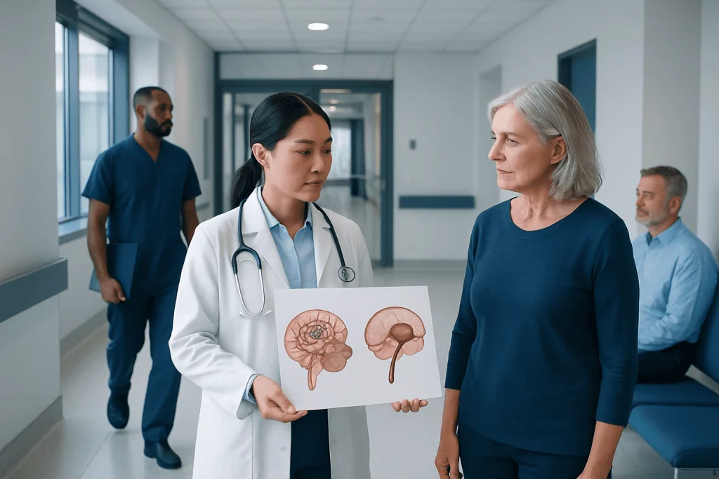
A brain AVM is an abnormal tangle of arteries and veins, while an aneurysm is a bulging weak spot in a blood vessel wall. Both conditions may cause no symptoms until they are found on imaging or bleed.
Key Takeaways
- A brain AVM is an abnormal tangle of arteries and veins, while an aneurysm is a bulging weak spot in a blood vessel wall.
- Both conditions may cause no symptoms until they are found on imaging or bleed.
- Sudden severe headache, seizure, weakness, speech trouble, or loss of consciousness need urgent medical attention.
- Diagnosis usually involves brain imaging such as CT, MRI, and specialized blood vessel imaging.
- Treatment depends on size, location, symptoms, bleeding risk, and overall health.
Brain AVMs and brain aneurysms both involve abnormal blood vessels in the brain, but they are different conditions with different patterns of symptoms, risk, and treatment. Understanding how they differ can help patients seek timely evaluation and make informed decisions with their care team.
Overview: How a Brain AVM Differs From an Aneurysm
A brain arteriovenous malformation, often called a brain AVM, is an abnormal connection between arteries and veins in the brain. Instead of blood flowing through tiny capillaries first, it passes directly from arteries to veins through a tangled network of vessels. This can place extra stress on blood vessels and surrounding brain tissue.
A brain aneurysm is different. It is a weak, balloon-like bulge in the wall of an artery. Over time, pressure from blood flow may cause the weakened section to enlarge. Some aneurysms remain stable for years, while others may leak or rupture and cause bleeding around the brain.
Although both conditions affect brain blood vessels, they are not the same disease. Their structure, causes, common symptoms, and treatment plans differ. In some cases, a person may have an AVM with an associated aneurysm, which can make diagnosis and management more complex.
Because both AVMs and aneurysms can lead to bleeding in or around the brain, they are taken seriously. Still, not every AVM or aneurysm requires the same approach. Care is individualized and often guided by specialists in neurology, neuroradiology, and neurosurgery.
Symptoms: What Each Condition May Feel Like
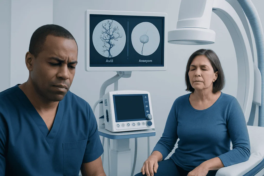
Both brain AVMs and aneurysms can be silent, meaning they cause no symptoms and are discovered only during imaging for another reason. When symptoms do occur, they may relate to pressure on nearby brain structures, changes in blood flow, or bleeding.
A brain AVM may cause headaches, seizures, weakness, numbness, vision changes, dizziness, or problems with speech and coordination. Symptoms often depend on the AVM’s size and location in the brain. Some people first learn they have an AVM after a seizure or a brain bleed.
An unruptured brain aneurysm may also cause no symptoms. If it becomes large or presses on nearby nerves, it may lead to headache, pain around the eye, double vision, drooping eyelid, or facial numbness. A ruptured aneurysm classically causes a sudden, very severe headache that may be described as the worst headache of a person’s life.
- Common warning signs of bleeding can include sudden severe headache
- Nausea and vomiting
- Seizure
- Confusion or fainting
- Weakness, trouble speaking, or sudden vision changes
Any sudden neurological symptom should be treated as an emergency. Rapid assessment is important because bleeding from either an AVM or an aneurysm can be life-threatening and may require urgent intervention.
Causes and Risk Factors

Brain AVMs are generally considered congenital, which means they are thought to form before or shortly after birth, although they may not be detected until later in life. The exact reason they develop is not always known. Most are not inherited, but rare genetic conditions can increase the likelihood of vascular malformations.
Brain aneurysms are more often linked to weakness in the artery wall that develops or becomes more apparent over time. Risk factors can include older age, high blood pressure, smoking, certain inherited connective tissue disorders, family history of aneurysm, and sometimes previous aneurysm in another blood vessel.
The pattern of bleeding also differs. AVMs tend to bleed into brain tissue itself because of the fragile abnormal vessels in the tangle. Aneurysms more commonly cause bleeding into the space around the brain, known as subarachnoid hemorrhage, when they rupture. Both types of bleeding can be serious and need prompt care.
Doctors look at more than whether a lesion is present. They also assess its size, shape, exact location, prior bleeding history, and relationship to important brain areas. These details help estimate risk and guide decisions about monitoring or treatment.
Diagnosis and Imaging Tests
Diagnosis usually begins with a clinical evaluation and brain imaging. If a person has sudden severe headache, seizure, or new neurological symptoms, emergency imaging is often needed right away. A CT scan is commonly used first because it can quickly detect many types of brain bleeding.
MRI can provide more detail about brain tissue and may help identify an AVM, prior small bleeds, or pressure on nearby structures. Specialized imaging of blood vessels, such as CT angiography or MR angiography, helps show whether the problem is a tangled vascular malformation or an aneurysm arising from an artery wall.
The most detailed test is often cerebral angiography, sometimes called digital subtraction angiography. In this procedure, contrast dye is used to map the brain’s blood vessels very precisely. It can show the shape of an aneurysm, the feeding arteries and draining veins of an AVM, and whether other vascular abnormalities are present.
In some situations, doctors may also evaluate related conditions such as stroke or bleeding complications. Patients undergoing assessment for these vascular problems may hear terms such as brain aneurysm or stroke during the diagnostic process, especially when imaging is being used to clarify the cause of symptoms.
Treatment Options for Brain AVM and Aneurysm
Treatment depends on whether the lesion has bled, what symptoms it is causing, and how risky treatment may be compared with careful observation. Not every AVM or aneurysm needs immediate intervention. In some cases, monitoring with periodic imaging is the safest option, especially if the lesion is small, stable, and not causing symptoms.
For brain AVMs, treatment options may include microsurgical removal, endovascular embolization, stereotactic radiosurgery, or a combination of these. Embolization involves guiding tiny materials through a catheter to block abnormal blood flow within the AVM. Radiosurgery uses focused radiation to gradually close off abnormal vessels over time.
For brain aneurysms, common treatments include surgical clipping and endovascular approaches such as coiling or flow-diverting devices. Clipping places a tiny clip across the aneurysm neck during surgery, while coiling fills the aneurysm from inside the vessel to reduce the risk of rupture. Endovascular care may be discussed as part of interventional neuroradiology treatment for selected patients.
When surgery is appropriate, patients may be evaluated for brain and nerve surgery. Some patients also benefit from Gamma Knife radiosurgery for carefully selected AVMs. Decisions are usually made by a multidisciplinary team, balancing the natural risk of the condition against the potential risks and benefits of treatment.
Recovery, Monitoring, and Daily Self-care
Recovery varies widely. A person treated for an unruptured aneurysm or AVM may recover relatively quickly, while someone who has had a brain hemorrhage may need a longer rehabilitation period. Recovery can involve rest, follow-up scans, medication review, and support from rehabilitation specialists if speech, movement, or memory have been affected.
Follow-up is important even after successful treatment. Imaging may be repeated to confirm that an aneurysm is fully secured or that an AVM has closed completely. Some treatments, especially radiosurgery for AVM, work gradually, so monitoring over time is part of standard care.
Self-care focuses on overall vascular health and avoiding factors that may increase risk. Patients are often advised to control blood pressure, stop smoking, take medicines exactly as prescribed, keep follow-up appointments, and ask their doctor when it is safe to return to exercise, travel, or work. It is also wise to discuss pregnancy planning or hormone-related questions with a specialist when relevant.
Emotional recovery matters too. Learning that there is a blood vessel abnormality in the brain can be stressful even when symptoms are mild. Clear communication with the care team, practical family support, and counseling when needed can help patients feel more confident during treatment and follow-up.
When to Seek Urgent Medical Care
Immediate medical attention is needed for a sudden severe headache, seizure, new weakness, numbness, facial drooping, difficulty speaking, confusion, collapse, or loss of consciousness. These symptoms can signal bleeding or another serious neurological event and should never be ignored.
People should also contact a doctor if they have persistent headaches, new vision changes, unexplained seizures, or neurological symptoms that come and go. Even when symptoms improve, evaluation is important because some vascular conditions can cause warning leaks or small bleeds before a larger hemorrhage.
Family history may also matter, especially for aneurysms. A person with close relatives who have had brain aneurysms or certain inherited vascular disorders may benefit from discussion about screening and risk assessment. A specialist can explain whether imaging is appropriate.
Near the end of the care journey, many patients benefit from coordinated specialist review. Acibadem International’s multidisciplinary teams in JCI-accredited hospitals diagnose and treat complex brain vascular conditions for international patients, with care plans tailored to imaging findings, symptoms, and overall health status.
Frequently asked questions
Is a brain AVM the same as a brain aneurysm?
No. A brain AVM is an abnormal tangle of arteries and veins, while a brain aneurysm is a bulge in the wall of an artery. Both can bleed, but they differ in structure, symptoms, and treatment.
Which is more dangerous: an AVM or an aneurysm?
Neither condition can be judged by name alone. Risk depends on factors such as size, location, prior bleeding, and the person's age and overall health. A specialist uses imaging and clinical history to estimate risk and recommend the safest plan.
Can a brain AVM or aneurysm exist without symptoms?
Yes. Many AVMs and aneurysms are found incidentally during scans performed for another reason. Even when there are no symptoms, follow-up may still be needed to assess bleeding risk and whether treatment would be beneficial.
What does an aneurysm headache feel like?
A ruptured aneurysm often causes a sudden, extremely severe headache that reaches maximum intensity quickly. It may be accompanied by nausea, vomiting, neck stiffness, confusion, or loss of consciousness. This is a medical emergency.
How are brain AVMs and aneurysms diagnosed?
Doctors usually use brain imaging such as CT, MRI, CT angiography, MR angiography, or cerebral angiography. These tests help show the shape of the blood vessels, whether bleeding has occurred, and which treatment options may be suitable.
Can these conditions be treated without open surgery?
Sometimes, yes. Many patients are candidates for endovascular treatment through blood vessels, and selected AVMs may be treated with focused radiosurgery. The best approach depends on the individual anatomy and the goals of treatment.
References
- World Health Organization
- National Institute of Neurological Disorders and Stroke
- American Stroke Association
- Brain Aneurysm Foundation
- National Health Service
This article is for general information only and is not a substitute for professional medical advice. Please consult a qualified doctor about your individual situation.
Explore treatments in Turkey — costs, top hospitals & a free quote
JCI-accredited · board-certified surgeons · reply within 24h
Add us as a Preferred Source to see more of our trusted health content across Google Search, AI Overviews and Discover.
Check your numbers in seconds
BMI, calories, due date, blood pressure and 30+ more clinical calculators — free, instant, doctor-reviewed ranges.