Breast Cancer Screening in Women: Mammograms, Ultrasound, and Who Needs Which Test
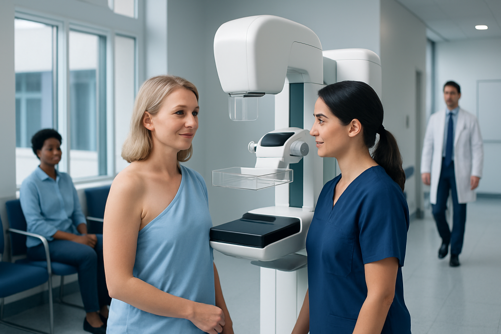
Mammograms are the main screening test for most women at average risk. Ultrasound is usually an additional test, not a replacement for mammography.
Key Takeaways
- Mammograms are the main screening test for most women at average risk.
- Ultrasound is usually an additional test, not a replacement for mammography.
- Women with dense breasts or higher cancer risk may need extra imaging or earlier screening.
- Any new breast lump, nipple discharge, or skin change should be assessed by a doctor, even after a normal screening test.
- Screening plans are most effective when tailored to personal and family history.
Breast cancer screening in women helps find cancer early, often before symptoms appear. The right test and timing depend on age, breast density, personal risk, and whether there are any breast symptoms.
Overview: what breast cancer screening is and why it matters
Breast cancer screening means checking the breasts for cancer before symptoms develop. The aim is to find cancer at an earlier stage, when treatment may be simpler and outcomes are often better. Screening is different from diagnostic testing, which is done when a woman has a symptom such as a lump, nipple discharge, or a change in breast skin.
For most women, the main screening test is a mammogram. A mammogram is a low-dose X-ray of the breast that can detect small changes that may not be felt on examination. In some situations, a breast ultrasound or breast MRI may be added to give more information, especially if the breasts are dense or if the woman has a higher-than-average risk of breast cancer.
There is no single screening schedule that fits every woman. Doctors consider age, family history, genetic risk, past breast conditions, breast density, and overall health when deciding which test is most suitable and how often it should be done. A personalized plan helps balance the benefits of early detection with the possibility of false alarms or unnecessary follow-up tests.
Mammograms: the main screening test for most women
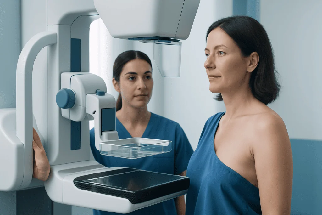
Mammography is the standard first-line screening tool for women at average risk of breast cancer. During the test, each breast is gently compressed for a few seconds to create clear images. This compression can be uncomfortable, but it is brief and helps improve image quality while keeping radiation exposure low.
A mammogram can show tiny calcium deposits, masses, or subtle distortions in breast tissue that may need closer review. Many findings on screening mammograms are not cancer, but they may lead to additional images or short-term follow-up. If the result is suspicious, the next steps may include targeted ultrasound, diagnostic mammography, or a biopsy.
Modern digital mammography, and in many centers 3D mammography called tomosynthesis, can improve visibility in certain women and reduce the chance of being called back for extra images. Even so, mammograms are not perfect. They can miss some cancers, especially in dense breast tissue, which is why some women need supplemental imaging.
Screening recommendations differ slightly between expert groups, but many women begin regular mammography in midlife. A doctor can help decide when to start, whether screening should be yearly or every two years, and when it may be reasonable to stop based on age, health status, and personal preferences.
When ultrasound is used and who may need it
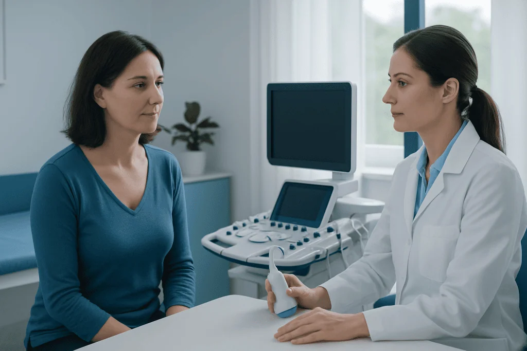
Breast ultrasound uses sound waves rather than radiation to create images of breast tissue. It is not usually the primary screening test for women at average risk, because it may miss some early cancers that mammography can detect, including certain calcifications. However, it is very useful as an additional test in the right setting.
Ultrasound is commonly used to evaluate a specific area seen on a mammogram or felt during an exam. It can help tell whether a breast lump looks like a fluid-filled cyst or a solid mass. It is also often helpful in women with dense breasts, where mammograms can be harder to interpret because both dense tissue and tumors can appear white on the image.
Some women may have ultrasound as a supplemental screening tool, especially if they have dense breasts and cannot have MRI, or if a doctor believes more imaging is needed. It is also widely used in younger women with breast symptoms and in pregnancy, when avoiding radiation is preferred. If needed, doctors may recommend breast ultrasound alongside mammography rather than instead of it.
Because ultrasound can find areas that turn out to be harmless, it may increase follow-up testing and biopsies. For that reason, it is best used selectively and as part of an individualized screening or diagnostic plan.
Who needs which test: age, breast density, and personal risk
The choice of screening test depends first on whether a woman is at average or increased risk. Average risk generally means no personal history of breast cancer, no known inherited cancer gene mutation, no strong family history, and no prior chest radiation at a young age. These women are usually screened with regular mammograms, with the timing decided in discussion with their doctor.
Women at increased risk may need screening to begin earlier and may need more than one imaging method. Higher risk can be related to BRCA1 or BRCA2 mutations, a strong family history, certain inherited syndromes, previous chest radiation, or some high-risk biopsy findings. In these cases, MRI may be added because it can detect some cancers not seen on mammography. Mammography remains important in many of these women as well.
Breast density also matters. Dense breasts are common and are not abnormal, but they can make mammograms harder to read. Dense tissue may also be linked with a somewhat higher risk of breast cancer. Women who are told they have dense breasts should ask whether additional imaging such as ultrasound or MRI might be helpful in their situation.
Age influences test choice too. Younger women with a new symptom often have ultrasound first because their breast tissue is usually denser, while women in standard screening age groups are generally evaluated with mammography. If a woman has a known breast condition or previous cancer, her doctor may recommend a tailored follow-up plan related to breast cancer.
What happens during diagnosis after an abnormal screening result
An abnormal screening result does not automatically mean cancer. It means that the images showed something that needs a closer look. The next step is often diagnostic mammography with extra views, targeted ultrasound, or both. These tests help radiologists study the area in more detail.
If the finding still looks uncertain or suspicious, a biopsy may be recommended. A biopsy removes a small tissue sample so it can be checked under a microscope. Many biopsies are done with a needle and image guidance, such as ultrasound guidance, and do not require major surgery.
In some situations, breast MRI is used to better define a finding or to assess the extent of disease once cancer is diagnosed. MRI is highly sensitive, but it can also detect noncancerous changes, so it is usually reserved for selected cases rather than routine screening in average-risk women. If recommended, advanced imaging such as breast MRI can provide additional detail.
Waiting for follow-up results can feel stressful, but most recalls after screening do not lead to a cancer diagnosis. It helps to ask the care team what the finding means, which test is being recommended next, and when results will be available.
Benefits, limits, and possible downsides of screening
The main benefit of breast screening is earlier detection. Cancers found on screening may be smaller and may not yet have caused symptoms, which can open the door to more treatment choices. Screening also helps identify some noncancerous breast changes that may need monitoring or reassurance.
At the same time, screening has limits. A mammogram can miss a cancer, especially in dense breasts. Screening can also find changes that look concerning but are not harmful. This can lead to extra imaging, short-interval follow-up, or biopsies that ultimately show benign results.
Another possible downside is overdiagnosis, meaning detection of some cancers that may never have caused illness during a woman’s lifetime. Because doctors cannot always know which findings will become dangerous, they investigate and treat cancers that are discovered. This is one reason screening decisions should reflect personal risk and values as well as general recommendations.
Understanding both the benefits and the trade-offs can help women feel more confident in their choices. A clear conversation with a doctor or breast imaging specialist is often the best way to decide on the most appropriate screening plan.
Prevention, self-awareness, and practical screening tips
Screening does not prevent breast cancer, but it supports early detection. Risk can sometimes be lowered by healthy lifestyle choices such as limiting alcohol, staying physically active, maintaining a healthy weight after menopause, and discussing hormone therapy carefully with a doctor. These steps cannot eliminate risk, but they support overall breast and general health.
Breast self-awareness is also important. Women do not need to perform a formal monthly self-exam in exactly the same way each time, but they should know what is normal for their own breasts. A new lump, thickening, nipple inversion, bloody discharge, persistent focal pain, or skin dimpling should be evaluated even if screening is up to date.
Practical steps can make screening easier. It may help to schedule a mammogram when the breasts are less tender, bring previous imaging records if changing centers, and tell the imaging team about implants, pregnancy, breastfeeding, or current symptoms. Women should also ask when and how they will receive results, and whether breast density was noted on the report.
Near the end of the screening pathway, some women may need additional procedures for diagnosis or treatment planning. In experienced centers, services such as mammography and multidisciplinary review can help guide care. Acibadem International’s multidisciplinary specialists and JCI-accredited hospitals diagnose and treat breast conditions for international patients.
When to see a doctor
A woman should see a doctor promptly if she notices a new breast lump, nipple discharge especially if it is bloody, skin thickening or dimpling, swelling, redness that does not improve, or a change in the size or shape of one breast. Persistent pain in one specific area can also deserve evaluation, even though breast pain alone is often not caused by cancer.
Medical advice is also important for women with a strong family history of breast or ovarian cancer, known genetic mutations, previous chest radiation, or earlier high-risk breast biopsy results. These factors may change the recommended age to start screening and the type of imaging used.
Even after a normal mammogram, new symptoms should not be ignored. Screening reduces risk but does not detect every cancer. A clinician can decide whether targeted imaging, a specialist consultation, or further assessment is needed.
Frequently asked questions
What is the best screening test for breast cancer in women?
For most women at average risk, a mammogram is the main and most established screening test. Other tests such as ultrasound or MRI may be added when breast density is high, symptoms are present, or overall risk is increased.
Can ultrasound replace a mammogram?
Usually no. Ultrasound is often used as an additional test because it helps examine specific areas and can be useful in dense breasts, but it does not replace the role of mammography for routine screening in most women.
Do women with dense breasts need extra screening?
Some may benefit from extra imaging, but not everyone needs the same approach. The decision depends on how dense the breasts are, the woman’s age, personal risk factors, and local screening recommendations.
At what age should breast cancer screening start?
The starting age varies among expert guidelines and depends on personal risk. Many women begin regular mammograms in midlife, while women at higher risk may need to start earlier and may need MRI in addition to mammography.
What happens if a mammogram is abnormal?
An abnormal result usually means more imaging is needed, not that cancer is certain. The next steps may include extra mammogram views, ultrasound, and sometimes a biopsy to clarify the finding.
Should women still be checked if they have no symptoms?
Yes. Screening is designed for women who feel well and have no breast symptoms, because some cancers can be found before they are noticeable. This is one of the main reasons screening can be helpful.
References
- World Health Organization
- American Cancer Society
- U.S. Preventive Services Task Force
- American College of Radiology
- National Cancer Institute
This article is for general information only and is not a substitute for professional medical advice. Please consult a qualified doctor about your individual situation.
Breast Cancer in Turkey — costs, top hospitals & a free quote
JCI-accredited · board-certified surgeons · reply within 24h
Add us as a Preferred Source to see more of our trusted health content across Google Search, AI Overviews and Discover.

Medical Oncology Department
Medical treatment of cancer with chemotherapy, immunotherapy and targeted therapies under a multidisciplinary tumor board.
60 specialists in this unit