

Breast Dense Tissue Ultrasound: Preparation, Procedure and Results

Dense breast tissue is common and can make some abnormalities less visible on mammograms. Ultrasound does not use ionizing radiation and is usually painless, noninvasive, and completed in a short outpatient visit.
Key Takeaways
- Dense breast tissue is common and can make some abnormalities less visible on mammograms.
- Ultrasound does not use ionizing radiation and is usually painless, noninvasive, and completed in a short outpatient visit.
- A breast ultrasound may be used after a mammogram, to assess a lump or symptom, or as supplemental imaging for selected patients.
- Most people can return to normal activities immediately after the examination.
- An ultrasound finding may be benign, may need follow-up imaging, or may occasionally require a biopsy for a definite diagnosis.
Breast dense tissue ultrasound is an imaging examination that uses sound waves to look at breast tissue that may be harder to assess on a mammogram. It can help clarify a specific area or provide additional information in selected people with dense breasts, but it does not replace routine mammography unless a clinician advises otherwise.
Overview: What Is a Breast Dense Tissue Ultrasound?
A breast dense tissue ultrasound is an imaging test that uses high-frequency sound waves to create pictures of breast tissue. It is often considered when mammography shows dense breast tissue, when a mammogram finding needs closer assessment, or when a person has a breast symptom such as a new lump. The test can distinguish many fluid-filled cysts from solid areas and can provide detail that complements a mammogram.
Breast density describes the amount of fibrous and glandular tissue compared with fatty tissue on a mammogram. Dense tissue is not something that can be felt by touch and is not determined by breast size or firmness. It is common, especially before menopause, and it can make mammogram images more difficult to interpret because both dense tissue and many breast cancers appear light on a mammogram.
Ultrasound is not automatically needed for every person with dense breasts. It can find some changes that are less visible on mammography, but it can also identify harmless areas that lead to additional scans or biopsy. A radiologist and the person’s clinician can decide whether supplemental imaging is appropriate based on breast density, personal risk factors, symptoms, and previous imaging results.
How Breast Ultrasound Works and Who May Benefit
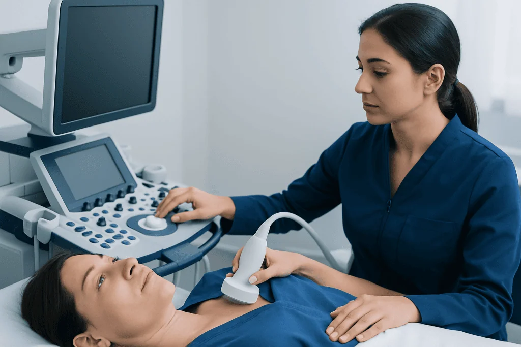
During breast ultrasound, a transducer placed on the skin sends sound waves into the breast. The waves reflect differently from fluid, fat, glands, and solid tissue. A computer converts these echoes into real-time images that a radiologist can review. Unlike mammography, ultrasound does not use X-rays or compress the breast.
A clinician may recommend a breast dense tissue ultrasound after an abnormal or incomplete mammogram, to investigate a localized concern, or to evaluate an area that can be felt during an examination. It is also commonly used to guide a needle during a breast biopsy when tissue sampling is needed.
Supplemental ultrasound may be discussed for some people with dense breasts, particularly when other factors raise breast cancer risk. These can include a strong family history, a known inherited genetic variant, prior chest radiation, certain prior breast biopsy findings, or a personal history of breast cancer. In higher-risk situations, breast MRI may also be considered; the most suitable plan should be individualized rather than based on density alone.
- Ultrasound can be useful for assessing a lump, focal pain, nipple changes, or an area seen on mammography.
- It can help characterize whether an area appears cystic or solid.
- It may provide additional information when dense tissue limits mammographic visibility.
- It cannot reliably detect every breast cancer and remains complementary to recommended mammography screening.
Preparing for the Examination

There is usually very little preparation for a breast ultrasound. People can normally eat, drink, and take their regular medications unless their healthcare team gives different instructions. It is helpful to bring previous mammogram, ultrasound, MRI, or biopsy records if they were performed at another imaging center, as comparisons with earlier images can be important.
On the day of the appointment, it is best to avoid applying lotion, powder, deodorant, or perfume to the breast and underarm area if possible. These products do not usually prevent ultrasound, but avoiding them may make the imaging visit simpler if mammography or another test is also planned. Wearing a two-piece outfit can make changing more convenient.
People should tell the imaging team about a new lump, skin change, nipple discharge, prior breast surgery, implants, pregnancy, breastfeeding, or any recent breast injury. They should also mention where symptoms are located and when they began. This information helps the technologist and radiologist focus the examination appropriately.
Step by Step: What Happens During the Procedure
A breast ultrasound is usually performed in an outpatient imaging department. After changing from the waist up, the person lies on an examination table, commonly with one arm raised above the head. A trained sonographer or radiologist applies a clear, water-based gel to the skin. The gel helps the transducer maintain contact and allows sound waves to travel effectively.
The transducer is moved gently across the breast and, when needed, the underarm area. The examination is generally comfortable, although light pressure over a tender area may cause temporary discomfort. Images are taken from multiple angles. If the ultrasound is targeted to a specific concern, the person may be asked to point to the exact location of a lump or other symptom.
The scan often takes about 15 to 30 minutes, although timing can vary depending on whether one or both breasts are examined and whether additional images are needed. In many centers, a radiologist reviews the images before the visit is complete. This may mean the technologist returns to obtain additional views, which is a routine part of obtaining clear diagnostic information.
If an area requires tissue sampling, the clinician may discuss an image-guided biopsy, which is a separate procedure or may be scheduled for another day. Image-guided breast biopsy can provide the tissue diagnosis needed when imaging alone cannot determine the nature of a finding.
Results, Benefits, Limits and Possible Risks
Results are commonly reported using the Breast Imaging Reporting and Data System, often called BI-RADS. A result may indicate no concerning finding, a benign condition, a probably benign area that should be rechecked after a defined interval, a suspicious finding requiring biopsy, or a known cancer. The report should be interpreted alongside mammography, symptoms, medical history, and clinical examination.
Possible ultrasound findings include a simple cyst, a fibroadenoma, normal dense glandular tissue, an inflamed area, or a solid mass that needs further evaluation. A result that recommends more imaging or a biopsy does not mean cancer is present. Follow-up is advised because imaging can identify changes that cannot be classified with certainty from one examination alone.
The main benefits of breast ultrasound are that it uses no radiation, provides real-time images, and can offer useful detail in dense tissue or in a focused area of concern. It can also guide procedures accurately. However, ultrasound may detect harmless changes that prompt further testing, and it can miss some abnormalities, including certain calcifications more readily seen on mammography.
The procedure itself has no known significant risks from diagnostic ultrasound. There are no injections, no radiation exposure, and no recovery period. Any risks associated with a subsequent biopsy depend on that procedure and will be discussed before it is performed.
Recovery, Follow-Up and Ongoing Breast Care
There is no recovery time after a diagnostic breast ultrasound. The gel is wiped away, and most people return to work, exercise, driving, and other usual activities immediately. No bandage, dietary restriction, or medication change is typically needed.
The next step depends on the imaging result. If the examination is normal or benign, the clinician may recommend continuing routine screening at the usual interval. If the report recommends short-term follow-up, it is important to arrange it on time, even when the finding is likely benign. If biopsy is recommended, timely discussion with a breast specialist can clarify the reason, process, and expected results timeline.
Breast health plans are individual. People should continue screening according to their clinician’s advice and local guidelines, and they should ask how breast density affects their own screening strategy. In some cases, other imaging such as breast MRI may be appropriate, particularly for people with a higher-than-average risk of breast cancer.
Acibadem International’s multidisciplinary breast care specialists and JCI-accredited hospitals assess breast concerns and coordinate imaging, biopsy, and treatment planning for international patients when needed.
When to Seek Medical Care
A person should arrange medical assessment for a new breast lump, a persistent area of thickening, skin dimpling, unexplained breast swelling, new nipple inversion, bloody or spontaneous nipple discharge, or changes that do not resolve after a menstrual cycle. Breast pain is common and is often not caused by cancer, but persistent focal pain should also be discussed with a clinician.
Prompt assessment is especially important for a new change in someone with a personal history of breast cancer, a strong family history, or a known high-risk genetic condition. A normal recent mammogram should not delay evaluation of a new persistent symptom, because no screening test detects every abnormality.
Urgent medical attention is appropriate if breast redness, warmth, swelling, fever, or severe pain develops, particularly during breastfeeding, because infection may need treatment. For non-urgent concerns, a primary care clinician, gynecologist, or breast clinic can determine whether clinical examination, diagnostic mammography, ultrasound, or another test is indicated.
Frequently asked questions
Does having dense breasts mean a person has breast cancer?
No. Dense breast tissue is common and is not cancer. It can slightly increase breast cancer risk and may make cancers harder to see on mammography, which is why individualized screening discussions are useful.
Is breast dense tissue ultrasound painful?
The examination is usually painless. Some gentle pressure is applied with the ultrasound probe, and this may feel uncomfortable if the breast is already tender or if a painful area is being examined.
Can ultrasound replace a mammogram for dense breasts?
Usually, no. Ultrasound and mammography provide different information, and ultrasound does not reliably identify all findings that mammography can show, including many calcifications. A clinician can advise whether ultrasound should complement mammography in an individual screening plan.
How soon are breast ultrasound results available?
Timing varies by imaging center. A radiologist often reviews diagnostic images on the day of the scan, while the formal report may be sent to the referring clinician soon afterward. The imaging team can explain when and how results will be communicated.
What does a cyst on breast ultrasound mean?
A simple cyst is a fluid-filled sac and is usually benign. Depending on its appearance and whether it causes symptoms, no treatment may be needed, although the clinician may recommend follow-up or further assessment for more complex findings.
Why might a biopsy be recommended after an ultrasound?
A biopsy may be recommended when an area has features that cannot be confirmed as benign on imaging alone. It is the most reliable way to examine cells or tissue under a microscope and determine the diagnosis.
References
- American College of Radiology
- American Cancer Society
- U.S. Food and Drug Administration
- National Cancer Institute
- Society of Breast Imaging
This article is for general information only and is not a substitute for professional medical advice. Please consult a qualified doctor about your individual situation.
Ultrasound in Turkey — costs, top hospitals & a free quote
JCI-accredited · board-certified surgeons · reply within 24h
Add us as a Preferred Source to see more of our trusted health content across Google Search, AI Overviews and Discover.

Check-up & Preventive Medicine
Comprehensive health screenings designed for early detection and prevention.
1 specialists in this unitMore from the Health Library


Related Specialists

Dr. Civan Kalafat
Gynecology & Obstetrics
Assoc. Prof. Dr. Yetkin Zeki Yılmaz
Otorhinolaryngology
Pdg. Gülşah Kızıl
Podiatry & Foot Health
Assoc. Prof. Dr. Sevgi Topal
Pediatric Intensive Care Unit