Breast Ultrasound Sonogram Explained: Causes, Management, and When to See a Doctor

A breast ultrasound sonogram uses sound waves, not radiation, to create images of breast tissue. It is often used to evaluate a lump, focal pain, nipple discharge, or an abnormal area seen on another imaging test.
Key Takeaways
- A breast ultrasound sonogram uses sound waves, not radiation, to create images of breast tissue.
- It is often used to evaluate a lump, focal pain, nipple discharge, or an abnormal area seen on another imaging test.
- Ultrasound can help distinguish cysts from solid masses, but it does not by itself diagnose cancer.
- Results may be normal, clearly benign, or may lead to follow-up imaging or a biopsy.
- The test is painless for most people, usually quick, and requires little preparation.
- New or changing breast symptoms should be assessed by a qualified doctor rather than self-diagnosed.
A breast ultrasound sonogram is an imaging test that uses sound waves to look at breast tissue, often to assess a lump, pain, or a finding seen on mammography or MRI. It does not use radiation and is commonly used to help tell whether an area looks fluid-filled, solid, or needs further evaluation.
Overview: What a Breast Ultrasound Sonogram Is
A breast ultrasound sonogram is a medical imaging test that uses high-frequency sound waves to produce pictures of the inside of the breast. It is commonly ordered when a person or clinician feels a lump, when there is breast pain in one spot, or when another test such as a mammogram shows an area that needs a closer look. Because it uses sound waves instead of X-rays, it does not expose the patient to radiation.
This test is especially helpful for looking at differences in breast tissue structure. For example, it can often show whether a lump is a simple fluid-filled cyst or a solid area that needs more attention. In people with dense breast tissue, ultrasound may provide added information alongside mammography, but it is not usually a complete replacement for routine screening mammograms.
A breast ultrasound sonogram can be performed as a focused test on one area of concern or as a more complete examination of one or both breasts. It may also be used to examine the underarm area if lymph nodes need evaluation. In some cases, ultrasound is used to help guide procedures such as needle biopsy or cyst drainage with precision.
Why a Doctor May Recommend It
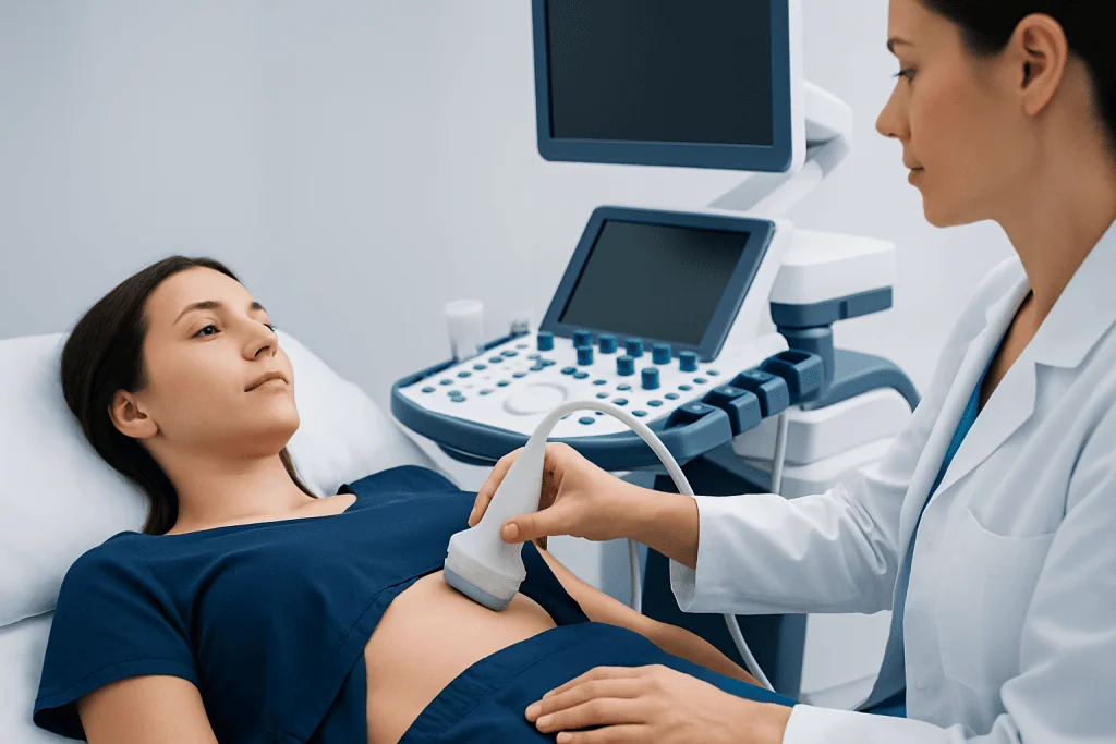
Doctors recommend a breast ultrasound sonogram for several practical reasons. One of the most common is to assess a breast lump found during self-awareness, a clinical breast exam, or on routine imaging. Ultrasound is also useful for checking focal breast pain, nipple discharge, skin thickening, swelling, or changes in the underarm area.
The test is often used as a follow-up rather than a stand-alone answer. For example, if a mammogram identifies a shadow, asymmetry, or mass, ultrasound may help clarify what that area represents. It may also be chosen for younger patients, pregnant patients, or breastfeeding patients when avoiding radiation is preferred or when breast tissue is naturally denser.
Ultrasound can support the evaluation of a wide range of breast conditions, from harmless cysts to infections and suspicious masses. If symptoms suggest a benign breast problem such as a breast cyst, ultrasound can be one of the most useful first tests. If imaging shows an area that cannot be confidently labeled benign, the next step may involve additional imaging or a breast biopsy to confirm the diagnosis.
What Symptoms or Findings It Can Help Assess
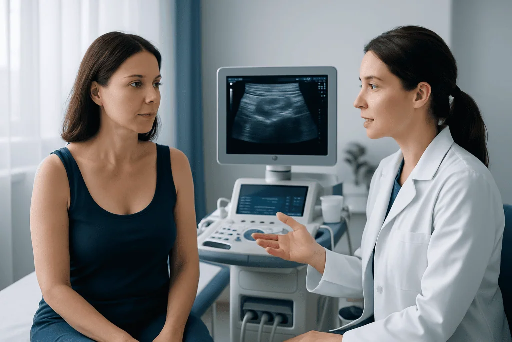
A breast ultrasound sonogram does not treat symptoms, but it can help explain them. It is commonly used when someone notices a new lump, thickening, or a firm area in the breast. It may also be helpful when there is tenderness in a specific location, especially if the discomfort is new, one-sided, or linked to a physical finding.
Other reasons for ultrasound include nipple discharge, redness, swelling, or skin changes over the breast. During breastfeeding, ultrasound can help evaluate blocked ducts, fluid collections, or signs of infection such as mastitis. It can also be used if a clinician wants to look at enlarged lymph nodes in the armpit.
Still, not every breast symptom requires ultrasound, and not every ultrasound finding is serious. Many causes of breast concerns are benign, including cysts, fibroadenomas, and hormone-related changes. The purpose of the sonogram is to add clarity and help decide whether reassurance, follow-up, or further testing is most appropriate.
- New breast lump or thickened area
- Focal breast pain
- Nipple discharge
- Abnormal mammogram or MRI finding
- Skin redness or swelling
- Assessment of lymph nodes or guidance for procedures
How the Test Is Done and How to Prepare
A breast ultrasound sonogram is usually performed in an imaging center, radiology department, or breast clinic. During the test, the patient lies on an examination table, often with one arm raised. A clear gel is placed on the skin, and a handheld device called a transducer is moved over the breast to collect images. The gel helps the sound waves travel effectively.
The examination is generally well tolerated and usually takes a short time, though timing can vary depending on whether one targeted area or a broader exam is needed. Most people feel little more than mild pressure from the transducer. If the breast is already sore, some temporary discomfort can occur, but the test itself is not typically painful.
Preparation is simple. Patients are often advised not to apply powders, lotions, or deodorants on the chest or underarm area on the day of the test, because these can interfere with imaging in some settings. Wearing a two-piece outfit may be more convenient. Bringing prior breast imaging results can also help the radiologist compare findings over time.
If an abnormal area is seen, the same ultrasound session may help plan the next step. In some cases, a doctor may recommend image-guided sampling using ultrasound-guided biopsy so the suspicious tissue can be examined in the laboratory.
Understanding the Results
Breast ultrasound results are interpreted by a radiologist, who looks at the shape, borders, internal pattern, and behavior of a finding on the scan. Some results are clearly benign. A simple cyst, for example, usually has a very characteristic appearance and may need no treatment unless it is painful or large. Other findings may look probably benign and only require repeat imaging after a period of observation.
When a mass appears solid, irregular, or otherwise suspicious, ultrasound alone cannot confirm whether it is cancerous. In that situation, the radiologist may recommend a biopsy. This does not mean cancer is certain; it means tissue analysis is the safest way to make a definite diagnosis. Doctors may also coordinate ultrasound findings with mammography, breast MRI, physical examination, and medical history.
Results are often reported using standardized language to help guide next steps. Patients may hear terms such as benign, probably benign, suspicious, or incomplete assessment. If a report is unclear, asking the referring doctor to explain what was seen, what level of concern exists, and whether follow-up is needed can be very helpful.
If a concerning lesion is identified, the care team may discuss further evaluation in a dedicated breast cancer pathway. This can include biopsy, staging tests if appropriate, and consultation with breast specialists.
Management and Treatment After an Ultrasound
Management after a breast ultrasound sonogram depends entirely on what the test shows and on the patient’s symptoms. If the scan is normal and the clinical exam is reassuring, the doctor may simply advise observation and routine age-appropriate screening. If a benign cyst is found, no treatment may be needed unless it causes discomfort, in which case drainage may be considered.
Solid but likely benign lesions, such as some fibroadenomas, may be followed with repeat imaging to confirm stability. If there are signs of infection, treatment may include antibiotics and close follow-up. If a milk-filled cyst or fluid collection is present during breastfeeding, ultrasound can help guide aspiration when necessary.
When ultrasound findings are uncertain or suspicious, further testing is often the next step. This may include biopsy, mammography, MRI, or consultation with a breast surgeon or breast radiologist. The goal is not to over-treat, but to make sure important conditions are not missed while avoiding unnecessary procedures whenever possible.
For international patients who need coordinated evaluation, Acibadem International’s multidisciplinary specialists and JCI-accredited hospitals diagnose and treat breast conditions using modern imaging and pathology pathways. Care plans are individualized based on symptoms, imaging findings, age, and overall health.
Limitations, Prevention, and Breast Self-care
A breast ultrasound sonogram is very useful, but it has limits. It can miss certain abnormalities that mammography is better at detecting, especially tiny calcium deposits known as microcalcifications. It is also more operator-dependent than some other imaging methods, meaning image quality and interpretation rely on skilled technique and expert review.
Because of these limits, ultrasound is usually one part of breast care rather than the only test. Routine screening recommendations vary by age, personal history, family history, breast density, and risk level. A doctor can advise whether a person needs mammography alone, added ultrasound, MRI, or a high-risk screening plan.
There is no guaranteed way to prevent all breast conditions, but practical self-care can support breast health. This includes being familiar with what is normal for the breasts, attending recommended screening visits, reporting new changes promptly, and discussing personal risk factors with a clinician. General healthy habits such as regular physical activity, limiting alcohol, and maintaining a healthy weight may also support overall breast health.
- Keep up with recommended breast screening
- Report new lumps, skin changes, or nipple discharge
- Discuss family history and personal risk factors with a doctor
- Do not rely on ultrasound alone for routine screening unless specifically advised
When to Seek Medical Care
Medical care should be sought promptly for any new breast lump, a persistent area of thickening, or a breast change that does not go away after one menstrual cycle. A doctor should also assess nipple discharge that is spontaneous, bloody, or from one breast only, as well as dimpling, redness, swelling, or changes in the nipple or breast skin.
More urgent evaluation is appropriate if breast symptoms are accompanied by fever, significant redness, rapidly increasing swelling, or severe pain, since these may suggest infection or an abscess. People who are pregnant, breastfeeding, have a strong family history of breast cancer, or have had prior breast cancer should not ignore new symptoms.
Even when symptoms seem minor, it is safest not to guess the cause at home. Many breast changes are benign, but only a qualified clinician can determine whether reassurance, imaging, or further testing is needed. Early assessment often leads to clearer answers and more appropriate care.
Frequently asked questions
What is a breast ultrasound sonogram used for?
A breast ultrasound sonogram is used to look more closely at breast tissue when there is a lump, pain, nipple discharge, or an abnormality on another imaging test. It helps show whether an area is fluid-filled, solid, or in need of further evaluation.
Can a breast ultrasound sonogram detect cancer?
It can detect areas that look suspicious and may need more testing, but it does not diagnose cancer by itself. If the appearance is concerning, a biopsy is usually needed to confirm what the tissue is.
Is a breast ultrasound sonogram better than a mammogram?
They serve different purposes and often work best together. Mammography is the standard screening test for many adults, while ultrasound is commonly used to clarify specific findings or evaluate symptoms, especially in dense breast tissue.
Does a breast ultrasound sonogram use radiation?
No. It uses sound waves to create images, so there is no ionizing radiation exposure during the test. This is one reason it is often chosen in pregnancy or for younger patients when appropriate.
How should someone prepare for a breast ultrasound?
Preparation is usually minimal. Patients may be asked to avoid lotions, powders, or deodorant on the chest and underarm area and to wear clothing that is easy to remove from the waist up.
What happens if the ultrasound shows a cyst?
Many simple cysts are harmless and do not need treatment if they are not causing symptoms. If a cyst is painful, large, or uncertain in appearance, the doctor may suggest monitoring, drainage, or further assessment.
When should someone see a doctor for breast symptoms?
A doctor should evaluate any new lump, persistent focal pain, unusual nipple discharge, or visible change in the breast or nipple. Symptoms with fever, marked redness, or rapidly worsening swelling should be assessed more urgently.
References
- American College of Radiology
- Radiological Society of North America
- National Cancer Institute
- American Cancer Society
- World Health Organization
This article is for general information only and is not a substitute for professional medical advice. Please consult a qualified doctor about your individual situation.
Ultrasound in Turkey — costs, top hospitals & a free quote
JCI-accredited · board-certified surgeons · reply within 24h
Add us as a Preferred Source to see more of our trusted health content across Google Search, AI Overviews and Discover.

Check-up & Preventive Medicine
Comprehensive health screenings designed for early detection and prevention.
3 specialists in this unitMore from the Health Library



Related Specialists

Prof. Dr. Ahmet Alponat
General Surgery
Dr. Oğuz Aslan
Radiology
Dr. Melisa Ulufi
Inpatient Clinic Physicians Clinical Service
Dr. Munara Bolatkhan
Inpatient Clinic Physicians Clinical Service