Can Prostate Cancer Be Detected by Ultrasound: Preparation, Procedure and Results

Ultrasound is useful for viewing the prostate and guiding biopsy needles, but it does not diagnose prostate cancer by itself. Many prostate cancers are not clearly visible on standard ultrasound, while non-cancerous conditions can also look abnormal.
Key Takeaways
- Ultrasound is useful for viewing the prostate and guiding biopsy needles, but it does not diagnose prostate cancer by itself.
- Many prostate cancers are not clearly visible on standard ultrasound, while non-cancerous conditions can also look abnormal.
- A raised or changing PSA level, an abnormal digital rectal examination, or suspicious MRI findings may lead to further assessment.
- Biopsy is the only reliable way to confirm prostate cancer by examining prostate tissue under a microscope.
- Transrectal and transperineal ultrasound procedures are usually brief, with mild temporary discomfort or bleeding possible afterward.
- Prompt medical review is important for urinary changes, blood in urine or semen, persistent bone pain, or unexplained weight loss.
Can prostate cancer be detected by ultrasound? Ultrasound can show changes in the prostate and help guide a biopsy, but it cannot confirm or rule out prostate cancer on its own. Diagnosis usually combines symptoms, PSA blood testing, examination, MRI when appropriate, and biopsy results.
Overview: can prostate cancer be detected by ultrasound?
Can prostate cancer be detected by ultrasound? Ultrasound can identify features that may be suspicious and can accurately guide a needle during a prostate biopsy, but it cannot diagnose prostate cancer on its own. A normal ultrasound does not reliably exclude cancer, and an unusual-looking area may be caused by a non-cancerous condition.
Prostate ultrasound uses sound waves to create real-time images of the prostate gland. It may be performed through the rectum, called transrectal ultrasound (TRUS), or through the skin between the scrotum and anus, called transperineal ultrasound. It is often part of an assessment after an elevated prostate-specific antigen (PSA) blood test, a change in PSA over time, an abnormal digital rectal examination, or a concerning MRI result.
The definitive diagnosis comes from pathology: a specialist examines tissue samples taken during a biopsy. Depending on the findings, a urology team may discuss further tests, monitoring, or prostate cancer care options.
What ultrasound shows—and what it cannot show
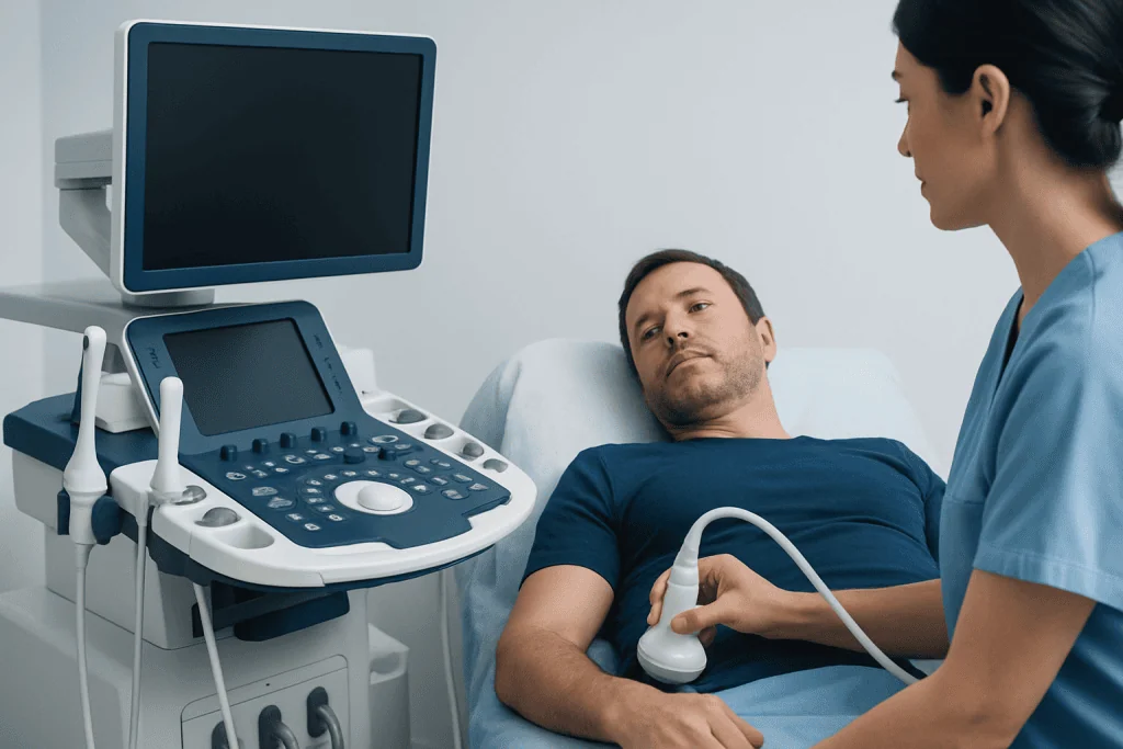
Ultrasound can estimate prostate size, assess its overall shape, and sometimes reveal areas with a different texture or appearance. It can also help identify issues that may contribute to urinary symptoms, such as prostate enlargement, cysts, inflammation, or retained urine in the bladder. These findings are valuable, but they are not specific for cancer.
Some cancers appear as darker areas on transrectal ultrasound, but many do not have a distinctive appearance. Conversely, benign prostatic hyperplasia, prostatitis, scarring, and normal variation can create changes that resemble a suspicious area. For this reason, clinicians interpret ultrasound alongside a person’s age, family history, PSA results, examination findings, and MRI results when indicated.
Multiparametric MRI has become an important test for many people being considered for prostate biopsy because it can better identify areas that may need targeted sampling. Ultrasound remains especially important because it provides real-time guidance during biopsy, including MRI-ultrasound fusion biopsy in suitable cases.
Can you tell prostate cancer from ultrasound?

No. An ultrasound image alone cannot reliably tell whether a prostate abnormality is cancer. It may raise or lower clinical suspicion, but it cannot distinguish every cancer from benign enlargement, inflammation, or other harmless changes.
If the overall assessment suggests a possible cancer, the doctor may recommend an MRI, repeat PSA testing, or a biopsy. During biopsy, ultrasound helps the clinician see the prostate and place the sampling needle in planned locations. In some cases, MRI findings are overlaid on live ultrasound images so that suspicious regions can be sampled more precisely.
Pathology results from biopsy tissue are required to confirm cancer and describe its grade. This information helps the care team determine whether active surveillance, surgery, radiation therapy, hormone therapy, or another approach may be appropriate.
Who may need a prostate ultrasound or biopsy?
A prostate ultrasound is not usually a routine screening test for people without symptoms or risk factors. It is more commonly used when a urologist needs more information after a PSA result, examination, MRI, or symptom review. The decision is individualized because PSA can rise for several reasons other than cancer, including benign enlargement, inflammation, infection, recent ejaculation, and some urinary procedures.
People may be referred for assessment if they have a persistently elevated PSA, a PSA that is rising over time, a firm or irregular area found during a digital rectal examination, or an MRI lesion that needs tissue sampling. Risk may be higher for those with a close relative who has had prostate or certain related cancers, and for people with inherited genetic changes associated with prostate cancer.
Urinary symptoms such as a weak stream, frequency, urgency, or getting up at night are common and often result from benign prostate enlargement rather than cancer. However, they deserve medical assessment when new, worsening, or troublesome. Related conditions, including benign prostatic hyperplasia, can be evaluated at the same time.
How the procedure works: preparation, steps and recovery
Preparation depends on the type of ultrasound and whether a biopsy is planned. For an imaging-only transrectal ultrasound, the clinic may ask the person to use an enema beforehand so the rectum is relatively clear. Before a biopsy, the team reviews medicines, allergies, bleeding risks, and any history of urinary infection. Blood-thinning medication should never be stopped without instructions from the prescribing clinician and procedural team.
For transrectal ultrasound, the person usually lies on their side with knees bent. A slim, lubricated probe is gently inserted into the rectum and images are obtained in a few minutes. For a transperineal approach, imaging and biopsy are performed through the skin between the scrotum and anus, commonly using local anesthesia, sedation, or general anesthesia depending on the planned procedure and local practice.
When biopsy is needed, ultrasound guides a spring-loaded needle to collect small tissue cores. The procedure itself is usually short, although check-in, preparation, anesthesia, and recovery take longer. Many people return home the same day. Mild soreness, a small amount of blood in urine or semen, and light rectal bleeding after a transrectal procedure can occur temporarily; the care team provides individualized aftercare instructions.
For people advised to have tissue sampling, prostate biopsy information can help explain the evaluation process and expected preparation. Results are often discussed at a follow-up appointment after pathology has reviewed the samples.
How effective is ultrasound for prostate cancer?
Ultrasound is highly effective as a practical, real-time tool for measuring the prostate and guiding biopsy safely and accurately. Its effectiveness as a stand-alone test for finding or excluding prostate cancer is limited, because some cancers are invisible or appear similar to non-cancerous tissue on ultrasound.
Its value is strongest when used as one part of a structured diagnostic pathway. PSA testing and digital rectal examination can identify people who may need further investigation. Multiparametric MRI may help locate suspicious areas and reduce unnecessary biopsy in some patients, while ultrasound then guides systematic or MRI-targeted tissue sampling.
Biopsy can still miss a small cancer, particularly when a lesion is difficult to reach or cancer is scattered in small areas. If concern remains despite negative biopsy results, the clinician may recommend follow-up PSA monitoring, repeat MRI, or repeat biopsy based on the individual situation.
What is the first hint of prostate cancer?
Early prostate cancer often causes no symptoms. The first hint may be an unexpected PSA result, a PSA level that rises over time, or an abnormal area found during a digital rectal examination. These findings do not prove cancer, but they can prompt further testing.
When symptoms occur, they may include difficulty starting urination, a weaker stream, frequent urination, urgency, or waking at night to pass urine. Because these symptoms are more commonly linked to benign enlargement or other urinary conditions, they should not be used to diagnose cancer without appropriate assessment.
Less commonly, blood in urine or semen, persistent pain in the back, hips, or bones, unintentional weight loss, or worsening fatigue can require prompt medical review. These symptoms have many possible causes, but a clinician can assess them carefully and arrange the right tests.
What is the 2 week rule for prostate cancer?
The “2 week rule” is a term commonly used in the United Kingdom for an urgent suspected-cancer referral pathway. It aims for a person with concerning symptoms, examination findings, or test results to be offered a specialist appointment within two weeks of referral. It is an administrative target for timely assessment, not a rule that confirms cancer or predicts how serious a condition is.
Referral criteria and waiting-time standards differ between countries and health systems. A person does not need to wait for a particular time threshold if they have alarming symptoms, such as inability to pass urine, fever with urinary symptoms after a prostate procedure, heavy bleeding, or severe pain; they should seek urgent care according to local medical guidance.
After a specialist review, the next step may be repeat testing, MRI, ultrasound-guided biopsy, or monitoring. The exact sequence depends on risk factors, prior results, clinical examination, and shared decision-making.
When to seek medical care
A person should arrange a routine medical appointment for persistent or worsening urinary symptoms, changes in urine flow, recurrent urinary infections, blood in urine or semen, or concerns about prostate cancer risk. They should also discuss screening and PSA testing with a qualified clinician, particularly if they have a close family history of prostate cancer.
Urgent medical advice is appropriate for inability to urinate, fever or chills with urinary symptoms, severe pelvic pain, or significant bleeding. After a prostate biopsy, fever, worsening pain, difficulty urinating, or feeling generally unwell should be reported promptly because infection and urinary retention need timely assessment.
Acibadem International’s multidisciplinary specialists and JCI-accredited hospitals evaluate prostate concerns and coordinate diagnosis and treatment for international patients. Individual recommendations should always be made after a consultation with an experienced urologist.
Frequently asked questions
Is ultrasound enough to diagnose prostate cancer?
No. Ultrasound can show prostate size and areas that may look unusual, but it cannot confirm whether the finding is cancer. A biopsy, in which tissue is examined by a pathologist, is needed to make a diagnosis.
Can a normal prostate ultrasound rule out cancer?
No. Some prostate cancers are not visible on standard ultrasound, especially when they are small or do not create a clear change in tissue appearance. Doctors consider ultrasound together with PSA results, examination findings, MRI when appropriate, and follow-up assessment.
Is a transrectal prostate ultrasound painful?
Most people describe pressure or mild discomfort rather than significant pain during an imaging-only transrectal ultrasound. If biopsy is performed, local anesthesia, sedation, or another anesthetic plan may be used depending on the approach and individual needs.
How long does it take to recover after a prostate biopsy?
Many people can return home on the day of the procedure and resume light activities within a short period, following their clinician’s advice. Mild soreness and small amounts of blood in urine, semen, or stool may occur temporarily. Fever, chills, worsening pain, heavy bleeding, or inability to urinate require prompt medical attention.
Does a high PSA mean prostate cancer?
Not necessarily. PSA may be elevated because of benign prostate enlargement, inflammation, infection, recent sexual activity, or medical procedures, as well as cancer. A clinician evaluates the PSA level in context and may recommend repeat testing, MRI, or biopsy.
Which is better for prostate cancer, MRI or ultrasound?
MRI and ultrasound have different roles. MRI can help identify areas that are more suspicious for clinically significant cancer, while ultrasound is commonly used for real-time imaging and to guide biopsy. They are often used together rather than as competing tests.
References
- European Association of Urology
- American Urological Association
- National Cancer Institute
- NHS
- Prostate Cancer Foundation
This article is for general information only and is not a substitute for professional medical advice. Please consult a qualified doctor about your individual situation.
Prostate Cancer in Turkey — costs, top hospitals & a free quote
JCI-accredited · board-certified surgeons · reply within 24h
Add us as a Preferred Source to see more of our trusted health content across Google Search, AI Overviews and Discover.

Check-up & Preventive Medicine
Comprehensive health screenings designed for early detection and prevention.
3 specialists in this unitMore from the Health Library



Related Specialists

Dr. Nuri Soysal
Aesthetic Plastic & Reconstructive Surgery
Dr. Begüm Öykü Kesim
Oral & Dental Health
Dr. Sema Çetin
Physical Medicine & Rehabilitation
Dr. Uğur Kaya
Cardiovascular Surgery