

Cancerous Moles: How to Spot Warning Signs Early

Most moles are harmless, but new or changing moles should be checked by a doctor. The ABCDE rule can help identify suspicious features of a mole.
Key Takeaways
- Most moles are harmless, but new or changing moles should be checked by a doctor.
- The ABCDE rule can help identify suspicious features of a mole.
- Bleeding, itching, crusting, or rapid growth are important warning signs.
- People with fair skin, heavy sun exposure, or a family history of melanoma may have higher risk.
- Dermatology review and biopsy are the main ways to confirm whether a mole is cancerous.
Cancerous moles are skin lesions that may develop into or signal melanoma, the most serious type of skin cancer. Knowing what changes to look for can help people seek medical advice early, when treatment is often simpler and more effective.
Overview
Cancerous moles are moles or pigmented skin spots that may represent melanoma or another form of skin cancer. While many moles are completely benign, a small number can become abnormal over time. Melanoma is the main concern because it can grow deeper into the skin and spread if it is not diagnosed early.
Moles are common and can appear anywhere on the body. They may be flat or raised, light brown or dark, and can change slowly with age. A cancerous mole is not always easy to identify at first, which is why regular self-checks and skin examinations are important.
Early detection makes a meaningful difference. When suspicious lesions are found early, treatment is often more straightforward and outcomes are generally better. Learning the warning signs can help people decide when a professional skin assessment is needed.
What Do Cancerous Moles Look Like?
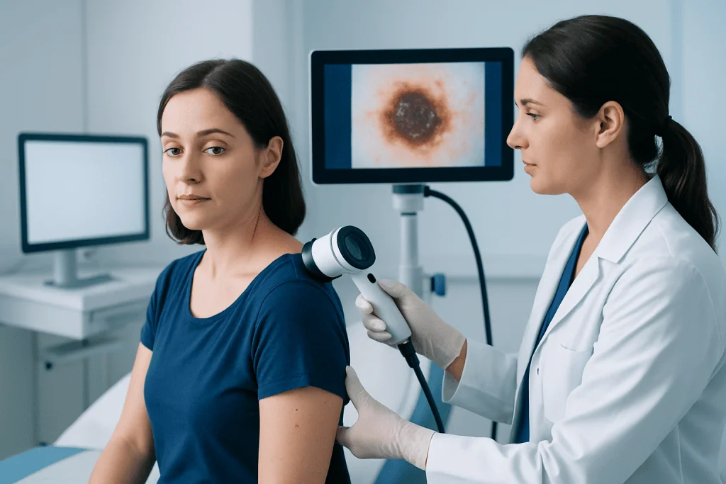
Doctors often use the ABCDE rule to help assess whether a mole may be concerning. This rule does not diagnose melanoma on its own, but it offers a practical way to notice changes that deserve medical attention.
The ABCDE features include:
- A – Asymmetry: one half of the mole does not match the other.
- B – Border: edges are irregular, blurred, notched, or ragged.
- C – Color: the mole has several shades, such as brown, black, red, white, or blue.
- D – Diameter: the spot is larger than about 6 mm, although smaller melanomas can occur.
- E – Evolving: the mole changes in size, shape, color, height, or symptoms.
Another helpful clue is the “ugly duckling” sign. This means a mole looks noticeably different from a person’s other moles. Some melanomas do not fit the usual pattern, so any lesion that stands out, grows quickly, or simply seems unusual should be evaluated.
People should also pay attention to symptoms such as itching, tenderness, bleeding, crusting, or a sore that does not heal. These signs do not always mean cancer, but they do justify a medical review.
Causes and Risk Factors
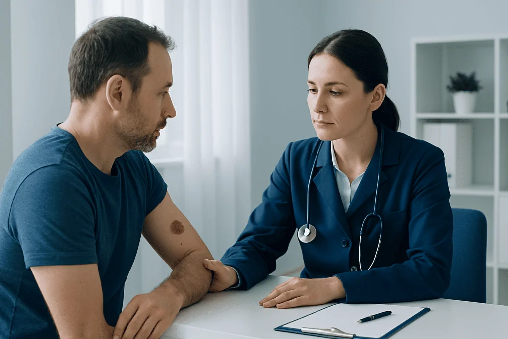
Cancerous moles develop when pigment-producing skin cells, called melanocytes, begin to grow abnormally. This process can be influenced by both environmental exposure and inherited risk. Ultraviolet radiation from the sun or tanning beds is one of the most important contributors.
Several factors can raise the likelihood of melanoma or other suspicious mole changes:
- Fair skin, light eyes, or light hair
- A history of repeated sunburns, especially in childhood
- Frequent or intense sun exposure
- Use of tanning beds
- Having many moles or atypical moles
- A personal or family history of melanoma
- A weakened immune system
Not everyone with these risk factors will develop skin cancer, and melanoma can also occur in people without obvious risk. It may appear on sun-exposed skin, but it can also arise in less expected areas such as the scalp, soles of the feet, under nails, or between the toes.
Because risk varies from person to person, a dermatologist may recommend different screening intervals based on skin type, mole pattern, family history, and previous biopsy results.
How Doctors Diagnose a Suspicious Mole
Diagnosis usually begins with a clinical skin examination. A doctor will ask when the lesion appeared, whether it has changed, and whether there are symptoms such as bleeding or itching. They will also check the rest of the skin, since people can have more than one suspicious lesion.
A dermatoscope, a handheld magnifying tool with specialized light, may be used to examine the mole in greater detail. This can help a dermatologist see pigment patterns and structures that are not visible to the naked eye. Some clinics also use total body photography or digital mole mapping for people with many moles or higher melanoma risk.
If the mole appears suspicious, the doctor usually recommends a biopsy. This means removing all or part of the lesion so it can be examined under a microscope by a pathologist. Biopsy is the only way to confirm whether a mole is cancerous and, if so, what type of skin cancer it is.
When melanoma is diagnosed, the next steps depend on how deep the lesion is and whether there are signs of spread. This evaluation may guide staging and treatment planning for melanoma.
Treatment Options
Treatment depends on the biopsy result. If the mole is benign, no further treatment may be needed beyond observation or routine follow-up. If it shows atypical cells, a doctor may advise complete removal to reduce uncertainty and prevent future problems.
For confirmed melanoma or another skin cancer, surgery is usually the main treatment. The aim is to remove the lesion completely along with a margin of normal-looking skin. In many cases, this can be done with a focused skin cancer treatment plan centered on surgical excision.
If the melanoma is deeper or has features that suggest higher risk, additional tests or procedures may be considered. These can include sentinel lymph node evaluation, imaging in selected cases, or consultation with oncology specialists. More advanced disease may require systemic treatments such as immunotherapy or targeted therapy as part of comprehensive oncology treatment.
Care is individualized according to the type of cancer, its stage, the lesion’s location, and the person’s overall health. In complex cases, dermatology, pathology, surgery, and oncology teams often work together. For international patients, Acibadem International’s multidisciplinary specialists and JCI-accredited hospitals diagnose and treat skin cancers with coordinated care.
Prevention and Self-Care
Not every skin cancer can be prevented, but sun protection can lower risk. Practical steps include using a broad-spectrum sunscreen, seeking shade during peak sunlight hours, wearing protective clothing and a hat, and avoiding tanning beds. Sunscreen should be reapplied as directed, especially after swimming or sweating.
Monthly skin self-exams can help people notice changes earlier. It is helpful to check the entire body in good light, including the scalp, back, buttocks, soles, and between the toes. A mirror or help from a partner can make hidden areas easier to inspect.
People who have many moles may benefit from taking clear reference photos to track change over time. The goal is not to diagnose at home, but to notice anything new, changing, or unusual. Any suspicious finding should be shown to a doctor rather than monitored for long periods without advice.
Those with a past history of skin cancer or very atypical moles may need regular dermatology follow-up. A doctor can advise how often these skin checks should happen and whether more detailed surveillance is appropriate.
When to See a Doctor
A doctor should assess any new mole that appears unusual or any existing mole that changes. The most important warning signs are rapid growth, asymmetry, irregular borders, multiple colors, bleeding, crusting, or persistent itching. A dark streak under a nail or a sore that does not heal also deserves prompt attention.
People sometimes delay evaluation because a mole is painless or because they assume skin cancer only develops after heavy sun exposure. In reality, melanoma can be subtle in the early stages and may occur in places that receive little sun. It is safer to have a suspicious spot examined than to wait for clearer symptoms.
Urgent assessment is especially important for lesions that bleed repeatedly, change quickly over weeks to months, or look very different from other moles. If there is a personal history of skin cancer, even small changes should be discussed with a dermatologist.
Seeking care early does not mean a lesion is definitely cancerous. It simply allows timely examination, biopsy when needed, and reassurance or treatment based on accurate diagnosis.
Frequently asked questions
Are all changing moles cancerous?
No. Many moles change slowly over time for harmless reasons, and some noncancerous skin growths can also look different with age. However, any noticeable change in size, shape, color, or symptoms should be checked by a doctor.
What is the most important warning sign of a cancerous mole?
Change is often the most important clue. A mole that evolves in appearance or starts itching, bleeding, or crusting should be professionally assessed, especially if it looks different from a person’s other moles.
Can melanoma develop in a mole that was normal before?
Yes. Melanoma can arise in an existing mole or appear as a completely new spot. That is why both old and new lesions should be included in regular skin checks.
Do cancerous moles always hurt or itch?
No. Many suspicious moles cause no pain or discomfort at all. Some may itch or bleed, but the absence of symptoms does not rule out melanoma.
How is a cancerous mole confirmed?
A suspicious mole is confirmed with a biopsy. The tissue is examined under a microscope by a pathologist, which shows whether cancer cells are present and helps guide treatment.
Who should have regular skin examinations?
People with fair skin, many moles, atypical moles, a family history of melanoma, heavy sun exposure, or a previous skin cancer often benefit from regular checks. A dermatologist can recommend the right follow-up schedule based on individual risk.
References
- World Health Organization
- American Academy of Dermatology
- National Cancer Institute
- American Cancer Society
- National Health Service
This article is for general information only and is not a substitute for professional medical advice. Please consult a qualified doctor about your individual situation.
Oncology care in Turkey — second opinion and treatment plan
JCI-accredited · board-certified surgeons · reply within 24h
Add us as a Preferred Source to see more of our trusted health content across Google Search, AI Overviews and Discover.

More from the Health Library



Related Specialists

Prof. Dr. Erkan Vardareli
Nuclear Medicine
Prof. Dr. Süleyman Özyalçin
Treatment of Pain Algology
Prof. Dr. Sinan Zeren
Urology
Dr. Gizem Akça
Gynecology & Obstetrics