Carcinoma in Situ: Diagnosis, Outlook, and Modern Treatment Approaches

Carcinoma in situ means abnormal cells are limited to the surface layer of tissue and have not spread into nearby tissue. It can occur in different organs, including the cervix, breast, skin, bladder, and other epithelial tissues.
Key Takeaways
- Carcinoma in situ means abnormal cells are limited to the surface layer of tissue and have not spread into nearby tissue.
- It can occur in different organs, including the cervix, breast, skin, bladder, and other epithelial tissues.
- Many people have no symptoms, so screening tests and follow-up of abnormal results are important.
- Treatment is tailored to the location and extent of the lesion and may include local procedures, surgery, or careful monitoring in selected cases.
- Outlook is often favorable when carcinoma in situ is found and managed before invasion develops.
Carcinoma in situ is an early form of cancer in which abnormal cells remain confined to the tissue where they started and have not invaded deeper layers. It is not all one disease, so diagnosis, treatment, and outlook depend on the organ involved, but timely evaluation often leads to very good outcomes.
What carcinoma in situ means
Carcinoma in situ is a medical term for abnormal cells that look like cancer under the microscope but are still confined to the layer of cells where they began. In simple terms, the cells have not broken through the natural boundary that separates the surface lining from deeper tissue. Because of this, carcinoma in situ is often described as a very early or preinvasive cancer.
This term does not describe one single disease. Instead, it is a pattern that can appear in different organs lined by epithelial cells, such as the cervix, breast ducts, skin, bladder, or parts of the digestive and respiratory tracts. The exact behavior, preferred treatment, and long-term outlook depend on where the abnormal cells are found and how extensive they are.
For patients, one of the most important points is that carcinoma in situ is different from invasive cancer. In invasive cancer, abnormal cells have grown beyond the surface layer and entered surrounding tissue, where they have greater potential to spread. In carcinoma in situ, that invasion has not happened, which usually creates more treatment options and a more favorable outlook.
Common types and where it can occur
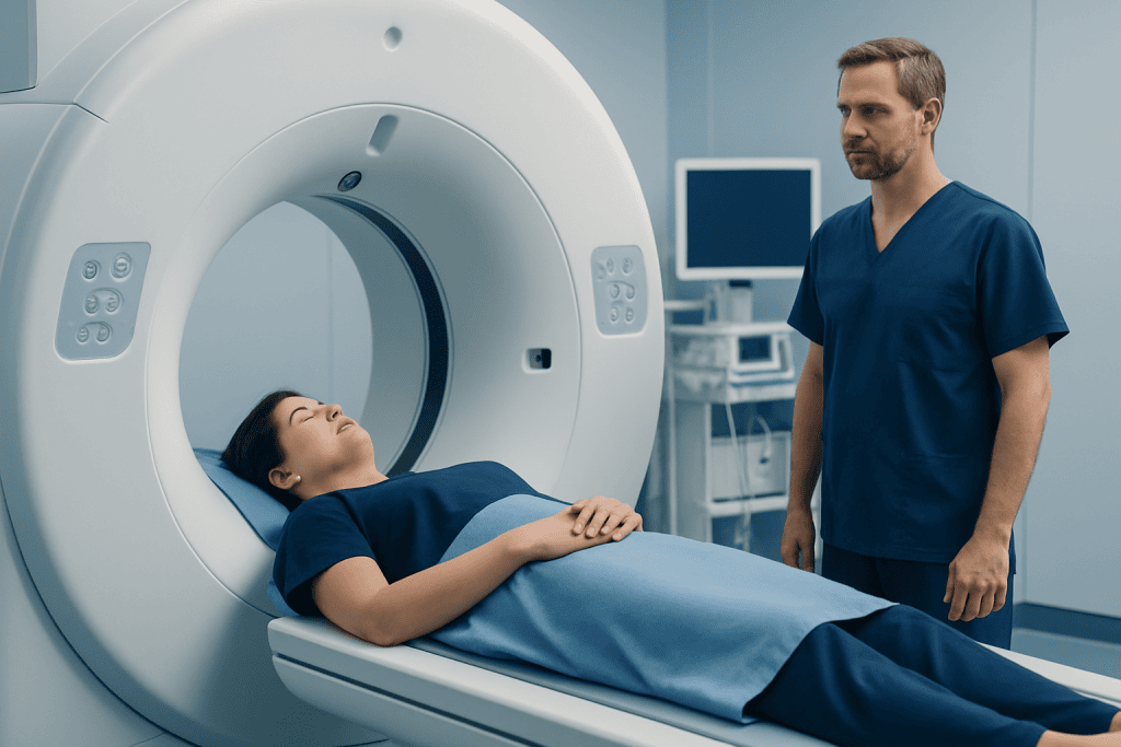
Several well-known conditions fall under the broad concept of carcinoma in situ. One example is ductal carcinoma in situ of the breast, often called DCIS, in which abnormal cells are limited to the milk ducts. Another is cervical carcinoma in situ, which may be detected through screening after changes caused by persistent human papillomavirus infection. Skin specialists may also use related terms for very early squamous cell changes confined to the epidermis.
Other organs can be affected as well. Urologists may diagnose carcinoma in situ in the bladder, where the abnormal cells are usually flat and limited to the bladder lining. Less commonly, similar in situ changes may be found in the mouth, larynx, or gastrointestinal tract during evaluation of suspicious lesions or abnormal screening findings.
Because different organs behave differently, doctors usually explain the diagnosis using both the general term and the organ name. For example, a patient may hear about breast cancer when discussing ductal carcinoma in situ, or cervical cancer when discussing cervical in situ disease. That organ-specific detail is what guides treatment planning and follow-up.
Symptoms and how it is often found

Many people with carcinoma in situ have no symptoms at all. The condition is frequently discovered through screening tests, routine checkups, or investigations for another concern. This is especially true for the cervix and breast, where Pap tests, HPV testing, mammography, and follow-up biopsies may detect cell changes before they cause noticeable problems.
When symptoms do occur, they depend on the site involved. Breast ductal carcinoma in situ usually does not cause a lump, but some people may have a breast change that leads to imaging. Cervical in situ disease often causes no warning signs, though some patients may report unusual bleeding or discharge that prompts assessment. Bladder carcinoma in situ may be associated with urinary irritation or blood in the urine, but these symptoms can also have many noncancerous causes.
Visible skin lesions may appear as a scaly, red, crusted, or slowly changing patch. In the mouth or throat, a persistent sore, white or red patch, or hoarseness may trigger further examination. Because symptoms are not specific, diagnosis cannot be made from symptoms alone; proper testing and biopsy are needed to confirm whether a lesion is carcinoma in situ or another condition.
Causes and risk factors
Carcinoma in situ develops when genetic and cellular changes build up in surface-lining cells, causing them to grow abnormally. These changes can happen for different reasons depending on the organ. In some cases, long-term irritation, inflammation, ultraviolet light exposure, viral infection, smoking, hormonal influences, age, or inherited susceptibility can play a role.
Examples help make this clearer. Persistent high-risk HPV infection is strongly linked to cervical precancer and carcinoma in situ. In the breast, age, hormonal exposure, dense breast tissue, and certain family history patterns may increase the chance of ductal changes. In the skin, chronic sun exposure and fair skin can raise risk for in situ squamous lesions. Smoking is an important risk factor for several epithelial cancers, including those affecting the bladder and airways.
Having a risk factor does not mean a person will definitely develop carcinoma in situ, and some patients diagnosed with it have few obvious risks. Doctors look at the whole picture, including age, family history, personal medical history, lifestyle factors, and any prior abnormal screening results. This broader assessment helps decide which tests are needed and how closely follow-up should be planned.
How diagnosis is confirmed
The diagnosis of carcinoma in situ is usually made in stages. It often begins with a screening test, physical examination, or imaging finding that shows an abnormal area. The next steps may include closer imaging, endoscopy, colposcopy, cystoscopy, or a minor procedure to sample the tissue. A pathologist then examines the cells under a microscope to determine whether they are benign, dysplastic, in situ, or invasive.
Biopsy is the key test because it shows whether abnormal cells remain confined to the surface layer. The pathology report may also describe the grade of the cells, whether the abnormality is focal or more widespread, and whether the edges of a removed sample are clear. These details help doctors understand how likely the lesion is to persist, recur, or progress if left untreated.
Additional tests depend on the site. Breast lesions may be assessed with diagnostic mammography, ultrasound, or MRI in selected cases, followed by breast biopsy if needed. Cervical changes may require colposcopy-guided sampling, and bladder lesions may be evaluated by cystoscopy and targeted biopsy. In centers caring for international patients, multidisciplinary review can be useful when pathology is complex or treatment choices are not straightforward.
Modern treatment approaches and outlook
Treatment for carcinoma in situ aims to remove, destroy, or carefully monitor abnormal cells before they become invasive. The best approach depends on the organ involved, the size and location of the lesion, pathology features, the patient’s age and general health, and personal priorities such as fertility preservation or cosmetic outcomes. In many cases, treatment is local rather than systemic because the cells are confined to the tissue where they started.
Common options include excision, ablation, endoscopic removal, local therapies, or organ-specific surgery. In the breast, management of DCIS may include breast-conserving surgery or mastectomy in selected situations, sometimes followed by radiation or hormone-related treatment depending on pathology and recurrence risk. Cervical in situ disease may be managed with procedures that remove the abnormal area while preserving the uterus when appropriate. Bladder carcinoma in situ may require local bladder treatments and close cystoscopic surveillance.
The outlook is often very good when carcinoma in situ is diagnosed early and treated appropriately. Even so, follow-up matters because some lesions can recur or coexist with nearby invasive disease that was not obvious at first. Doctors usually recommend an individualized surveillance plan, such as repeat imaging, screening, exams, or endoscopic checks. If a patient needs further evaluation or treatment planning for a suspicious lesion, oncology care may involve surgeons, pathologists, radiologists, and other specialists working together.
Near the end of the care journey, patients often want reassurance about where to go for coordinated treatment. Acibadem International’s multidisciplinary specialists in JCI-accredited hospitals diagnose and treat carcinoma in situ and related cancers for international patients, with care plans tailored to the organ involved and the pathology findings.
Living with the diagnosis and reducing future risk
Hearing the word carcinoma can be unsettling, even when the diagnosis is in situ rather than invasive. Many patients find it helpful to ask for a plain-language explanation of the pathology report, what the lesion can and cannot do at its current stage, and whether treatment is intended to cure, prevent progression, or reduce recurrence risk. Understanding these points often makes decision-making less overwhelming.
After treatment, self-care focuses on keeping follow-up appointments and supporting overall health. The exact plan depends on the organ involved, but patients are commonly advised to attend regular screening, report new symptoms promptly, and follow site-specific guidance for wound care or recovery after procedures. Smoking cessation, sun protection, maintaining a healthy weight, limiting alcohol when advised, and staying physically active can support general cancer prevention.
Some patients may benefit from discussing family history or inherited cancer risk, especially if there are multiple relatives with related cancers or diagnosis at younger ages. Emotional support is also important. Speaking with a clinician, counselor, or support group can help people cope with uncertainty while they move through treatment and surveillance.
When to seek medical care
Medical advice should be sought promptly after any abnormal screening result, because carcinoma in situ often causes few or no symptoms. Early follow-up gives doctors the best chance to confirm the diagnosis and discuss treatment before any deeper invasion develops. Delaying evaluation can make later management more complicated.
A person should also arrange medical review for persistent warning signs related to the affected organ. These may include a new or changing breast finding, abnormal vaginal bleeding, unexplained urinary blood, a skin patch that does not heal, a mouth sore lasting more than two weeks, or ongoing hoarseness or swallowing difficulty. Most of these symptoms have noncancerous causes, but they still deserve assessment.
Urgent care may be appropriate if symptoms are severe, such as heavy bleeding, significant pain, inability to pass urine, or rapid worsening of a suspicious lesion. A qualified doctor can decide which tests are needed and whether referral to a specialist is appropriate.
Frequently asked questions
Is carcinoma in situ considered cancer?
Carcinoma in situ is generally considered a very early, noninvasive form of cancer or a preinvasive cancer, depending on the organ and the classification system used. The key point is that the abnormal cells have not invaded deeper tissues.
Can carcinoma in situ spread to other parts of the body?
By definition, carcinoma in situ has not invaded nearby tissue, so it does not have the same ability to spread as invasive cancer. However, it can progress over time in some cases, which is why follow-up and treatment are important.
Does carcinoma in situ always need treatment?
Not always in exactly the same way, but it always needs proper specialist evaluation. Many cases are treated to prevent progression, while some selected lesions may be managed with careful surveillance depending on the organ, pathology, and patient factors.
What is the outlook for someone with carcinoma in situ?
The outlook is often favorable because the abnormal cells are still confined to the surface layer of tissue. Outcomes depend on the site, extent of disease, pathology findings, and whether appropriate follow-up and treatment are completed.
How is carcinoma in situ different from dysplasia?
Dysplasia describes abnormal cell growth that can range from mild to severe, while carcinoma in situ usually refers to the most advanced preinvasive stage before invasion occurs. The exact terminology can vary by organ and pathology system.
Can carcinoma in situ come back after treatment?
Yes, recurrence is possible in some organs, even after successful treatment. That is why doctors usually recommend scheduled follow-up visits, repeat imaging, or repeat screening tests based on the site involved.
References
- National Cancer Institute
- American Cancer Society
- World Health Organization
- National Comprehensive Cancer Network
- American Society of Clinical Oncology
This article is for general information only and is not a substitute for professional medical advice. Please consult a qualified doctor about your individual situation.
Oncology care in Turkey — second opinion and treatment plan
JCI-accredited · board-certified surgeons · reply within 24h
Add us as a Preferred Source to see more of our trusted health content across Google Search, AI Overviews and Discover.